
Ischemic heart disease
What aspect of ischemic heart disease are you most interested in? · What level of detail do you need?
ischemic heart disease coronary artery atherosclerosis diagram
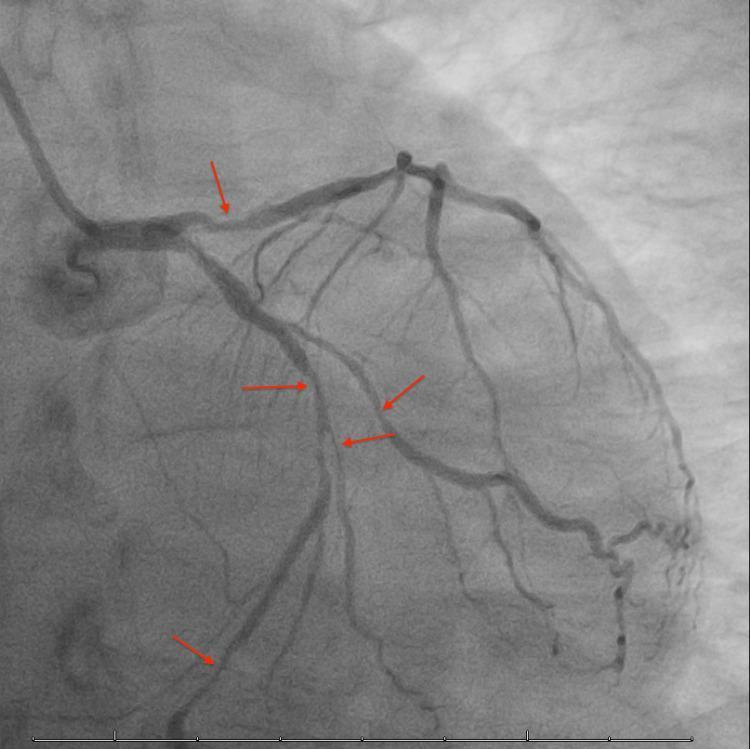
This diagnostic image is a coronary angiogram (left heart catheterization) showing the left coronary artery system. The visual focus is on the Left Anterior Descending (LAD) artery and the Left Circumflex (LCx) artery along with their diagonal and marginal branches. Multiple red arrows indicate areas of multifocal pathology, characterized by significant luminal narrowing and abrupt terminations of contrast opacification. Specifically, the image demonstrates diffuse atherosclerosis with severe segmentary stenoses (ranging from 60% to 90% in clinical context) and distal vessel occlusions. These findings are consistent with severe multivessel coronary artery disease (CAD), manifesting as compromised distal blood flow and reduced vessel diameter in both the LAD and LCx territories. This imaging is typically used in cardiology to evaluate ischemic heart disease and plan revascularization strategies.
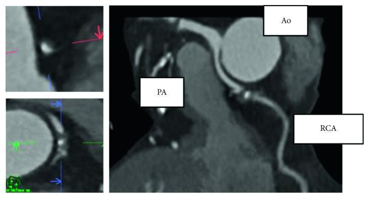
This diagnostic image consists of a large multiplanar reconstruction (MPR) and two smaller magnified views from a coronary computed tomography angiography (CCTA). The primary image demonstrates the spatial relationship between the aorta (Ao), pulmonary artery (PA), and the right coronary artery (RCA). The RCA is shown originating and coursing between the large circular aorta and the adjacent pulmonary artery. Pathological findings characteristic of atherosclerosis are highlighted within the RCA, specifically showing spotty calcification—indicated by focal, high-attenuation (bright) deposits within the vessel wall—and evidence of mild positive remodeling, characterized by a localized outward expansion of the arterial wall diameter at the site of the lesion. The magnified panels on the left provide detailed cross-sectional and longitudinal views of these atherosclerotic changes, emphasizing the eccentric plaque and calcified elements. This clinical image is used in cardiology to evaluate coronary artery anomalies and the morphological characteristics of coronary plaques in the context of ischemic heart disease.
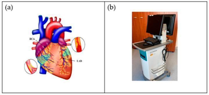
The visual content is composed of two educational panels focused on coronary artery disease (CAD) and intravascular ultrasound (IVUS) technology. Panel (a) is an anatomical diagram of a human heart illustrating the coronary circulation. It highlights the Right Coronary Artery (RCA) and the Left Anterior Descending (LAD) artery. Two inset circular cross-sections demonstrate the pathophysiology of atherosclerosis: one shows a healthy, patent vessel with a smooth lumen, while the other depicts a diseased vessel with yellow atherosclerotic plaque causing significant luminal narrowing (stenosis). The large vessels, including the aorta (red) and pulmonary artery (blue), provide anatomical context. Panel (b) is a clinical photograph of an IVUS acquisition system. The medical equipment is a mobile workstation featuring a high-resolution monitor for real-time cross-sectional vessel imaging, a keyboard/input console, and a wheeled base. This dual-panel image illustrates the clinical application of imaging technology in diagnosing and managing coronary atherosclerosis and guiding percutaneous coronary interventions (PCI).
Ischemic Heart Disease (IHD) - Clinical Summary
Definition
- In >90% of cases, IHD results from obstructive atherosclerotic coronary artery disease (CAD).
- The remaining cases involve increased demand (tachycardia, hypertension), reduced blood volume (shock), reduced oxygenation (pneumonia, CHF), or reduced oxygen-carrying capacity (anemia, CO poisoning).
Clinical Syndromes
| Syndrome | Key Feature |
|---|---|
| Stable angina | Chest pain predictably at exertion; >70% stenosis ("critical stenosis") |
| Unstable angina | Chest pain at rest or with less exertion; plaque disruption + non-occlusive thrombus |
| Vasospastic (Prinzmetal) angina | Coronary spasm, often at rest; ST elevation that resolves |
| NSTEMI | Troponin elevation, no ST elevation; subendocardial infarction |
| STEMI | Full-thickness (transmural) infarction; ST elevation + new LBBB |
| Sudden cardiac death (SCD) | Ventricular fibrillation from acute ischemia |
| Chronic IHD / ischemic cardiomyopathy | Progressive pump failure from cumulative ischemic insults |
Pathophysiology
-
Fixed atherosclerotic stenosis - plaques gradually narrow the lumen. Lesions <70% are typically asymptomatic. >70% causes stable angina. >90% may cause rest pain. If slow progression occurs, collaterals can form and protect against infarction.
-
Acute plaque change (most ACS) - rupture or erosion of a plaque exposes thrombogenic material, triggering rapid thrombus formation:
- Plaque disruption → platelet adhesion → thromboxane A2, ADP, serotonin release → platelet aggregation + vasospasm
- Tissue factor activation → coagulation cascade → occlusive thrombus
- Angiography within 4 hours of MI shows thrombotic occlusion in ~90% of cases
- Aerobic metabolism stops within seconds → ATP drops, lactate accumulates
- Contractility lost within minutes
- Irreversible necrosis after 20-40 minutes of sustained ischemia
- After 4-6 hours: gross infarct visible; peak cell death at 12-24 hours
Diagnosis
| Test | Key Finding |
|---|---|
| ECG | ST elevation (STEMI), ST depression/T-wave inversion (NSTEMI/UA), new Q waves (old MI) |
| High-sensitivity troponin (hs-cTnI/T) | Gold standard biomarker; rises within 1-3 h, peaks 12-24 h, returns to baseline in 7-10 days |
| CK-MB | Rises in 4-8 h, returns to normal in 48-72 h; useful for reinfarction detection |
| Echocardiography | Regional wall motion abnormalities; assesses LV function |
| Coronary angiography | Definitive anatomy; guide to PCI or CABG |
| CT coronary angiography | Non-invasive assessment of stenosis, plaque burden |
- Type I MI - spontaneous from plaque rupture (includes STEMI and NSTEMI)
- Type II MI - supply-demand mismatch (e.g., tachyarrhythmia, anemia)
- Myocardial injury - troponin elevation without ischemic mechanism
Risk Factors
- Modifiable: Smoking, hypertension, dyslipidemia, diabetes mellitus, obesity, sedentary lifestyle
- Non-modifiable: Age, male sex, family history
Management
Acute ACS (STEMI/NSTEMI)
| Drug | Role |
|---|---|
| Aspirin (162-325 mg load, then 81 mg daily) | Antiplatelet; immediate, indefinite |
| P2Y12 inhibitor (clopidogrel, ticagrelor, prasugrel) | Dual antiplatelet therapy (DAPT) |
| Anticoagulant (UFH, LMWH, fondaparinux) | All ACS patients unless active bleeding |
| Beta-blocker (metoprolol 25-50 mg PO) | Within 24 h; reduces ischemia and MI risk |
| Nitrates (sublingual / IV) | Symptomatic relief; avoid with PDE-5 inhibitors |
| Statin (atorvastatin 40-80 mg) | Immediately; indefinitely |
| ACE inhibitor/ARB | Post-MI, especially with reduced EF |
- STEMI: Primary PCI (goal door-to-balloon <90 min) is preferred; thrombolysis if PCI unavailable within 120 min
- High-risk NSTEMI: Early invasive strategy (PCI within 24-48 h); high-sensitivity troponin guides risk stratification
Stable Angina (Chronic IHD)
- Beta-blockers - first-line anti-anginal
- Long-acting nitrates - symptom control (nitrate-free interval to prevent tolerance)
- Calcium channel blockers (diltiazem, verapamil) - alternative or add-on
- Revascularization (PCI or CABG) - for refractory angina, left main disease, or 3-vessel disease with reduced EF
Complications of MI
| Complication | Timing |
|---|---|
| Ventricular fibrillation | First hours ("electrical storm") |
| Pump failure / cardiogenic shock | Days 1-3 |
| Papillary muscle rupture (mitral regurgitation) | Days 2-7 |
| Free wall rupture / tamponade | Days 3-7 |
| Ventricular septal defect (VSD) | Days 3-7 |
| Pericarditis (Dressler syndrome) | Weeks to months |
| LV aneurysm | Weeks (anterior MI, LAD territory) |
| Mural thrombus / embolism | Weeks |
Coronary Artery Anatomy Affected