
Write the Physiotherapy management of brown sequard syndrome refer sullivan
Brown Sequard syndrome physiotherapy management Sullivan Physical Rehabilitation
Brown Sequard syndrome spinal cord hemisection diagram sensory motor deficits
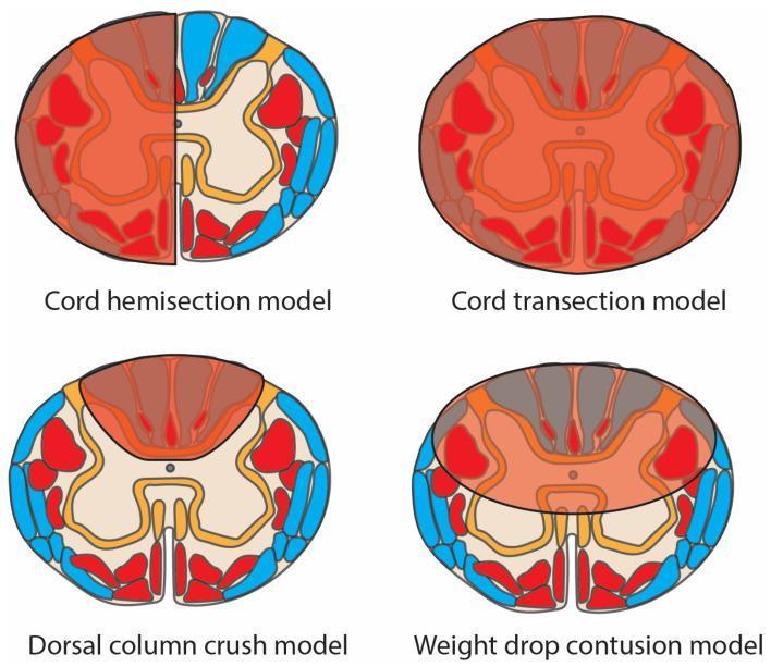
This medical schematic diagram illustrates four pre-clinical spinal cord injury (SCI) models in axial cross-section, highlighting damaged regions (translucent red overlay), motor tracts (opaque red), and sensory tracts (blue). 1. Cord Hemisection Model: Shows damage isolated to one lateral half of the spinal cord, characteristic of Brown-Séquard syndrome, affecting both motor and sensory tracts on the ipsilateral side. 2. Cord Transection Model: Depicts complete injury across the entire cord diameter, resulting in total disruption of all motor and sensory pathways. 3. Dorsal Column Crush Model: Features localized damage concentrated in the posterior (dorsal) funiculus, primarily affecting ascending sensory tracts. 4. Weight Drop Contusion Model: Displays a diffuse, central injury zone impacting the grey matter and surrounding white matter tracts, simulating clinical traumatic contusion. The diagram serves as an educational tool for comparing injury patterns across experimental models used in neurotrauma research and their resulting impact on anatomical tracts.
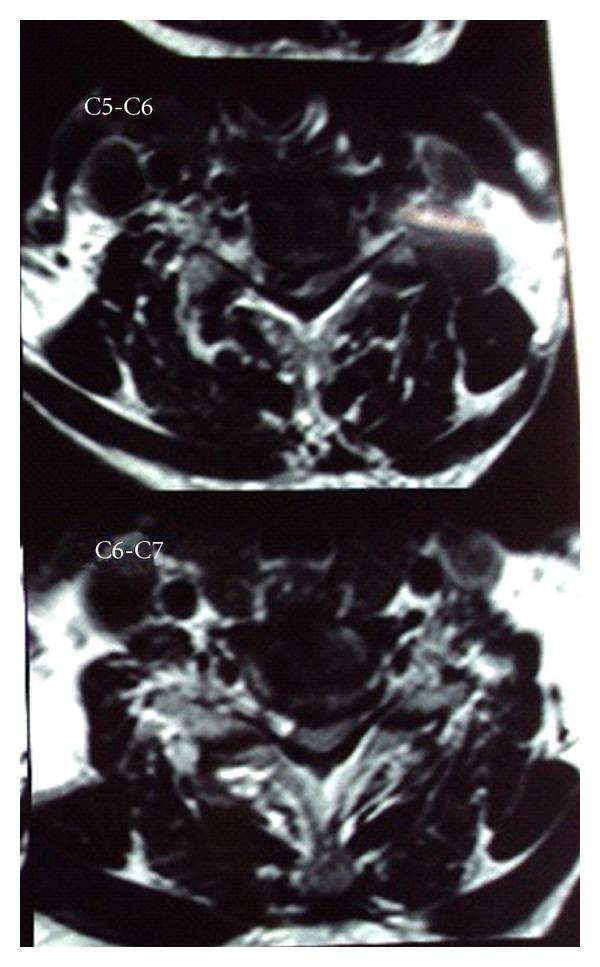
This diagnostic image consists of two axial T2-weighted magnetic resonance imaging (MRI) slices of the cervical spine, labeled at the C5-C6 and C6-C7 levels. The images demonstrate significant right paracentral disc herniation at both levels, resulting in pronounced hemicord compression. At the C5-C6 level, the herniated disc material is seen extruding into the spinal canal, causing asymmetric flattening and displacement of the spinal cord toward the left. Similar pathological findings are observed at the C6-C7 level, though the compression at C5-C6 appears more severe. The vertebral bodies and surrounding soft tissues show signs of degenerative spondylosis. The visual evidence of unilateral cord compression is a classic radiological correlate for Brown-Séquard syndrome, illustrating the mechanical etiology of ipsilateral motor loss and contralateral sensory deficits. This material is suitable for intermediate to advanced medical learners focusing on neuroradiology, orthopedics, or neurology.
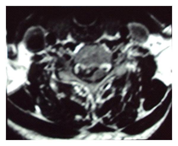
This diagnostic image is an axial T2-weighted MRI of the cervical spine at the C4-C5 level. The scan demonstrates a significant right paracentral disc herniation protruding into the spinal canal. The herniated disc material causes focal mass effect on the right side of the spinal cord, resulting in evident hemicord compression and distortion of the cord's normal elliptical contour. Despite the mechanical compression, the signal intensity within the spinal cord remains relatively uniform, with no definitive T2 hyperintensity to suggest acute myelomalacia or cord edema at this level. The vertebral body and posterior elements are visualized, though anatomical detail is slightly limited by image graininess. This clinical finding correlates with Brown-Séquard Syndrome, characterized by ipsilateral motor deficits and contralateral sensory loss due to unilateral cord compression. This image serves as a key educational example of how a focal disc protrusion can cause significant mechanical deformation of the spinal cord and lead to specific neurological deficits.
Physiotherapy Management of Brown-Sequard Syndrome
1. Review of the Syndrome (Clinical Basis for PT Planning)
| Side | Deficit |
|---|---|
| Ipsilateral (same side as lesion) | UMN lesion - spastic paresis/paralysis; loss of proprioception, vibration, discriminative touch (dorsal column) |
| Contralateral (opposite side) | Loss of pain and temperature sensation (spinothalamic tract, 1-2 levels below lesion) |
2. Goals of Physiotherapy Management (Sullivan's Framework)
- Prevent secondary complications
- Maintain/improve ROM and prevent contractures
- Strengthen intact/recovering musculature
- Promote functional independence in ADLs
- Achieve highest level of mobility (ambulation where possible)
- Patient and caregiver education
3. Phases of Physiotherapy Management
Phase I - Acute/Immobilization Phase
- Prevent complications: pressure ulcers, DVT, pneumonia, contractures, muscle atrophy
- Maintain cardiorespiratory function
- Prevent muscle shortening on the spastic side
- Regular repositioning every 2 hours to prevent pressure ulcers
- Anti-spasticity positioning: avoid prolonged hip and knee flexion
- Careful alignment of the paretic limbs using pillows/foam wedges
- Elevation of limbs to prevent dependent oedema
- Full ROM to all joints of the paretic limbs at least twice daily
- Special attention to shoulder (flexion, abduction, external rotation), wrist extension, hip extension, knee extension, and ankle dorsiflexion
- Splinting/serial casting may be used for the paretic ankle to prevent equinus contracture
- Diaphragmatic breathing exercises
- Assisted coughing if cervical lesion compromises respiratory muscles
- Incentive spirometry
- Postural drainage if secretions accumulate
- Tactile and proprioceptive stimulation to the ipsilateral paretic limb to promote cortical re-mapping
- Awareness training: educating the patient about the contralateral sensory loss (pain/temperature) to prevent burns and injuries
Phase II - Subacute Rehabilitation Phase
- Develop trunk stability and balance
- Progress from bed mobility to sitting and transfers
- Strengthen recovering muscles
- Introduce mat activities and developmental sequence
-
Rolling - Begin with log rolling; progress to segmental rolling using head/neck/upper trunk initiation. The patient learns to use the stronger contralateral limb to assist the weak ipsilateral side.
-
Prone on elbows - Develops shoulder girdle stability, scapular control, and weight-bearing through the paretic upper limb.
-
Quadruped/Crawling position - Promotes bilateral limb weight-bearing; trains trunk and proximal limb stability.
-
Sitting (short-sitting and long-sitting) - Trunk balance training in gravity-eliminated and gravity-resisted positions. Dynamic sitting balance is practiced with perturbations and reach activities.
-
Kneeling and half-kneeling - Progressively loads the paretic lower limb; develops hip extensor and quadriceps strength in weight-bearing posture.
-
Standing - Progressed from parallel bars, to free standing, to stepping.
- Ipsilateral paretic side: Progressive resistive exercises as motor return begins. Neuromuscular facilitation techniques (PNF) - particularly rhythmic initiation and repeated contractions for weak muscles.
- Contralateral intact side: Maintain and strengthen as it will take on compensatory functional roles.
- Trunk: Core stabilization exercises - bridging, pelvic tilts, and dead bug exercises in supine; progressed to sitting and standing trunk exercises.
- D1 and D2 upper extremity patterns for shoulder, elbow, wrist
- D1 and D2 lower extremity patterns for hip, knee, ankle
- Techniques used:
- Rhythmic initiation - for initiation of movement in severely weak muscles
- Repeated contractions - for strengthening throughout range
- Hold-relax active movement - for improving ROM and reducing spasticity
- Rhythmic stabilisation - for improving trunk and proximal joint stability
- Slow passive stretching held for 20-30 seconds to the spastic muscles (typically hip flexors, knee flexors, ankle plantar flexors on the ipsilateral side)
- Weight-bearing through the paretic limb in standing to inhibit spasticity via prolonged stretch
- Neutral warmth and cold modalities as adjuncts
- Inhibitory casting or splinting if necessary
- Coordination with physician for pharmacological management (baclofen, tizanidine)
- Because pain and temperature are lost contralaterally, the patient must learn compensatory strategies - visual inspection of the limbs, using the ipsilateral side for temperature testing
- Graded sensory input for proprioceptive deficits on the ipsilateral side: texture discrimination, two-point discrimination, vibration awareness training
Phase III - Ambulation Training
- Adequate trunk control and sitting balance
- Sufficient proximal lower limb strength (minimum Grade 3/5 in key muscle groups)
- Adequate cardiovascular endurance
- Weight shifting side to side and anterior-posterior
- Step-through and step-to gait pattern
- Bilateral limb weight acceptance
- Ipsilateral side: Spastic gait pattern - hip circumduction, knee stiffness, foot drop (equinovarus); reduced stride length
- Contralateral side: Ataxic/sensory gait pattern due to loss of pain/temperature feedback
- Foot drop: Ankle-foot orthosis (AFO) on the ipsilateral paretic side
- Circumduction: Strengthen hip flexors and dorsiflexors; functional electrical stimulation (FES) may assist
- Sensory ataxia (contralateral): Compensatory visual strategies; surface progression (from even to uneven terrain)
- Parallel bars → Walker → Forearm crutches → Quad cane → Single-point cane → No device (when possible)
- The walking aid prescription depends on upper limb strength and balance
- Walking on inclines, stairs (step-over-step technique)
- Walking on uneven terrain
- Community ambulation - crossing roads, shopping, transportation
- Treadmill training with partial body weight support (BWSTT) - recommended by current SCI guidelines (intensity: moderate to high); promotes neuroplasticity via repetitive stepping
- For upper extremity: Restraint of the stronger contralateral arm forces the use of the weaker ipsilateral arm
- Massed practice of 6 hours/day over 2 weeks promotes cortical reorganization
Phase IV - Functional Activities and Discharge Planning
- Bed-to-wheelchair, wheelchair-to-toilet, wheelchair-to-car
- Sliding board transfers initially; progress to stand-pivot transfers as lower limb strength returns
- Propulsion, pressure relief lifts, ascending/descending curbs
- Dressing, grooming, feeding
- Adaptive equipment as needed
- Independent exercise program for stretching, strengthening, and walking
- Patient and family education: skin inspection, bowel/bladder management, fall prevention due to contralateral sensory loss
- Removal of throw rugs, adequate lighting
- Grab bars in bathroom, ramp access if needed
- Caution regarding hot water scalding and sharp objects (due to contralateral pain/temperature loss)
4. Special Considerations in BSS
| Issue | PT Approach |
|---|---|
| Neuropathic pain | TENS, heat/cold (carefully - sensory deficits), graded motor imagery, desensitization |
| Bladder/bowel | Timed voiding, pelvic floor exercises, coordination with nursing |
| Sexual dysfunction | Education, referral to specialist |
| Psychological | Motivation, goal-setting, peer support groups |
| DVT prevention | Early mobilization, compression stockings, ankle pumps |
| Orthostatic hypotension | Tilt-table training, abdominal binders, gradual head-up tilt |
5. Outcome Measures Used (Sullivan)
- ASIA Impairment Scale - Baseline and follow-up neurological classification
- FIM (Functional Independence Measure) - Functional progress
- Berg Balance Scale - Balance assessment
- 10-Meter Walk Test / 6-Minute Walk Test - Gait performance
- Spinal Cord Independence Measure (SCIM) - Self-care, respiration, mobility
- Modified Ashworth Scale - Spasticity grading
- Manual Muscle Testing (MMT) - Muscle strength grading
6. Prognosis
- BSS has the best prognosis of all incomplete SCI syndromes
- ~90% of patients regain functional ambulation within 6 months if motor return begins early
- Bowel and bladder continence is typically regained
- Upper extremity function recovers well, especially when the upper limbs are not at the level of injury
- Neurological recovery may continue for up to 2 years post-injury