
Gustofon method for age determination
The Gustafson method (1950) estimates age from dental changes in extracted teeth by scoring six morphological criteria on a scale of 0-3, then applying a regression formula. The six features are: attrition, secondary dentin, periodontosis, cementum apposition, root resorption, and root transparency.
Searching sources...
Reading File
Searching the Web
Gustafson method age determination teeth forensic dentistry
Finding Sources
Searching PubMed
Gustafson method dental age estimation forensic
Finding Sources
Finding Sources
Finding Sources
I found content in two forensic medicine textbooks. Let me read both sections fully.
Reading File
Reading File
Reading File
Reading File
Now let me get the regression formula and the remaining details from the Dikshit textbook, plus the Essentials of Forensic Medicine MCQ answers.
Reading File
Finding Sources
Now let me fetch the Gustafson diagram image from the textbook.
Loading Image
I now have comprehensive information from two forensic medicine textbooks plus the classic diagram. Here is the full answer:
Gustafson's Method of Age Determination
Introduced by Gösta Gustafson, a Swedish stomatologist, in 1950 (published in J Am Dent Assoc 41:45-54), this is the standard forensic method for estimating age in adults by studying progressive degenerative changes in an individual extracted tooth.
This method is applicable only to dead bodies. It requires extraction and ground sectioning of the tooth (destructive method).
The Six Criteria (A-P-S-R-C-T)
Each criterion is scored on a scale of 0 to 3 based on degree of change:
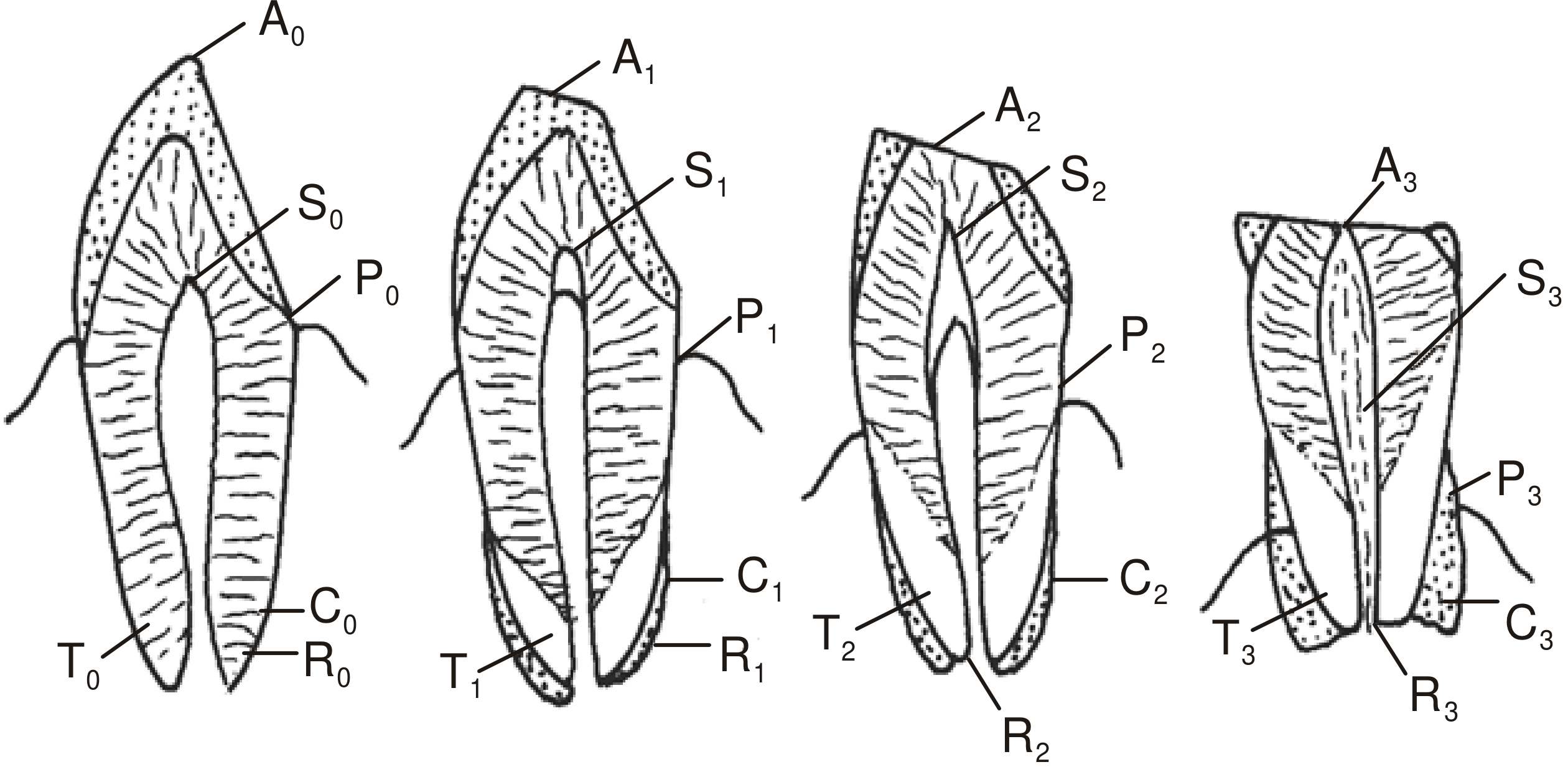
1. Attrition (A) - Wearing of tooth surface
Wear on the opposing mastication surfaces due to continuous friction:
| Grade | Meaning |
|---|---|
| A0 | No attrition |
| A1 | Attrition within enamel only |
| A2 | Attrition reaches dentine |
| A3 | Attrition exposes soft pulp |
2. Periodontosis (P) - Gum/periodontal recession
Regression of gums with root exposure due to poor maintenance:
| Grade | Meaning |
|---|---|
| P0 | No periodontosis / no root exposure |
| P1 | Exposure of less than 1/3rd of root near crown |
| P2 | Exposure >1/3rd but <2/3rd of root |
| P3 | Periodontosis beyond 2/3rd of root length |
3. Secondary Dentine (S) - Pulp cavity reduction
With age, secondary dentine deposits progressively fill the pulp cavity. In mandibular teeth it starts from above; in maxillary teeth from below:
| Grade | Meaning |
|---|---|
| S0 | No deposition |
| S1 | Slight deposition |
| S2 | Deposition in half of pulp cavity (from above) |
| S3 | Almost entire pulp cavity involved |
4. Root Resorption (R) - Decay at root apex
Involves both cementum and dentine; starts at apex and extends upward:
| Grade | Meaning |
|---|---|
| R0 | No root resorption |
| R1 | Seen in only some parts |
| R2 | Seen over a large area |
| R3 | Involves both cementum and dentine |
5. Cementum Apposition (C) - Incremental cementum layers
Cementum deposition increases on the root surface with age, forming incremental lines:
| Grade | Meaning |
|---|---|
| C0 | Only normal layer of cementum present |
| C1 | Slightly greater than normal |
| C2 | Thick layer present |
| C3 | Heavy layer present |
6. Root Transparency (T) - Dentinal tubule obliteration
Occurs due to gradual mineralization of peritubular dentine obliterating dentinal tubules. In mandibular teeth it progresses root-apex upward; in maxillary teeth from above downward. Typically does not appear before age 18:
| Grade | Meaning |
|---|---|
| T0 | No transparency anywhere |
| T1 | Transparency mostly over apical region |
| T2 | Transparency up to 1/3rd from apex |
| T3 | Transparency up to 2/3rd of root length from apex |
Scoring & Age Formula
All six criteria are summed:
An + Pn + Sn + Rn + Cn + Tn = Total points
Total points range from 0 (young) to 18 (very old). These are plotted on a regression graph comparing the score to teeth of known age.
Gustafson's original regression equation:
Age = 11.43 + 4.56 × (total points)
The estimated age is usually within an error of ±3.5 years.
Gustafson found a strong correlation (r = 0.98) between dental microstructural features and age.
Most Reliable Criterion
Root transparency (T) is the single most reliable indicator for age estimation. Multiple authors have confirmed it is the feature most closely correlated to chronological age. Secondary dentine deposits are the second best indicator.
The MCQ answer from Essentials of Forensic Medicine & Toxicology confirms: root transparency = most reliable criterion.
Procedure
- Before extraction, record the degree of periodontosis
- Examine tooth with the naked eye
- Grind tooth on a glass slab to ~1 mm - observe transparency macroscopically
- Further grind to ~0.25 mm (1/4 mm) for microscopic examination
- Score all six criteria
- Sum the points and apply the regression formula or graph
Preferred Teeth
Anterior teeth (incisors, canines) are most suitable because they erupt early and show changes earlier than posterior teeth.
Advantages
- Considers multiple features simultaneously
- Uses information from several teeth
- High correlation with actual age (r = 0.98)
Limitations
- Destructive method (tooth must be sectioned)
- Requires thorough knowledge of dental histology
- Poor oral health overestimates age (gives higher age-at-death estimates)
- Not applicable to living persons
- Pathological conditions can alter each factor independently
Modifications
Several authors have proposed improvements to Gustafson's original statistical methodology, including Johanson (1971), Kashyap and Koteswara Rao (1990), Lamendin et al. (1992), and Prince and Ubelaker (2002), who developed non-destructive methods based on root translucency measured on intact teeth.
Sources: PC Dikshit Textbook of Forensic Medicine and Toxicology; The Essentials of Forensic Medicine and Toxicology, 36th ed. (2026); Forensic Anthropology: A Comprehensive Introduction, 2nd ed.
Various methods of age identication
Age identification (estimation) uses physical, skeletal, dental, and radiological markers. Broadly: in children, dental eruption and bone ossification are most reliable; in adults, skeletal changes, pubic symphysis morphology, and dental wear are used. Methods span clinical examination, radiology, and forensic autopsy.
Searching sources...
Finding Sources
Finding Sources
Finding Sources
Finding Sources
Finding Sources
Finding Sources
Finding Sources
Finding Sources
Let me now read the main forensic medicine textbook sections on age estimation comprehensively.
Reading File
Reading File
Reading File
Reading File
Reading File
Reading File
Reading File
Reading File
Reading File
Reading File
Reading File
Reading File
I now have comprehensive data from both textbooks. Let me compile the complete answer.
Methods of Age Identification (Estimation)
Age identification in forensic medicine uses a combination of clinical, anthropological, dental, radiological, and biochemical methods. They are broadly divided by the age group being assessed.
I. GENERAL / EXTERNAL CLINICAL METHODS
A. Body Measurements
- Height/weight/head circumference - compared to standard growth charts in children
- Crown-rump length - used in fetuses (1 month = 1 cm, up to 3 months; thereafter length in cm = gestational age in months × 5)
B. Secondary Sexual Characteristics (Tanner Staging)
Hair development (both sexes):
| Feature | Male | Female |
|---|---|---|
| Pubic hair | 14-15 years | 13-14 years |
| Axillary hair | 15-16 years | 14-15 years |
| Beard/moustache | 16-17 years | Absent |
| Graying of scalp hair | ~40 years | ~40 years |
| Graying of pubic hair | ~55 years | ~55 years |
Tanner Stages of Pubic Hair:
| Stage | Description | Age |
|---|---|---|
| 1 | Prepubertal - fine vellus hair | <12 years |
| 2 | Sparse, lightly pigmented | 12-13 years |
| 3 | Darker, coarse, spreading to mons | 13-14 years |
| 4 | Adult type, not reaching thighs | 14-15 years |
| 5 | Dense, extending to inner thighs | >15 years |
Other secondary characteristics:
- Female breast development (Tanner Stages 1-5): begins ~11-13 years, adult form by ~15-16 years
- Menarche - average 13-14 years; indicates female >14 years
- Menopause - around 45-50 years
II. DENTAL METHODS
A. Tooth Eruption (Children - most reliable in <14 years)
Deciduous dentition (20 teeth):
| Tooth | Eruption |
|---|---|
| Lower central incisor | 5-6 months |
| Upper central incisor | 6-7 months |
| Upper lateral incisor | 7-8 months |
| Lower lateral incisor | 8-9 months |
| Canines | 1½ years |
| First molar | 1 year |
| Second molar | 20-30 months |
Permanent dentition (32 teeth):
| Tooth | Age of Eruption |
|---|---|
| Lower central incisor | 7-8 years |
| Upper central incisor | 7-8 years |
| Lateral incisors | 8-9 years |
| Canines | 11-12 years |
| First premolar | 9-10 years |
| Second premolar | 10-11 years |
| First molar | 6-7 years |
| Second molar | 12-14 years |
| Third molar (wisdom) | 17-25 years |
Number of teeth at a given age:
- 5 years: 20 (all temporary)
- 6 years: 21-24 (mixed dentition, first permanent molar erupts)
- 7-11 years: 24 (mixed dentition)
- 12-14 years: 24-28 (second molar erupts)
- 14-17 years: 28 (no new eruption)
- 17-25 years: 32 (wisdom tooth erupts)
Teeth erupt about 1 year earlier in females. Eruption occurs first in the lower jaw (except lateral incisors, which erupt first in the upper jaw). First permanent molar erupts on the lower left first.
B. Gustafson's Method (Adults - for extracted teeth)
Used in adults when eruption changes are complete. Based on 6 degenerative changes (each scored 0-3):
- A - Attrition
- P - Periodontosis
- S - Secondary dentine deposition
- R - Root resorption
- C - Cementum apposition
- T - Root transparency (most reliable single indicator)
Formula: Age = 11.43 + 4.56 × (A+P+S+R+C+T)
Accuracy: ±3.5 years. Applicable only to dead bodies (destructive method).
C. Root Transparency Method (Miles, 1963)
Anterior teeth ground to 1 mm, placed on dotted paper. Dots visible through transparent root counted. Percentage used to calculate age.
D. Tooth Calcification (Nolla's / Demirjian's Method)
Radiographic staging of tooth mineralization - particularly useful in children aged 3-14 years. Less affected by systemic factors than eruption.
III. SKELETAL / OSSIFICATION METHODS
A. Fetal Age - Ossification Centers
Key ossification centers in late fetal development:
| Center | Gestational Week/Month |
|---|---|
| Calcaneum, ischium | End of 5th month |
| Talus | End of 7th month |
| Last sacral vertebra | End of 8th month |
| Lower end of femur (Béclard's point) | End of 9th month (36 weeks) |
| Upper end of tibia, cuboid | End of 10th month (full term) |
Béclard's point (lower epiphysis of femur) = present at full term birth = important sign of a mature born-alive fetus.
B. Epiphyseal Union (Children and Young Adults - up to ~25 years)
- At 11-12 weeks intrauterine life: 806 ossification centers; at birth: ~450
- Adult has 206 bones
- Epiphyseal union is studied up to 20-22 years
- Radiological union precedes anatomical union by ~3 years
- In females, union occurs 1-2 years earlier than males
- Ossification occurs earlier in tropical climates vs temperate zones
Key late-fusing sites (for young adult estimation):
- Medial clavicle epiphysis - last growth plate to fuse (16-30 years; well-defined flake 16-21 yrs; complete fusion 22-30 yrs)
- S1-S2 sacral fusion - late fusion, useful for young adults (~18-30 yrs)
- Third molar eruption - ~18 years indicator
McKern & Stewart Maturation Scoring (1957): Scores 7 combinations of epiphyseal segments; total fed into prediction equation for more accurate estimation.
C. Post-22 Years - Skull Suture Closure
Studied when ossification centers can no longer be used. Used up to ~50 years but less precise:
- Spheno-occipital synchondrosis (basilar suture) - fuses around puberty (recent studies); located between sphenoid and occipital at skull base
- Vault sutures (coronal, sagittal, lambdoid) - assessed for degree of fusion (open → beginning fusion → >50% bridging → obliteration)
- Generally unreliable alone; used in combination with other methods
D. Pubic Symphysis (Adults)
One of the most commonly used adult skeletal aging methods:
Todd's Method (1920) - Original 10-phase system
Suchey-Brooks Method (1990) - 6 phases based on 1225 known individuals from Los Angeles. Separate standards for males and females. Most widely used today.
Hartnett Method (2010) - 7 phases, narrower age ranges, uses tactile characteristics
Morphological changes tracked:
- Surface texture (granular → smooth → porous)
- Rim and margin formation
- Dorsal and ventral arc changes
- Marginal lipping (osteophytes in old age)
E. Auricular Surface of Sacroiliac Joint
Lovejoy et al. (1985) - 8 phases, each covering a 5-year range. Significant variation makes it less reliable alone.
F. Sternal End of Right 4th Rib (İşcan Method)
Progressive pit formation and trabecular changes at the costo-sternal junction. Staged into phases correlated with age.
G. Changes in Specific Bones
Assessed in older age (>40 years):
- Mandible: Angle becomes obtuse (>120°) with age and tooth loss; ramus height decreases
- Sternum: Union of segments; xiphoid ossification
- Vertebrae: Degenerative disc disease, osteophytes
- Skull vault sutures: Progressive obliteration
IV. RADIOLOGICAL METHODS
- X-ray of wrist and hand - bone age vs chronological age (Greulich-Pyle atlas)
- X-ray of elbow, shoulder, knee, hip - epiphyseal fusion staging
- Dental X-ray / OPG - tooth calcification stages (Nolla, Demirjian)
- CT scan of skull base - spheno-occipital synchondrosis
- Skeletal survey for multiple ossification centers simultaneously
V. BIOCHEMICAL / MOLECULAR METHODS (Advanced Forensic)
- Aspartic acid racemization in tooth enamel/dentin - highly accurate, used in skeletal remains; based on the L→D amino acid conversion rate that increases with age
- Cementum annuli (tooth rings) - thin sections of root cementum counted like tree rings; each annulus = 1 year. Accurate to ±2-3 years
- Telomere length - shorter with age; experimental
- DNA methylation (epigenetic clock) - increasingly used in forensic contexts
VI. FETAL AGE ESTIMATION SUMMARY
| Month | Length | Key Features |
|---|---|---|
| 1 | 1 cm | Eyes as dark spots |
| 2 | 4 cm | Placenta forms |
| 3 | 9 cm | Nails appear, pupillary membrane |
| 4 | 16 cm | Sex differentiated |
| 5 | 25 cm | Ossification of calcaneum, ischium |
| 6 | 30 cm | Eyelashes, eyebrows |
| 7 | 35 cm | Eyelids open, testes at external ring |
| 8 | 40 cm | Toes - nails reach tips; S4-S5 fused |
| 9 | 45 cm | Lower femoral epiphysis (Béclard's) |
| 10 | 50 cm | Full term; upper tibial + cuboid centers |
Summary Table: Methods by Age Group
| Age Group | Best Methods |
|---|---|
| Fetus | Crown-rump length, ossification centers, organ development |
| 0-5 years | Dental eruption (deciduous), ossification |
| 5-14 years | Dental eruption (permanent), epiphyseal appearance |
| 14-25 years | Epiphyseal union, third molar, medial clavicle, S1-S2 fusion |
| 25-50 years | Pubic symphysis, auricular surface, 4th rib, skull sutures, Gustafson's |
| >50 years | Skeletal degenerative changes, skull sutures, Gustafson's, cementum annuli |
| All adults | Gustafson's method (dead bodies only), aspartic acid racemization |
Sources: PC Dikshit Textbook of Forensic Medicine and Toxicology; Forensic Anthropology: A Comprehensive Introduction, 2nd ed.; The Essentials of Forensic Medicine and Toxicology, 36th ed. (2026)
This is a shared conversation. Sign in to Orris to start your own chat.