
Chloroma
chloroma myeloid sarcoma
Chloroma (Granulocytic Sarcoma / Myeloid Sarcoma)
Definition and Nomenclature
- Andrews' Diseases of the Skin, p. 3099
- Henry's Clinical Diagnosis and Management by Laboratory Methods
Associated Conditions (Four Clinical Settings)
- Known AML - the most common association; occurs in ~3% of patients with myelogenous leukemia
- CML or MDS - as a harbinger of an impending blast crisis
- De novo presentation - in a previously undiagnosed patient, as the first sign of AML
- Post-bone marrow transplantation (BMT) - as the initial sign of relapse
- Andrews' Diseases of the Skin, p. 3097
- Henry's Clinical Diagnosis and Management by Laboratory Methods
Epidemiology
- Incidence: 2.5-9.1% of AML cases on imaging series
- Orbital myeloid sarcoma in children typically presents around age 7 years
- AML overall has a median age of 65 years, but chloromas can occur at any age
Sites of Involvement
| Site Category | Examples |
|---|---|
| Bone / Periosteum | Skull, spine, ribs, sternum (most common on imaging) |
| Soft tissue | Subcutaneous nodules |
| Skin | 20-50% of reported cases |
| Orbit / Eye | Rapid-onset proptosis, bilateral, lid edema, ecchymosis |
| ENT / Head & Neck | Temporal bone, nasopharynx, upper respiratory tract |
| Lymph nodes | - |
| Viscera | Breast, ovary, perineural/epidural structures |
| Pleura | Pleural thickening mimicking pleural effusion |
- Cummings Otolaryngology Head and Neck Surgery
- Kanski's Clinical Ophthalmology, p. 6782
- Grainger & Allison's Diagnostic Radiology
Skin Lesions
- Red, mahogany, or violaceous firm nodules
- Predilection for the face, scalp, and trunk
- May be solitary or multiple
Microscopy and Pathology
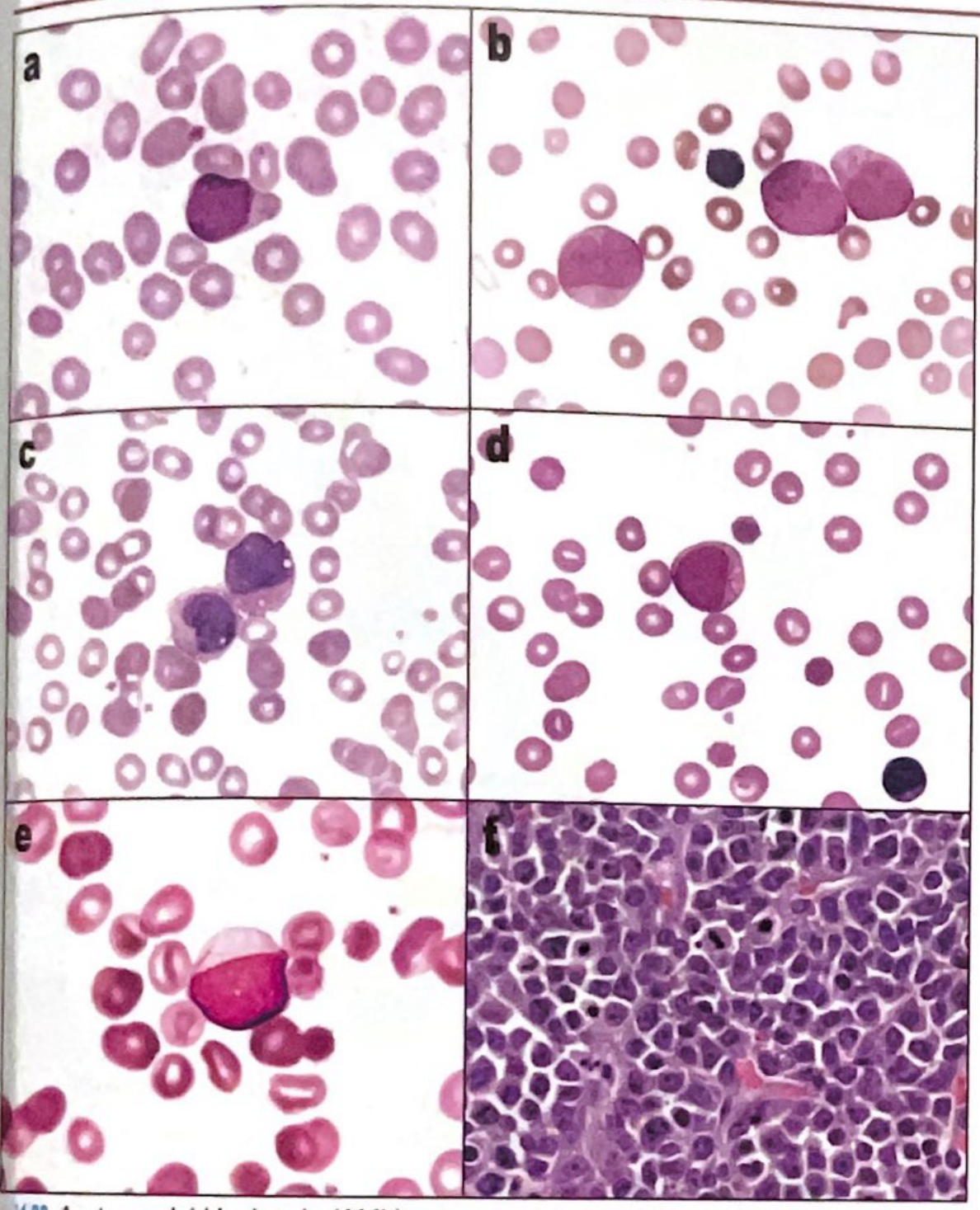
- Myeloblasts have more nuclear complexity and more cytoplasm (with granules and vacuoles) compared to lymphoblasts
- Auer rods - pathognomonic needle-like cytoplasmic inclusions; the only specific morphologic finding in AML
- Sheets of immature cells infiltrating extramedullary tissue
Diagnostic Pitfalls
- Non-Hodgkin lymphoma (most common error)
- Amelanotic melanoma
- Undifferentiated carcinoma
Immunophenotyping
- Anti-MPO (myeloperoxidase) - granulocytic lineage
- CD68 - monocytic lineage
- HLA-DR - positive in most AML subtypes (exception: acute promyelocytic leukemia/APL is HLA-DR negative)
- CD13, CD33 - myeloid markers
- CD19 - typically negative (anomalous expression in AML with t(8;21))
Radiology (MRI Features)
-
Chloroma manifests as an extra-osseous mass:
- Isointense to muscle on T1-weighted sequences
- Hyperintense to muscle on PD-weighted / T2-weighted sequences
- Avid enhancement following contrast administration
-
Most common skeletal locations in children: skull, spine, ribs, sternum
-
Pattern can mimic Ewing sarcoma (diffuse, permeative bone destruction)
-
FDG-PET shows focal/diffuse increased uptake
-
Grainger & Allison's Diagnostic Radiology
CML Blast Crisis Context
-
20% blasts in blood or marrow, OR
-
A tissue infiltrate of blasts (chloroma), OR
-
Prominent focal accumulation of blasts in the marrow biopsy
-
Quick Compendium of Clinical Pathology, 5th ed.
Management
- Local irradiation for symptomatic/localized disease
- Systemic chemotherapy as for AML (the diagnosis of a chloroma is considered equivalent to AML)
- Bone marrow transplantation is the definitive treatment for eligible patients
- Marrow involvement usually follows if not already antecedent - patients with de novo chloroma must be treated systemically
Recent Evidence
Gall bladder mass FNAC features with diagram
gallbladder carcinoma fine needle aspiration cytology
gallbladder carcinoma FNAC cytology features adenocarcinoma smear findings
Medical educational diagram showing FNAC (Fine Needle Aspiration Cytology) features of gallbladder adenocarcinoma. Show a cytology smear with labeled features: 1) Malignant epithelial cells in clusters and sheets, 2) Acinar/glandular arrangements, 3) Pleomorphic nuclei with coarse chromatin, 4) Prominent macronucleoli, 5) High N:C ratio, 6) Nuclear molding, 7) Dirty necrotic background (tumor diathesis), 8) Mucin vacuoles in cytoplasm, 9) Isolated atypical cells. Include a legend and labels. Clean medical illustration style with H&E stain appearance simulation.
https://cytojournal.com/gallbladder-carcinoma-an-attempt-of-…
Gallbladder Mass - FNAC Features
Background
- Grainger & Allison's Diagnostic Radiology
- Schwartz's Principles of Surgery, 11th ed.
Gross Pathology (for context)
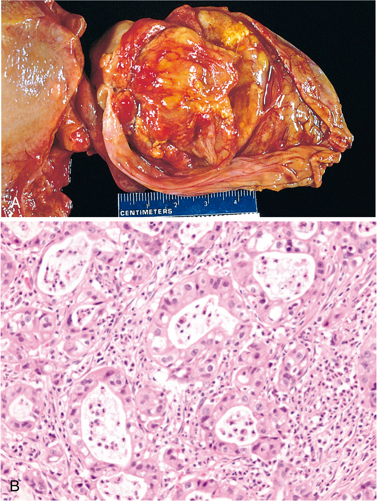
FNAC Diagram
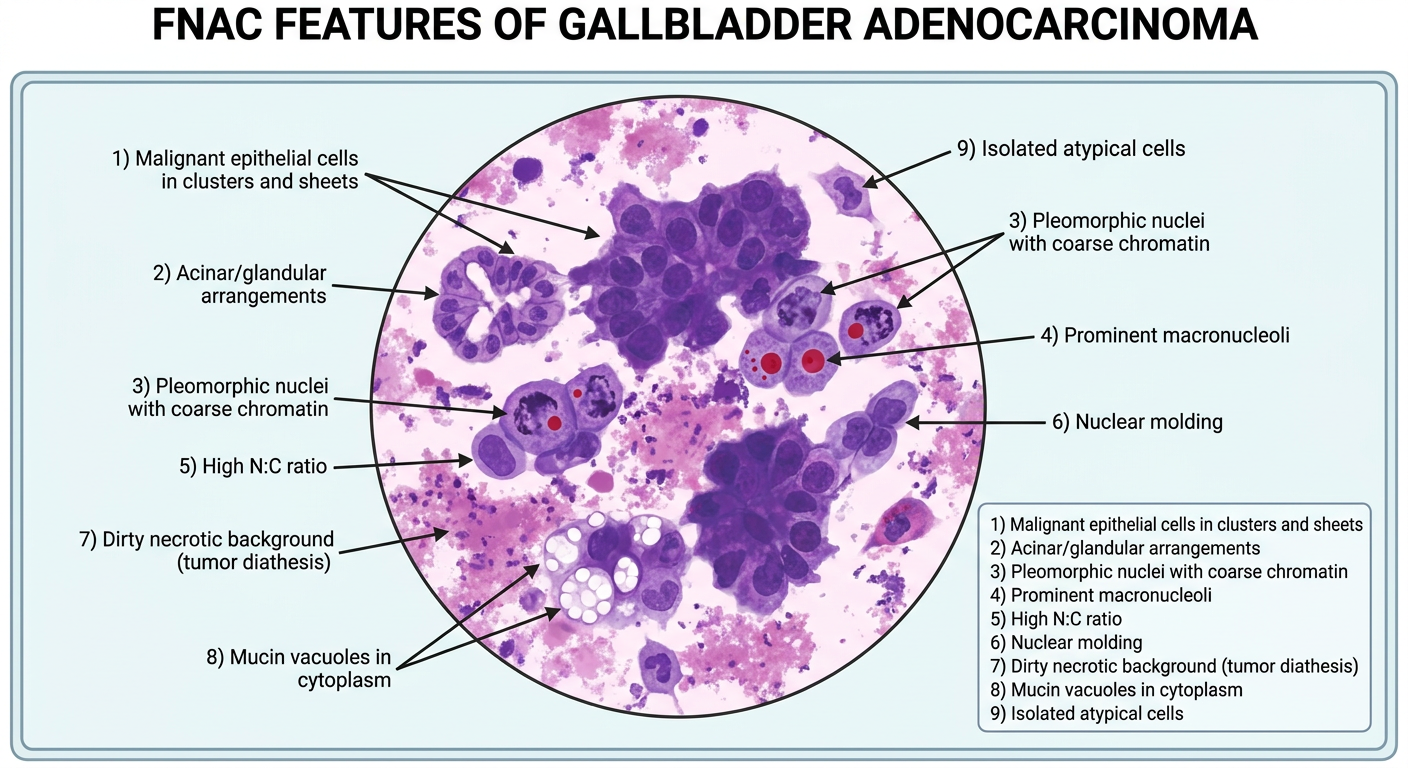
FNAC Cytological Features by Histological Subtype
1. Well-Differentiated Adenocarcinoma (Most Common)
| Feature | Description |
|---|---|
| Cellularity | Moderate to high |
| Cell arrangement | Clusters, sheets, acinar/glandular formations, papillary groups |
| Cell type | Columnar to cuboidal malignant epithelial cells |
| Nuclei | Enlarged, pleomorphic, eccentrically placed |
| Chromatin | Coarse, irregularly distributed |
| Nucleoli | Prominent single or multiple macronucleoli |
| Cytoplasm | Moderate, with intra-cytoplasmic mucin vacuoles |
| N:C ratio | Increased |
| Background | Mucin, mild inflammation |
2. Moderately Differentiated Adenocarcinoma
- Cell clusters with some acinar formation plus scattered isolated malignant cells
- Nuclear pleomorphism moderate to marked
- Focal tumor necrosis present
- Tumor giant cells may appear
- Intracellular and extracellular mucin seen
- Background: dirty with necrosis and inflammatory exudate ("tumor diathesis")
3. Poorly Differentiated Adenocarcinoma
- Predominantly dyscohesive (dissociated) tumor cells - few clusters/acini
- Abundant necrosis and inflammatory exudate in background
- Many tumor giant cells
- High N:C ratio with moderate to marked nuclear pleomorphism
- Multiple prominent nucleoli
- Stippled (salt-and-pepper) chromatin
- Few or no mucin-containing fragments
FNAC Features by Histological Type (WHO Classification)
| Carcinoma Type | Key FNAC Features |
|---|---|
| Adenocarcinoma NOS | Glandular/acinar clusters, columnar cells, mucin vacuoles, macronucleoli |
| Papillary adenocarcinoma | Papillary fronds with fibrovascular core, better differentiation, columnar cells |
| Mucinous adenocarcinoma | Abundant extracellular mucin pools, clusters and signet ring-like cells floating in mucin |
| Signet ring cell carcinoma | Single dissociated cells with intracytoplasmic mucin displacing nucleus to periphery |
| Adenosquamous carcinoma | Mixed: glandular clusters + atypical squamous cells with keratinization |
| Squamous cell carcinoma | Atypical squamous cells, keratin pearls/whorls, background of necrosis and inflammation |
| Small cell (neuroendocrine) | Hyperchromatic nuclei with nuclear molding, high N:C ratio, salt-and-pepper chromatin, scant cytoplasm |
| Undifferentiated (spindle/giant cell) | Spindle-shaped cells and bizarre multinucleated giant cells, abundant necrosis |
Key Diagnostic FNAC Features - Summary
MALIGNANT GALLBLADDER MASS ON FNAC:

1. CELLULARITY
   └── Usually high (moderately to highly cellular smear)

2. ARCHITECTURAL PATTERNS
   ├── Acinar / glandular arrangements (most specific)
   ├── Papillary clusters (papillary subtype)
   ├── Sheets and irregular clusters
   └── Dyscohesive single cells (poorly diff.)

3. NUCLEAR FEATURES (MALIGNANCY CRITERIA)
   ├── Nuclear enlargement + pleomorphism
   ├── Coarse/clumped or stippled chromatin
   ├── Prominent macronucleoli (single or multiple)
   ├── Irregular nuclear membrane
   ├── High N:C ratio
   └── Nuclear molding (small cell type)

4. CYTOPLASMIC FEATURES
   ├── Intracytoplasmic mucin vacuoles
   └── Signet ring cells (mucinous/signet ring type)

5. BACKGROUND
   ├── Extracellular mucin
   ├── Necrotic / dirty background (tumor diathesis)
   ├── Inflammatory exudate
   └── Tumor giant cells (poorly diff.)
FNAC Procedure Notes
- Needle size: 22G for cytology; 18G for histological core
- Guidance: US-guided (most common), CT-guided, or EUS-guided (preferred if resectable candidate - lower seeding risk)
- Sensitivity: ~62% for extrahepatic bile duct tumors; higher (~96.8%) for gallbladder mass in dedicated series
- Complication rate: <0.2% overall (bleeding, bile leak, pancreatitis, infection each <0.08%)
- Needle track seeding: ~0.05% - therefore percutaneous biopsy is avoided if surgical resection is planned; EUS-FNA preferred in such cases
- Biliary brushings: ~55% sensitivity; helpful if positive
Differential Diagnosis on FNAC
| Benign / Other | Distinguishing FNAC Feature |
|---|---|
| Reactive/inflammatory epithelium | Mild nuclear changes, preserved polarity, no macronucleoli, clean background |
| Cholecystitis | Inflammatory cells, foamy macrophages, bile pigment, no atypical cells |
| Metastatic adenocarcinoma | Clinical history + IHC needed (CK7+/CK20+/- profile) |
| Hepatocellular carcinoma extension | Trabecular pattern, bile pigment, polygonal cells, CD10/Hep-Par-1+ |
Immunocytochemistry (IHC) Profile
- CK7+, CK19+ (biliary type)
- CK20 - variable
- CEA - positive
- CA 19-9 - positive
- EMA - positive
- Negative for: HepPar-1, CD56 (except NE tumors), S100