External ear and tympanic membrane parts nerve supply blood supply
Reading File
External Ear and Tympanic Membrane: Parts, Nerve Supply, Blood Supply
1) External ear parts
A. Auricle (pinna)
- Helix
- Antihelix
- Tragus
- Antitragus
- Lobule
- Concha (leads into ear canal)
- Scaphoid fossa, triangular fossa (depressions)
B. External acoustic meatus (external auditory canal)
- Lateral 1/3 cartilaginous
- Medial 2/3 bony
- Ends at tympanic membrane
C. Tympanic membrane (eardrum)
- Pars tensa (large, taut, fibrous)
- Pars flaccida (small, superior, lax)
- Landmarks: handle of malleus, umbo, cone of light
2) Nerve supply
A. External ear (auricle + external canal)
- Auriculotemporal nerve (V3)
- Anterosuperior auricle, anterior wall/roof of canal
- Great auricular nerve (C2-C3)
- Most of inferoposterior auricle, lobule
- Lesser occipital nerve (C2)
- Upper posterior auricle
- Auricular branch of vagus (X) (Arnold nerve)
- Concha, posteroinferior canal (can cause cough reflex during cleaning)
- Facial nerve (VII) contribution (small sensory component around concha/meatus in some descriptions)
B. Tympanic membrane sensory innervation
- External (lateral) surface: mainly auriculotemporal nerve (V3) + auricular branch of vagus (X)
- Internal (medial) surface: glossopharyngeal nerve (IX) via tympanic plexus
3) Blood supply
A. External ear (auricle)
Mainly from branches of external carotid artery:
- Posterior auricular artery
- Superficial temporal artery (anterior auricular branches) (Occipital artery may contribute posteriorly)
B. External auditory canal
- Deep auricular artery (from maxillary artery) is key
- Contributions from posterior auricular/superficial temporal branches
C. Tympanic membrane
- Lateral surface: chiefly deep auricular artery (maxillary)
- Medial surface: vessels from middle ear mucosa, mainly anterior tympanic and related caroticotympanic/stylomastoid contributions (anastomotic network)
If you want, I can also give this as a quick exam-style table (very high-yield for anatomy viva/MCQ).
Diagram
Generating Image
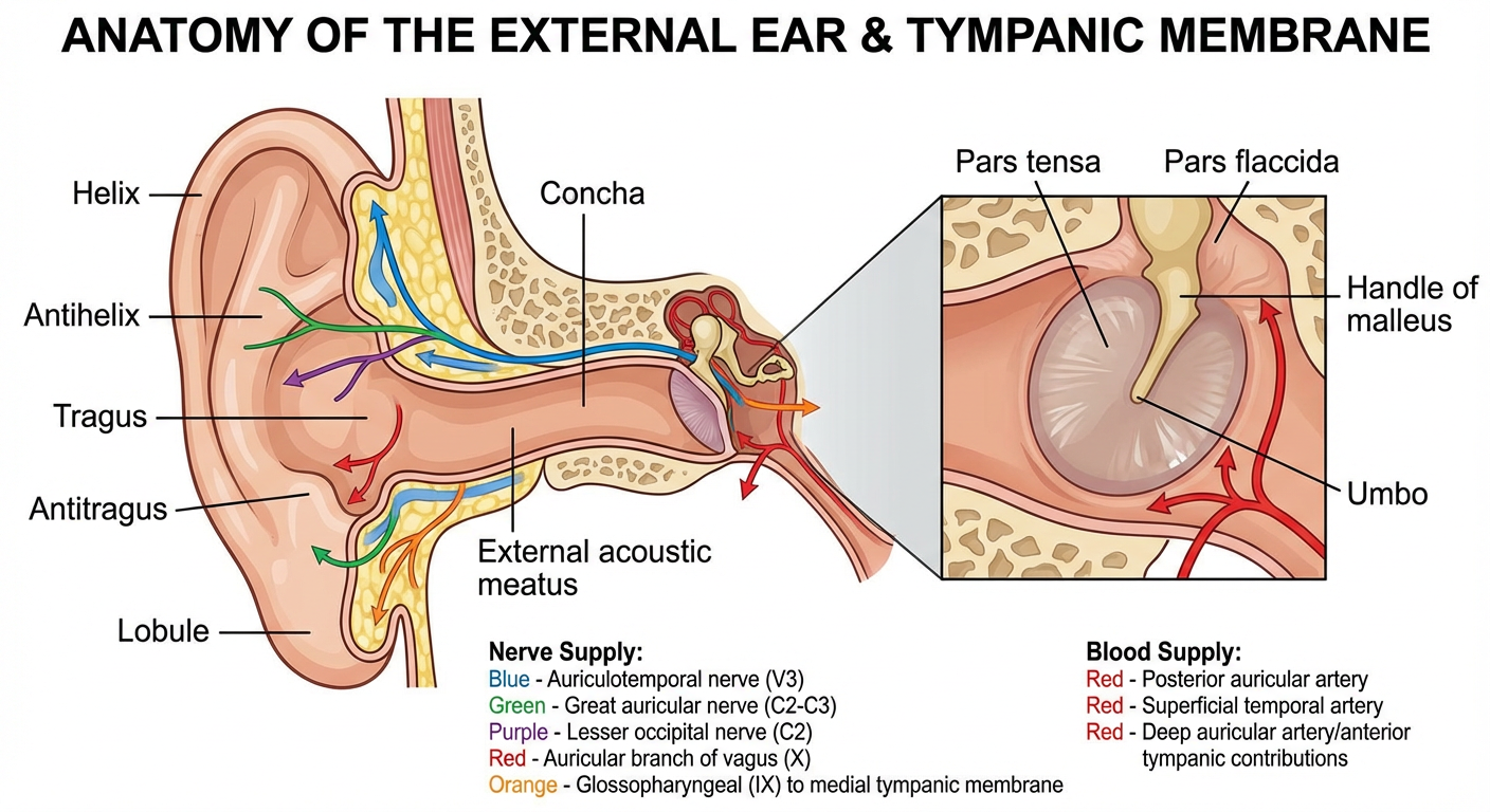
Labeled medical anatomy diagram of the external ear and tympanic membrane showing parts (helix, antihelix, tragus, antitragus, lobule, concha, external acoustic meatus, pars tensa, pars flaccida, handle of malleus, umbo) plus color-coded nerve supply (auriculotemporal nerve V3, great auricular nerve C2-C3, lesser occipital nerve C2, auricular branch of vagus X, glossopharyngeal IX to medial tympanic membrane) and blood supply (posterior auricular artery, superficial temporal artery, deep auricular artery/anterior tympanic contributions).
This is a shared conversation. Sign in to Orris to start your own chat.