
Femoral harnia
femoral hernia anatomy diagram
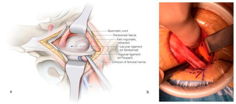
This composite visual contains an anatomical illustration (a) and a corresponding clinical intraoperative photograph (b) detailing the surgical repair of a femoral hernia. Panel (a) provides a labeled anatomical diagram of the right inguinal-femoral region. It depicts the spermatic cord and falx inguinalis retracted cranially, exposing the orificium of the femoral hernia. Key landmarks are identified: the transversalis fascia (opened), the inguinal ligament of Poupart (ventral border), and the lacunar ligament of Gimbernat (medial border). An instrument is shown passing through the femoral orifice to demonstrate the pathology. Panel (b) is a clinical photograph of the surgical field during an open repair. It demonstrates the clinical application of the anatomy shown in panel (a), with retractors maintaining exposure. A surgical instrument is seen incising the lacunar ligament to enlarge the femoral space for mesh placement. The image illustrates the relationship between the inguinal ligament, femoral vessels, and the hernia orifice. The content is intended for intermediate to advanced surgical education, specifically regarding open femoral hernia repair and the anatomy of the femoral canal.
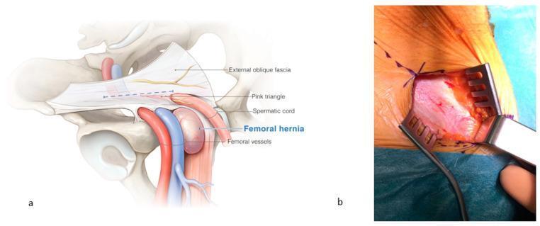
This dual-panel image provides an anatomical and clinical overview of a femoral hernia and its surgical exposure. Panel (a) is a detailed anatomical diagram of the right inguinal and femoral regions. It illustrates a femoral hernia as a distinct bulge emerging medially to the femoral vessels (artery and vein) and caudal to the inguinal canal. Labeled structures include the external oblique fascia, the 'pink triangle' (an area formed by the diversion of fascial vessels toward the external inguinal orifice), the spermatic cord, and femoral vessels. Panel (b) is a clinical photograph showing the intraoperative view after the incision of the external oblique fascia. The surgical field is exposed using three metal claw-style retractors, revealing the underlying fascia and fatty tissue in the femoral region. Sutures are visible along the superior aspect of the skin incision. This content is designed for medical education regarding hernia pathology and surgical repair, demonstrating the critical landmarks needed to differentiate femoral hernias from inguinal hernias and to safely navigate the femoral canal during surgery.
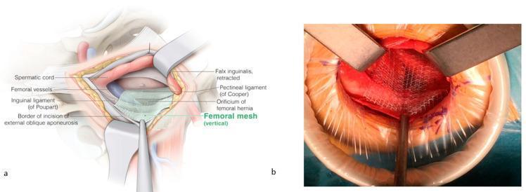
This composite image illustrates the surgical repair of a femoral hernia using a mesh-based approach. Figure (a) is an anatomical diagram of the inguinal and femoral regions during surgery. Key structures identified include the spermatic cord (superior-medial), femoral vessels (lateral), inguinal ligament of Poupart (ventral), and the pectineal ligament of Cooper (dorsal). The diagram shows the placement of a vertical femoral mesh covering the orificium of the femoral hernia, secured over Cooper’s ligament while maintaining an overlay medially and laterally over the femoral vessels. The falx inguinalis is shown in a retracted state. Figure (b) is an intraoperative clinical photograph corresponding to the diagram, showing the surgical field with metal retractors and a circular wound protector. A white, woven surgical mesh is visible, positioned over the femoral orifice and held in place with instruments. The image demonstrates the 'Lichtenstein-like' open technique for femoral hernia reinforcement, highlighting the spatial relationship between the prosthetic mesh and pelvic floor ligaments to prevent recurrence.
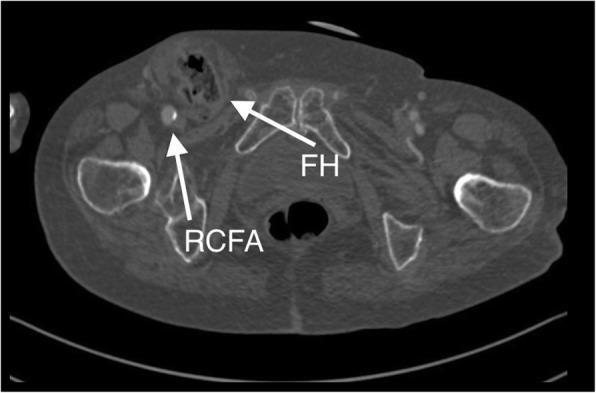
This diagnostic image is a transverse (axial) cross-section from a Computed Tomography Angiography (CTA) scan of the pelvic and groin region. The image illustrates the spatial anatomy of a right-sided femoral hernia (FH). A white arrow labeled 'RCFA' points to the right common femoral artery, which is visualized as a bright, contrast-enhanced circular structure. Medial to the RCFA, a second white arrow labeled 'FH' identifies the femoral hernia sac protruding through the femoral canal. The hernia sac contains convoluted soft tissue consistent with small bowel loops; the presence of contrast within these loops indicates maintained perfusion and the absence of acute strangulation at the time of imaging. Surrounding anatomical landmarks include the pelvic girdle bones (ilium and pubic symphysis) and the proximal femurs. This visual is clinically significant for demonstrating the relationship between vascular structures and hernial protrusions in the groin, serving as a critical diagnostic tool for pre-surgical planning in complex patients.
"femoral hernia"[MeSH Terms] AND repair
Femoral Hernia
Definition
- S Das Manual on Clinical Surgery, p.611 | Bailey and Love's Short Practice of Surgery, 28th Ed., p.1092
Anatomy of the Femoral Canal
| Border | Structure |
|---|---|
| Anterior (superoanterior) | Inguinal ligament (Poupart's) |
| Posterior (inferoposterior) | Pectineal (Cooper's / iliopectineal) ligament + pectineus muscle |
| Medial | Lacunar ligament (Gimbernat's ligament) - sharp, unyielding edge |
| Lateral | Femoral vein |
Epidemiology
- More common in women (2:1 female predominance) due to the wider female pelvis, which enlarges the femoral canal
- Even so, inguinal hernia remains the commonest groin hernia in women
- Rare before age 20; incidence rises with age, peaking after 50
- Right side is affected twice as often as the left
- Bilateral in ~20% of cases
- Thin, elderly women are the classic high-risk group
Clinical Features
- Swelling in the upper medial thigh, below and lateral to the pubic tubercle
- May extend upward above the inguinal ligament in advanced cases
- Cough impulse felt at the saphenous opening
- Symptoms are often less pronounced than inguinal hernia
- The hernia frequently becomes irreducible early and loses its cough impulse - making it easy to miss on routine examination
Why Strangulation is so Common
- 50% of femoral hernias present as emergencies with strangulation or bowel obstruction
- All patients with unexplained small bowel obstruction should be carefully examined for a femoral hernia
Differential Diagnosis
- Inguinal hernia - lies medial to pubic tubercle, above the inguinal ligament
- Saphena varix - saccular dilatation of the terminal long saphenous vein; disappears completely on lying down; fluid thrill on coughing (not an expansile impulse); Schwartz's test positive
- Enlarged inguinal lymph node - search for a focus of infection in the drainage area
- Femoral artery aneurysm - expansile pulsatile swelling
- Psoas abscess - points lateral to the femoral vessels; may have fluctuation; associated with spinal pathology
- Rupture of adductor longus with haematoma
Key Distinguishing Points (Femoral vs. Inguinal Hernia)
| Feature | Femoral Hernia | Inguinal Hernia |
|---|---|---|
| Position rel. to pubic tubercle | Lateral | Medial |
| Position rel. to inguinal ligament | Below (may later go above) | Above |
| Cough impulse location | Saphenous opening (~4 cm below & lateral to pubic tubercle) | Inguinal canal |
| Invagination test (inguinal canal) | Empty | Positive |
| Reduction by digital pressure | Over femoral canal / saphenous opening | Over deep inguinal ring |
Investigations
- Routine elective cases: no specific investigations needed
- Uncertainty: ultrasound or CT scan
- Emergency presentation: plain X-ray (dilated bowel loops), then CT to identify the hernia and exclude malignancy
Treatment - Surgery
Three Open Approaches:
1. Low Approach (Lockwood)
- Simplest approach; suitable only when bowel resection is not anticipated
- Can be done under local anaesthesia
- Transverse incision over the hernia, sac opened and contents reduced, defect closed with sutures between the inguinal ligament above and pectineal ligament below
- A small nick in the medial lacunar ligament may aid reduction (caution: an abnormal obturator artery branch may lie just deep to it - the "corona mortis")
- Some surgeons use a mesh plug for reinforcement
2. Inguinal Approach (Lotheissen)
- Incision identical to Bassini/Lichtenstein - into the inguinal canal
- Transversalis fascia opened, extraperitoneal space entered
- Hernia reduced from above; neck closed with sutures or mesh plug
- Allows simultaneous inguinal hernia repair
- More complex than Lockwood; suitable electively
3. High (Preperitoneal) Approach (McEvedy / Nyhus modification)
- Ideal in emergencies when bowel strangulation is suspected
- Transverse incision above the inguinal canal, anterior rectus sheath incised, rectus retracted, preperitoneal space entered
- Allows generous peritoneal incision, inspection of bowel, and bowel resection if needed (unlike the low approach, where a completed anastomosis cannot be returned through the narrow femoral canal)
- Can be extended to repair bilateral hernias through one incision (Henry's approach)
4. Laparoscopic Approach (TEP / TAPP)
- Both totally extraperitoneal (TEP) and transabdominal preperitoneal (TAPP) approaches are used
- Standard mesh inserted in the extraperitoneal plane
- Ideal for elective, reducible femoral hernias
- Increasing reports of use even in emergency cases
Special Variant: Pre-vascular Femoral Hernia
Key Clinical Pearls
- Femoral hernia has the highest rate of strangulation of all groin hernias because the femoral ring is rigid and inelastic
- It is often missed on clinical examination because the swelling is in the thigh, not the groin, and rapidly becomes irreducible
- In an obese or elderly woman with small bowel obstruction and no obvious cause, always examine for a femoral hernia
- Never make a nick in the lacunar ligament without being aware of an anomalous obturator artery (from the inferior epigastric artery), which lies just medial to the femoral ring in ~20-30% of people - historically known as the "corona mortis" or "crown of death"