
Bds first year essay Amelogenesis
Reading File
Now let me search the medical library for amelogenesis content.
Finding Sources
Finding Sources
Reading File
Finding Sources
Reading File
Reading File
Finding Sources
Reading File
Finding Sources
Reading File
Finding Sources
Reading File
Loading Image
Now I have comprehensive content. Let me also fetch the ameloblast detail diagram for visual support.
Loading Image
I now have all the content needed for a comprehensive BDS first year essay on Amelogenesis. Let me write the full essay.
Amelogenesis (Enamel Formation)
Introduction
Amelogenesis is the biological process by which dental enamel - the hardest and most highly mineralized tissue in the human body - is formed. It is a matrix-mediated biomineralization process carried out exclusively by a specialized epithelial cell called the ameloblast, derived from the inner enamel epithelium (ectoderm) of the enamel organ. Since ameloblasts degenerate at the time of tooth eruption, enamel has no capacity for regeneration or self-repair once fully formed.
Enamel Organ - The Structural Basis
Before amelogenesis begins, the enamel organ (derived from oral ectoderm) must be fully differentiated. In the bell stage of tooth development, the enamel organ consists of four layers:
| Layer | Description |
|---|---|
| Outer enamel epithelium | Convex outer surface; cuboidal cells |
| Inner enamel epithelium | Concave inner surface; gives rise to ameloblasts |
| Stratum intermedium | Adjacent to inner enamel epithelium; contains alkaline phosphatase, important for calcification |
| Stellate reticulum | Star-shaped cells; occupies the interior; provides support and nutrition |
The dental lamina degenerates just before dentinogenesis and amelogenesis begin, leaving the developing tooth primordium separated from its oral epithelial origin.
Cells of Amelogenesis - The Ameloblast
The inner enamel epithelial cells differentiate into ameloblasts under inductive signals from the underlying dental papilla mesenchyme (specifically the preodontoblasts/odontoblasts).
Key feature: Induction is reciprocal - the inner enamel epithelium first induces the mesenchymal cells to become odontoblasts and form dentin; the newly formed dentin then induces the inner enamel epithelial cells to differentiate into secretory ameloblasts. This means dentin is always formed first, before enamel deposition begins.
Secretory-stage ameloblasts are tall, polarized columnar cells (~40 µm) with:
- rER and Golgi apparatus (for protein synthesis)
- Secretory granules
- A distinctive apical cytoplasmic extension called the Tomes process
- A proximal terminal web (PTW) and distal terminal web (DTW) containing actin filaments
Stages of Amelogenesis
Stage 1: Matrix Production (Secretory Stage)
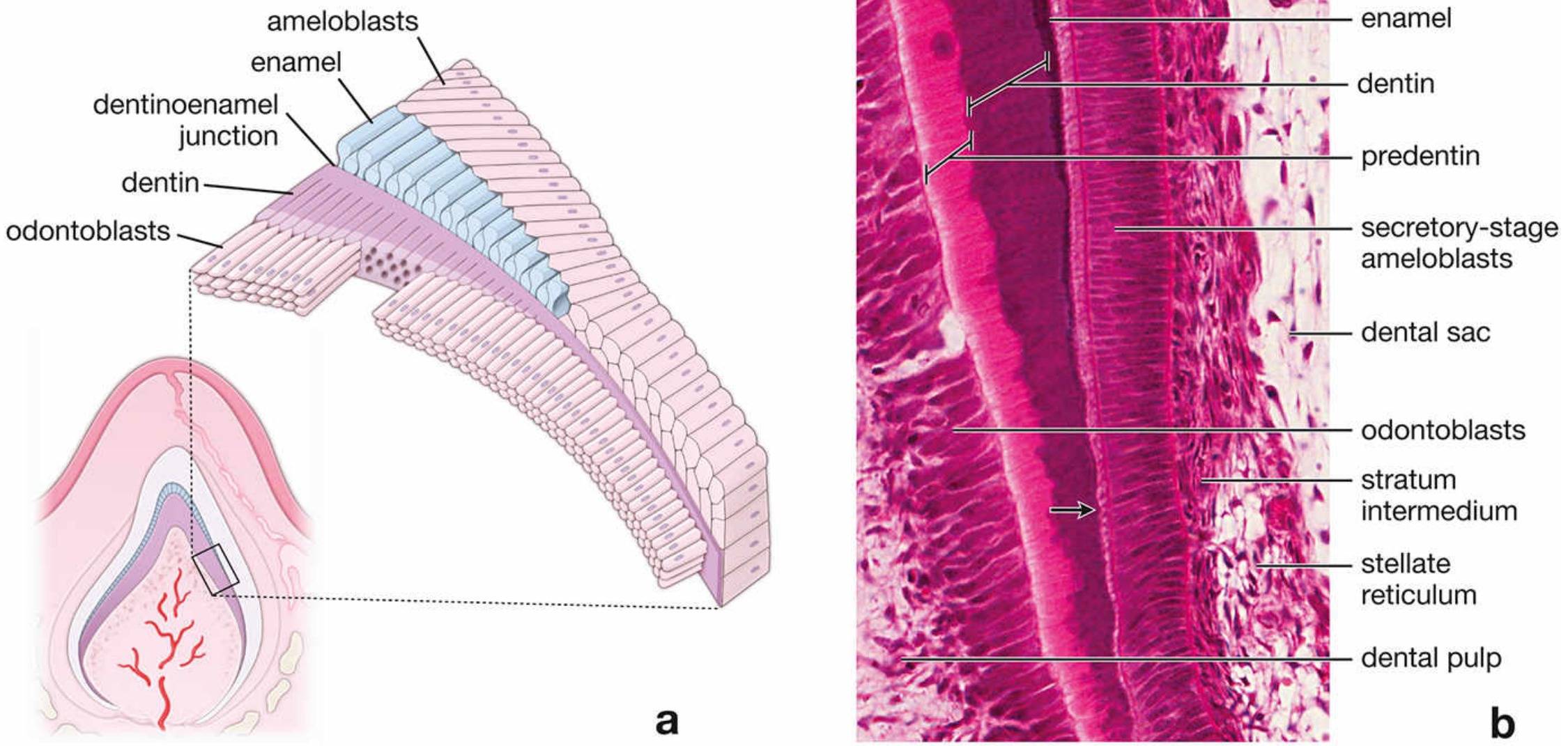
Figure: Enamel formation - secretory-stage ameloblasts depositing enamel matrix onto the previously formed dentin layer. Note the stratum intermedium adjacent to the ameloblasts. (Histology: A Text and Atlas, Lippincott)
- Secretory-stage ameloblasts produce an organic proteinaceous enamel matrix using their rER, Golgi apparatus, and secretory granules (the same mechanism osteoblasts use to produce bone matrix).
- The partially mineralized enamel matrix is deposited directly onto the surface of the previously formed dentin (at the dentinoenamel junction).
- The Tomes process of each ameloblast is surrounded by the developing enamel. The shape of this process determines the shape of the enamel rod (prism).
- Ameloblasts continue producing matrix until the full thickness of future enamel is reached.
- As enamel is laid down, ameloblasts move away from the dentin surface toward the outer enamel surface - so enamel formation proceeds from the DEJ outward.
- The direction of each enamel rod is a permanent record of the exact path taken by its ameloblast during secretion.
Stage 2: Matrix Maturation Stage
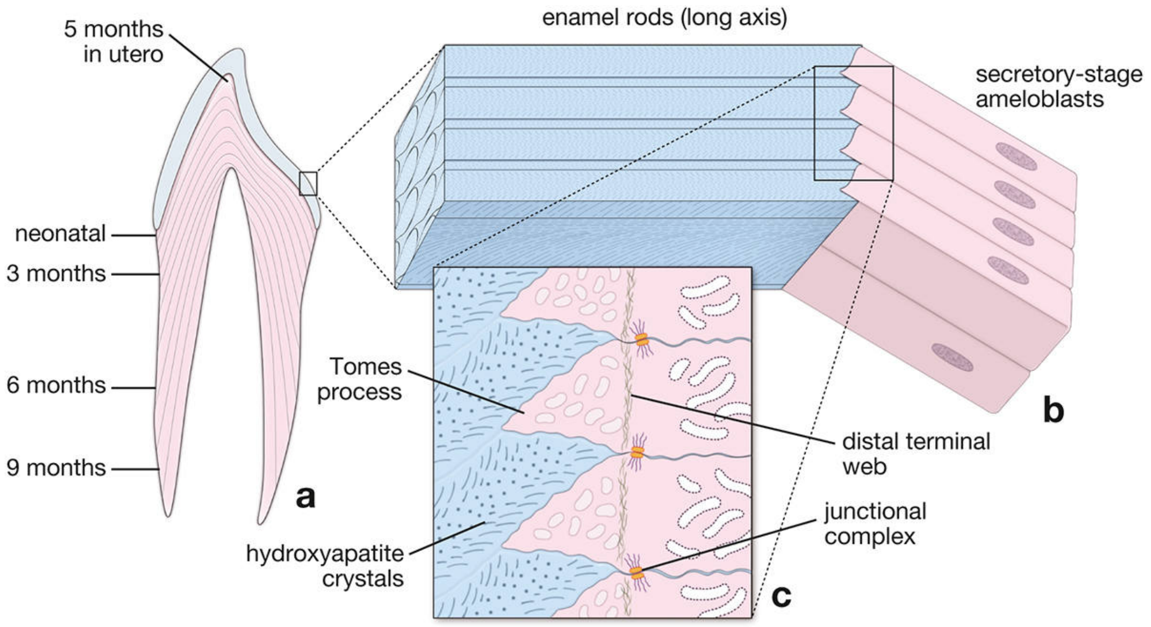
Figure: Schematic of amelogenesis showing enamel rods from DEJ to tooth surface, Tomes processes of ameloblasts, and hydroxyapatite crystal deposition. (Histology: A Text and Atlas, Lippincott)
- After full enamel thickness is achieved, the secretory-stage ameloblasts differentiate into maturation-stage ameloblasts.
- Maturation involves:
- Removal of organic material (amelogenins and ameloblastins are proteolytically degraded and reabsorbed)
- Continued influx of Ca²⁺ and phosphate to complete mineralization
- Maturation-stage ameloblasts function primarily as a transporting epithelium.
- Their hallmark histologic feature is a striated (ruffled) border on the apical surface.
- These cells undergo cyclic modulation - alternating between a striated border and a smooth border:
- Striated-border cells (~70%): secrete HCO₃⁻ ions and contain plasma membrane Ca²⁺-ATPases (PMCA) that pump Ca²⁺ into maturing enamel
- Smooth-border cells (~30%): secrete proteolytic enzymes to degrade and reabsorb the extracellular matrix
- The stratum intermedium disappears during this stage; instead, a papillary layer (formed from collapsed stellate reticulum, stratum intermedium, and outer enamel epithelium) provides support and vascularity to the maturation ameloblasts.
- Maturation-stage ameloblasts and adjacent papillary cells are rich in mitochondria, reflecting their high energy demand.
Stage 3: Post-Amelogenesis / Regression
- After enamel is fully formed, ameloblasts undergo apoptosis and degenerate - roughly at the time of tooth eruption through the gingiva.
- This is why enamel cannot be regenerated after formation is complete.
Enamel Matrix Proteins
Recent molecular biology has revealed the enamel extracellular matrix to be highly heterogeneous, containing proteins encoded by multiple genes:
| Protein | Role |
|---|---|
| Amelogenins | Most abundant (~90%); maintain spacing between enamel rods in early enamel development; removed during maturation |
| Ameloblastins | Produced from early secretory to late maturation stages; guide mineralization by controlling elongation of enamel crystals; form junctional complexes between individual enamel crystals |
| Enamelins | Distributed throughout the enamel layer; undergo proteolytic cleavage as enamel matures; low-molecular-weight cleavage products are retained in mature enamel, localized on crystal surfaces |
| Tuftelins | Earliest detected proteins; located near the dentinoenamel junction; acidic and insoluble - aid in nucleation of enamel crystals; present in enamel tufts; account for the hypomineralization seen in enamel tufts |
Fate of proteins in mature enamel: Amelogenins and ameloblastins are completely removed during maturation. Only enamelins and tuftelins are retained in mature enamel. This is why mature enamel is ~96% inorganic hydroxyapatite by weight.
Summary of Key Events
Dental lamina → Enamel organ (bell stage)
Inner enamel epithelium → Ameloblasts (under induction by odontoblasts)
Dentin formed FIRST by odontoblasts
↓
Secretory stage ameloblasts:
- Deposit partially mineralized enamel matrix via Tomes process
- Build full enamel thickness (DEJ → surface)
↓
Maturation stage ameloblasts:
- Remove organic matrix proteins (amelogenins, ameloblastins)
- Pump Ca²⁺ and phosphate via ruffled border
- Enamel becomes 96% hydroxyapatite
↓
Ameloblasts degenerate at eruption
→ No future enamel repair possible
Clinical Correlation: Amelogenesis Imperfecta
Amelogenesis imperfecta (AI) is a complex group of at least 14 clinical entities involving aberrations in enamel formation in the absence of any systemic disorder. It is an inherited ectodermal birth defect affecting enamel only.
- The enamel may be hypoplastic (thin), hypocalcified (soft), or hypomature (not fully developed)
- Depending on subtype: enamel may be hard or soft, pitted or smooth, thin or normal in thickness
- Incidence: 1 in 700 (Sweden) to 1 in 1,200 (USA)
- Inheritance: Multiple modes (autosomal dominant, autosomal recessive, X-linked)
- Genes involved: AMELX (amelogenin, X-linked), ENAM (enamelin), MMP20 (matrix metalloproteinase 20, involved in enamel matrix degradation during maturation)
- Classification is based on clinical and radiographic findings and mode of inheritance
Other conditions affecting enamel during amelogenesis:
- Tetracycline staining: Tetracyclines bind to hydroxyapatite, causing brown-yellow discoloration; risk period is from ~14 weeks fetal life to 8th postnatal year. Hence tetracyclines are contraindicated in pregnant women and children under 8 years.
- Fluorosis: Excess fluoride during amelogenesis disrupts maturation-stage ameloblasts, causing hypomineralization, white opacities, or pitting.
- Enamel hypoplasia: Any insult (nutritional deficiency, infection, prematurity) during the secretory stage can produce pits, lines, or missing enamel.
Conclusion
Amelogenesis is a tightly regulated, two-stage process - matrix secretion followed by matrix maturation - carried out entirely by ameloblasts derived from the inner enamel epithelium. The process is irreversible: once ameloblasts degenerate at eruption, enamel cannot be reformed. Understanding amelogenesis is fundamental to understanding enamel structure, the basis of enamel defects seen clinically, and the rationale for preventive dentistry.
Sources:
- Histology: A Text and Atlas with Correlated Cell and Molecular Biology (Lippincott Williams & Wilkins), Chapter 16 - Oral Cavity and Associated Structures
- The Developing Human: Clinically Oriented Embryology (Moore & Persaud), Chapter 19 - Dental Defects
This is a shared conversation. Sign in to Orris to start your own chat.