
Numbness in right leg with pain in nerve
Reading File
Finding Sources
Finding Sources
Searching PubMed
"lumbar radiculopathy" AND "leg pain numbness"
Reading File
Reading File
Reading File
Here is a structured overview of right leg numbness with nerve pain, based on authoritative neurology and anesthesiology textbooks.
Right Leg Numbness with Nerve Pain
Numbness and pain in the right leg almost always point to irritation, compression, or injury somewhere along the nervous pathway - from the lumbar spine down to the peripheral nerves in the leg itself.
1. Most Common Cause: Lumbar Radiculopathy (Pinched Nerve Root)
The most frequent culprit is a herniated intervertebral disc at L4-L5 or L5-S1 (90% of disc herniations occur at these two levels). The nucleus pulposus bulges out and compresses an adjacent nerve root, causing pain that radiates down the leg along a specific pattern called a dermatome.
| Disc Level | Nerve Root | Pain Pattern | Weakness | Reflex Lost |
|---|---|---|---|---|
| L3-L4 | L4 | Anterolateral thigh, anteromedial calf to ankle | Quadriceps (knee extension) | Knee jerk |
| L4-L5 | L5 | Lateral thigh, anterolateral calf, top of foot (1st/2nd toe area) | Foot dorsiflexion (foot drop) | None typically |
| L5-S1 | S1 | Buttock, posterior thigh, posterolateral calf, outer foot (4th/5th toes) | Plantar flexion (push off) | Ankle jerk |
Key clinical features:
- Pain worsened by bending, lifting, prolonged sitting, coughing, or sneezing
- Relieved by lying down
- Straight leg raise test - raising the leg with knee extended at the angle that reproduces the pain is a key diagnostic sign; pain on raising the opposite leg is an even more reliable sign of nerve compression
- Numbness or weakness alongside the pain confirms radiculopathy (rather than simple disc bulging)
- Morgan and Mikhail's Clinical Anesthesiology, 7e
2. Sciatica
Sciatica refers to painful paresthesias (burning, shooting, electric pain) in a sciatic distribution - down the back of the leg. The most common cause is compression of lumbosacral roots (L4-S1) by disc material or bone spurs (osteophytes).
Sciatic neuropathy itself (injury to the sciatic nerve trunk) can occur from:
- Posterior hip dislocation or acetabular fracture
- Hip surgery
- Intramuscular injection placed too medially/inferiorly in the buttock
Findings: weakness of all foot/ankle muscles and knee flexion, loss of ankle reflex, sensory loss in the foot and lateral leg below the knee.
- Neuroanatomy through Clinical Cases, 3rd Ed.
3. Common Fibular (Peroneal) Nerve Palsy
If the numbness and pain are on the outer shin and top of the foot, with foot drop (difficulty lifting the foot), this suggests compression of the common fibular nerve as it wraps around the fibular head just below the knee. Causes include:
- Crossing legs habitually
- Tight cast or stockings
- Direct trauma to the outer knee
Most cases recover spontaneously when the cause is removed.
- Neuroanatomy through Clinical Cases, 3rd Ed.
4. Femoral Nerve Injury
If pain and numbness are at the front of the thigh and inner calf, with weakness lifting the knee or extending the leg, the femoral nerve (L2-L4) may be affected. The patellar (knee jerk) reflex may be reduced or absent.
5. Tarsal Tunnel Syndrome
Entrapment of the tibial nerve behind the inner ankle causes burning pain and numbness in the sole of the foot and toes. Percussion over the nerve behind the medial malleolus often reproduces symptoms.
6. Other Important Causes to Consider
| Cause | Key Feature |
|---|---|
| Meralgia paresthetica | Burning/numbness on the outer thigh only; lateral femoral cutaneous nerve compression (often from tight clothing or obesity) |
| Diabetic neuropathy | Stocking-pattern numbness; bilateral; gradual onset |
| Multiple sclerosis | Unilateral sensory changes in a young person; may have other neurological symptoms |
| Spinal stenosis | Leg pain/numbness brought on by walking, relieved by sitting or bending forward |
| Cauda equina syndrome | Emergency - bilateral leg weakness/numbness + bladder/bowel problems after disc herniation |
Red Flag Symptoms - Seek Immediate Care
- Numbness in the inner thighs/groin (saddle area)
- Loss of bladder or bowel control
- Rapidly worsening weakness in the leg
- Pain following a significant fall or injury
These may indicate cauda equina syndrome, a surgical emergency.
Diagnosis
- MRI of the lumbar spine is the gold standard for disc pathology and nerve root compression
- Nerve conduction studies (NCS) + EMG help localize peripheral nerve lesions and assess severity
- Imaging should be reserved for cases with neurological deficits or suspected serious cause - up to 30-40% of symptom-free people have abnormal spine MRI findings
Bottom line: Right leg numbness with nerve pain most often reflects compression of an L4, L5, or S1 nerve root from a herniated disc - or irritation of the sciatic nerve. A clinical examination with straight leg raise testing, reflex testing, and muscle strength testing can usually point to the level affected. Imaging confirms the diagnosis. If you are experiencing these symptoms, it is best to see a doctor who can examine you and determine the exact cause.
More details lumber radiculopathy
Finding Sources
Finding Sources
Reading File
Reading File
Searching PubMed
"lumbar radiculopathy" AND treatment
Reading File
Finding Sources
Finding Sources
Finding Sources
Finding Sources
Reading File
Loading Image
Reading File
Excellent - I now have comprehensive textbook content. Let me compile the full detailed answer.
Lumbar Radiculopathy - Detailed Overview
What Is It?
Lumbar radiculopathy is compression or irritation of one or more nerve roots in the lumbar spine, producing pain, numbness, and/or weakness that radiates along the path of that nerve root into the leg. The most common cause is a herniated (prolapsed) intervertebral disc, but degenerative changes (spondylosis), facet hypertrophy, and spinal canal narrowing (stenosis) are also frequent culprits.
Anatomy - How Disc Herniation Compresses Nerves
The intervertebral disc has a tough outer ring (annulus fibrosus) and a soft gel-like center (nucleus pulposus). When the annulus weakens, the nucleus can herniate posterolaterally into the spinal canal, directly compressing the adjacent nerve root.
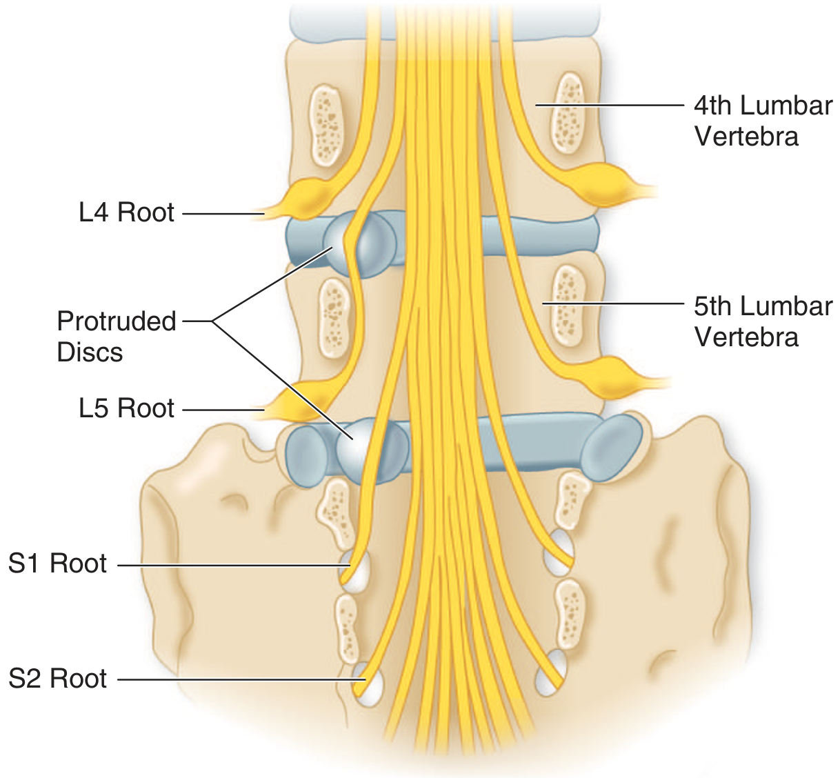
Fig: Protruded discs at L4-L5 and L5-S1 compressing the L5 and S1 nerve roots. A lateral protrusion at L4-L5 compresses L5; at L5-S1, it compresses S1. A more medially placed protrusion at L4-L5 can also involve the S1 (or even S2-S3) roots.
- 90% of disc herniations occur at L4-L5 or L5-S1
- The root is squeezed between the posterior vertebral body, the facet joint (laterally), and the ligamentum flavum (posteriorly)
Nerve Root Levels and Their Patterns
This is the most clinically important table for diagnosing which level is affected:
| Disc Level | Root | Pain Distribution | Weakness | Reflex Lost | Key Tip |
|---|---|---|---|---|---|
| L2-L3 | L3 | Anterior thigh, over knee | Thigh adductors, quadriceps | Knee jerk (absent/diminished) | |
| L3-L4 | L4 | Anterolateral thigh, medial foreleg to ankle | Anterior tibialis (partial foot drop possible) | Knee jerk (diminished or normal) | |
| L4-L5 | L5 | Posterolateral gluteal region, lateral thigh, anterolateral calf, dorsal foot, great toe or 2nd/3rd toes | Extensor hallucis longus, extensor digitorum brevis; foot drop possible | Usually unaffected | Pain with straight-leg raise; tenderness over L4 lateral process |
| L5-S1 | S1 | Midgluteal, posterior thigh, posterolateral leg, lateral foot, heel, lateral toes | Plantar flexion and hamstring weakness | Ankle jerk (absent/diminished) | Tenderness over lumbosacral joint and sciatic notch; discomfort walking on heels |
- Adams and Victor's Principles of Neurology, 12th Ed.
Symptoms
Pain characteristics:
- Radiates from the lower back down the leg along the dermatome ("sciatica" when in sciatic distribution)
- Described as shooting, stabbing, burning, or electric
- Worsened by: bending forward, lifting, prolonged sitting, coughing, sneezing, or straining (any maneuver that raises intra-abdominal/spinal pressure)
- Relieved by: lying flat
Sensory symptoms:
- Numbness, tingling, or paresthesias along the affected dermatome
- L5 involvement: lateral calf and top of foot/great toe
- S1 involvement: back of calf, lateral foot, and heel
Motor symptoms:
- L5: difficulty lifting the foot (foot drop) - cannot walk on heels
- S1: difficulty pushing off the foot (plantar flexion) - cannot walk on tiptoe
- L4: weak knee extension (quad weakness)
Clinical Examination
Straight Leg Raise (SLR) Test
With the patient lying flat, the affected leg is raised with the knee fully extended. Reproduction of radicular leg pain (not just back or hamstring tightness) between 30-70° is positive.
- Sensitivity: ~80%, Specificity: ~40% for disc herniation
- Crossed SLR (pain in the affected leg when raising the opposite leg): lower sensitivity but much higher specificity - a very reliable sign of nerve root compression
Other Examination Findings
- Reduced or absent deep tendon reflexes (ankle jerk for S1, knee jerk for L3-L4)
- Sensory loss to pinprick in the dermatome
- Muscle weakness in myotome distribution
- Paravertebral muscle tenderness and spasm
Imaging
MRI of the lumbar spine (L3-S1) is the investigation of choice:
- Shows disc herniation, nerve root compression, canal dimensions
- Preferred over CT because sagittal images clearly show disc-root relationships
- Also excludes other causes (tumor, infection)
Important caveat: In asymptomatic adults:
-
50% have disc bulging on MRI
- 27% have disc protrusion
- Only 1% have disc extrusion
This means MRI findings must always be interpreted alongside clinical symptoms - do not treat the scan, treat the patient.
CT myelography is used when MRI is unavailable or inconclusive.
Electrodiagnostic Studies (EMG / Nerve Conduction)
EMG is useful for confirming and localizing the nerve root lesion:
- Fibrillation potentials in muscles of the affected root appear 1-2 weeks after onset, in >90% of cases with compressive radiculopathy
- Sensory nerve action potentials remain normal (because the compression is proximal to the dorsal root ganglion, where the sensory cell body sits)
- H-reflex loss is a useful marker of S1 radiculopathy (mirrors the absent ankle jerk)
- Denervation in paraspinal muscles confirms root-level pathology (rather than a peripheral nerve problem)
- EMG is not routinely required - it provides corroborating data when clinical findings are uncertain
Treatment
Conservative (First-line - Works in ~85-90% of cases)
- Activity modification - avoid heavy lifting, prolonged sitting, flexion movements during the acute phase; bed rest beyond 1-2 days is not recommended
- NSAIDs (ibuprofen, naproxen) - first-line analgesic for both pain and inflammation
- Muscle relaxants - for associated paraspinal spasm
- Physical therapy - core strengthening, posture correction, McKenzie exercises (extension-based exercises often help L4-L5 and L5-S1 herniations)
- Oral corticosteroids - short course may reduce acute inflammation and pain
- Gabapentinoids (gabapentin, pregabalin) - for neuropathic/radicular pain component
Interventional
Lumbar Epidural Steroid Injection (LESI):
- Injected into the epidural space near the affected nerve root
- Provides fast and effective pain relief when conservative care fails, provided the nerve is not severely compressed
- Benefit is typically temporary (weeks to months); does not change long-term natural history
- A 2025 AAN Systematic Review (PMID 39938000) reviewed evidence for epidural steroids in lumbar radicular pain and spinal stenosis
Radiofrequency ablation - used for facet-related radiculopathy
Surgical
Indications for surgery:
- Moderate to severe focal neurological deficit (significant foot drop, progressive weakness)
- Bowel or bladder dysfunction (possible cauda equina syndrome - emergency)
- Severe pain lasting >3 months despite aggressive conservative management, with confirmed disc herniation on MRI/CT
Procedure: Microdiscectomy (surgical removal of the herniated disc fragment) is the standard procedure. It relieves leg pain faster than conservative management, though outcomes at 1-2 years are similar.
Complications of surgery: CSF leak, infection, nerve injury, and failure to relieve pain ("failed back surgery syndrome").
Special Scenario: Lumbar Spinal Stenosis and Neurogenic Claudication
In older patients, degenerative spondylotic changes narrow the spinal canal, compressing multiple nerve roots. This produces neurogenic claudication:
- Aching and leg weakness/numbness that comes on with standing or walking
- Forces the patient to sit down for relief
- Relief by bending forward (flexing the spine opens the canal)
- Distinguished from vascular claudication by: preserved leg pulses, onset in standing (not just walking), relief with spinal flexion rather than simple rest
When to Seek Emergency Care
Cauda equina syndrome is a surgical emergency caused by central disc herniation compressing the cauda equina (nerve bundle below L1):
- Bilateral leg pain and weakness
- Saddle anesthesia (numbness in the inner thighs and groin)
- Urinary retention or incontinence
- Fecal incontinence
Requires immediate MRI and urgent surgical decompression.
Recent Evidence Update
A 2024 systematic review (PMID 38629664) reviewed medication recommendations from clinical practice guidelines for lumbosacral radiculopathy, and a 2025 AAN systematic review (PMID 39938000) specifically evaluated epidural steroids for lumbar radicular pain - both support current standard-of-care approaches but note that evidence for individual drug classes remains moderate-quality.
Sources: Adams and Victor's Principles of Neurology, 12th Ed. | Bradley and Daroff's Neurology in Clinical Practice | Morgan and Mikhail's Clinical Anesthesiology, 7th Ed.
This is a shared conversation. Sign in to Orris to start your own chat.