I now have all the information needed. Let me update todos and compile the comprehensive response.---
Hypersensitivity Reactions - Complete Reference
Definition
Hypersensitivity refers to exaggerated or inappropriate immune responses to antigens (exogenous or self) that lead to tissue injury and disease. The term arose because an individual previously sensitized to an antigen exhibits a detectable reaction (sensitivity) on re-exposure - when this reaction is injurious and excessive, it is called hypersensitivity.
Normal immune responses eradicate pathogens without serious tissue injury, but hypersensitivity occurs when responses are:
- Inadequately controlled
- Inappropriately targeted against self-tissues (autoimmunity)
- Triggered by harmless environmental antigens (allergy)
- Caused by commensal organisms
Robbins & Kumar Pathologic Basis of Disease - Classification of Hypersensitivity Reactions
Classification - Gell and Coombs (1963)
| Type | Name | Mediator | Timing |
|---|
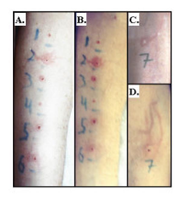
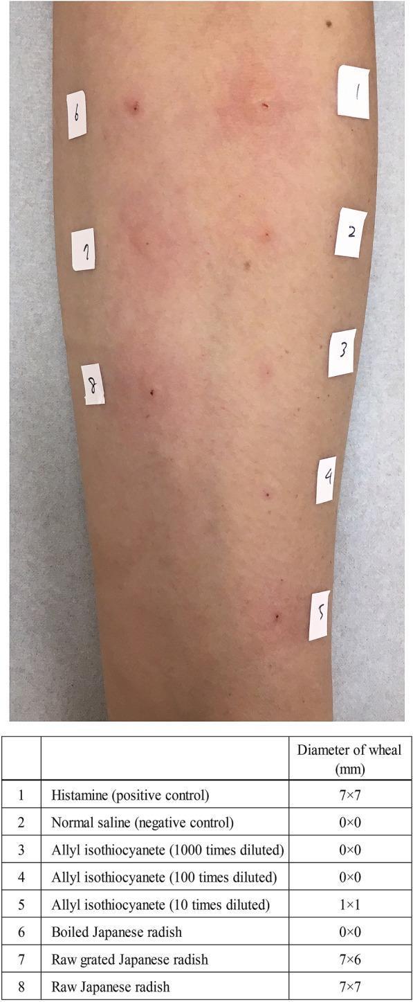
| Type I | Immediate / Anaphylactic / IgE-mediated | IgE + Mast cells | Minutes |
| Type II | Antibody-mediated cytotoxic | IgG / IgM on cell surface | Hours |
| Type III | Immune complex-mediated | Soluble IgG/IgM + Complement | Hours - days |
| Type IV | Delayed-type / Cell-mediated | T lymphocytes (CD4+/CD8+) | 24-72 hours |
Robbins & Kumar Basic Pathology, Table 5.2
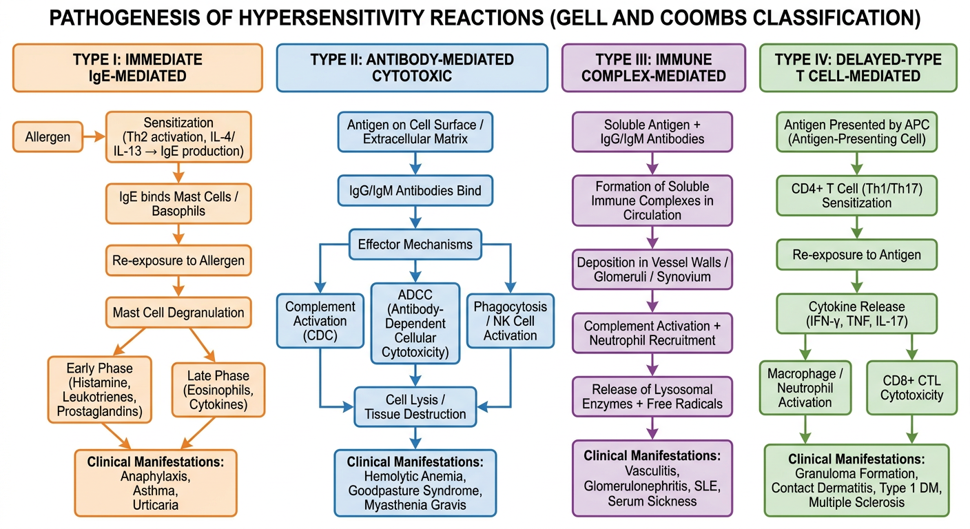
Pathogenesis Flowchart
TYPE I - Immediate (IgE-Mediated) Hypersensitivity
Definition
A tissue reaction occurring within minutes of antigen contact with IgE bound to mast cells. Also called allergy or atopy.
Pathogenesis
Phase 1 - Sensitization
FIRST EXPOSURE TO ALLERGEN
        ↓
Allergen processed by Dendritic Cells / APCs
        ↓
Th2 cell activation
        ↓
Cytokine release: IL-4, IL-5, IL-13
        ↓
IL-4 + IL-13 → B cell class switching → IgE production
IL-5 → Eosinophil activation and recruitment
        ↓
IgE binds to FcεRI receptors on MAST CELLS and BASOPHILS
        ↓
Sensitized mast cells distributed in tissues
(Individual now "sensitized")
Phase 2 - Activation (Re-exposure)
SECOND EXPOSURE TO SAME ALLERGEN
        ↓
Allergen cross-links 2 IgE molecules on mast cell surface
        ↓
FcεRI receptor clustering → intracellular signaling
        ↓
MAST CELL DEGRANULATION
Cellular Mediators
Primary (Preformed, released in seconds-minutes):
| Mediator | Effect |
|---|
| Histamine | Vasodilation, ↑ vascular permeability, smooth muscle contraction, mucus secretion |
| Heparin | Anticoagulant in granules |
| Neutral proteases (tryptase, chymase) | Tissue damage, kinin activation |
| Chemotactic factors (ECF, NCF) | Recruit eosinophils and neutrophils |
Secondary (Newly synthesized - Lipid mediators, hours):
| Mediator | Effect |
|---|
| Leukotrienes C4, D4, E4 | Prolonged bronchospasm, ↑ mucus, ↑ vascular permeability (1000x more potent than histamine) |
| Prostaglandin D2 | Bronchospasm, vasodilation |
| PAF (Platelet Activating Factor) | Platelet aggregation, bronchoconstriction |
| Thromboxane A2 | Vasoconstriction, bronchoconstriction |
Cytokines (Late phase, hours):
| Cytokine | Effect |
|---|
| IL-4, IL-13 | Sustain Th2 responses, IgE production |
| IL-5 | Eosinophil survival and activation |
| TNF, IL-1 | Inflammation |
| IL-8 (CXCL8) | Neutrophil recruitment |
Biphasic Response
- Early phase (0-30 min): Histamine-driven - vasodilation, edema, bronchospasm
- Late phase (2-24 hrs): Eosinophils, neutrophils, basophils - tissue damage, sustained inflammation
Clinical Examples
- Anaphylaxis (systemic - bee sting, penicillin, peanuts)
- Bronchial asthma (atopic)
- Allergic rhinitis / hay fever
- Urticaria (hives)
- Atopic dermatitis (eczema)
- Food allergies
Treatment & Prophylaxis
| Category | Agents | Mechanism |
|---|
| Emergency | Epinephrine (adrenaline) | α1: vasoconstriction; β2: bronchodilation; reverses anaphylaxis |
| Antihistamines | Cetirizine, fexofenadine, diphenhydramine | H1 receptor blockade |
| Bronchodilators | Salbutamol, terbutaline | β2 agonist |
| Corticosteroids | Prednisolone, hydrocortisone | Anti-inflammatory, suppress late phase |
| Mast cell stabilizers | Cromoglycate, nedocromil | Prevent degranulation (prophylaxis) |
| Anti-IgE | Omalizumab | Binds free IgE, blocks FcεRI binding |
| Anti-IL-5 | Mepolizumab | Blocks eosinophil activation |
| Anti-IL-4R | Dupilumab | Blocks IL-4/IL-13 signaling |
| Leukotriene antagonists | Montelukast, zafirlukast | Block LTC4/LTD4/LTE4 receptors |
| Immunotherapy (desensitization) | Allergen-specific SIT | Shift Th2 → Th1, induce regulatory T cells, IgG4 blocking antibodies |
Prophylaxis:
- Avoidance of known allergens
- Pre-treatment with antihistamines and corticosteroids for known triggers
- Epinephrine auto-injector (EpiPen) for at-risk individuals
- Allergen immunotherapy (desensitization)
TYPE II - Antibody-Mediated Cytotoxic Hypersensitivity
Definition
Injury caused by IgG or IgM antibodies directed against antigens present on cell surfaces or in the extracellular matrix.
Pathogenesis
ANTIGEN ON CELL SURFACE / ECM
        ↓
IgG or IgM antibodies produced (B cell activation)
        ↓
Antibodies bind to cell-surface antigen
        ↓
        ├──→ COMPLEMENT ACTIVATION (Classical pathway)
        │           ↓
        │    C3b opsonization → Phagocytosis (macrophages)
        │    C5b-9 (MAC) → Direct cell lysis (CDC)
        │    C3a, C5a → Inflammation, mast cell activation
        │
        ├──→ ADCC (Antibody-Dependent Cellular Cytotoxicity)
        │           ↓
        │    NK cells + macrophages bind Fc region of IgG
        │    → Release perforin, granzymes → Cell death
        │
        ├──→ PHAGOCYTOSIS
        │           ↓
        │    Fc receptors on macrophages bind opsonized cells
        │    → Engulfment and destruction
        │
        └──→ ANTIBODY-MEDIATED DYSFUNCTION (no lysis)
                    ↓
             Antibody blocks receptor function
             OR mimics ligand (stimulatory)
Cellular Mediators
- Complement proteins: C1q, C3b (opsonin), C3a/C5a (anaphylatoxins), C5b-9 (MAC)
- Macrophages: FcγR-mediated phagocytosis
- NK cells: ADCC via FcγRIII (CD16)
- Neutrophils: Recruited by C5a; tissue damage via enzymes
- Autoantibodies (functional): Anti-AChR (MG), anti-TSH-R (Graves'), anti-GBM (Goodpasture)
Clinical Examples
| Disease | Antigen | Mechanism |
|---|
| Autoimmune hemolytic anemia | RBC surface proteins | Complement + phagocytosis → RBC destruction |
| Immune thrombocytopenia (ITP) | Platelet glycoproteins | Platelet phagocytosis |
| Goodpasture syndrome | Type IV collagen (GBM) | Complement-mediated glomerulonephritis + pulmonary hemorrhage |
| Myasthenia Gravis | Acetylcholine receptor | Antibody blocks AChR → muscle weakness (no lysis) |
| Graves' disease | TSH receptor | Antibody stimulates TSH-R → hyperthyroidism |
| Transfusion reactions | Blood group antigens | Complement-mediated RBC lysis |
| Pemphigus vulgaris | Desmoglein (desmosome) | Disruption of skin adhesion → blisters |
| Rhesus incompatibility | Rh antigen on fetal RBCs | Hemolytic disease of the newborn |
Treatment & Prophylaxis
| Approach | Examples |
|---|
| Corticosteroids | Reduce antibody production, suppress inflammation |
| IV Immunoglobulin (IVIG) | Fc receptor blockade, immune modulation |
| Rituximab (anti-CD20) | Depletes B cells → reduces antibody production |
| Plasma exchange | Removes circulating antibodies |
| Splenectomy | Removes site of phagocytosis and antibody production (ITP) |
| Acetylcholinesterase inhibitors | Pyridostigmine - symptomatic (MG) |
| Anti-Rh immunoglobulin | Rh prophylaxis to prevent sensitization in Rh-negative mothers |
TYPE III - Immune Complex-Mediated Hypersensitivity
Definition
Injury caused by deposition of soluble antigen-antibody complexes (IgG/IgM) in vascular walls and tissues, with subsequent complement activation and inflammation.
Pathogenesis
PERSISTENT / EXCESS ANTIGEN
        ↓
IgG / IgM antibodies formed
        ↓
Soluble antigen-antibody complexes formed in circulation
        ↓
NORMALLY: cleared by phagocytes (large complexes)
DISEASE: small/intermediate complexes escape clearance
        ↓
Complexes deposit in:
- Vessel walls (vasculitis)
- Glomerular basement membrane (nephritis)
- Synovial membranes (arthritis)
        ↓
COMPLEMENT ACTIVATION (Classical pathway)
        ↓
C3a + C5a (anaphylatoxins) →
  - Mast cell degranulation → histamine → ↑ vascular permeability
  - MORE complex deposition
C5a →
  - Chemotaxis → Neutrophil and monocyte recruitment
        ↓
NEUTROPHIL ACTIVATION
        ↓
Release of:
- Lysosomal enzymes (proteases, collagenase)
- Reactive oxygen species (ROS)
- Inflammatory cytokines
        ↓
TISSUE INJURY: Vasculitis, fibrinoid necrosis, glomerulonephritis
Cellular Mediators
- Complement fragments: C3a, C5a (anaphylatoxins + chemotaxins), C5b-9
- Neutrophils: Primary effectors - release enzymes and ROS
- Macrophages/Monocytes: Recruited via Fc receptors and C5a
- Mast cells: Activated by C3a/C5a - amplify vascular permeability
- Platelets: Aggregation within vessels - microthrombi
Factors Determining Severity
- Size: Small complexes are most pathogenic (not cleared by phagocytes)
- Charge: Cationic antigens bind basement membranes more avidly
- Amount: Excess antigen over antibody favors small complex formation
- Site: Kidneys and joints are especially vulnerable
Classical Model - Serum Sickness
- Day 0: Foreign protein (horse serum) injected
- Day 6-8: Antibodies produced; complexes form in antigen excess
- Day 10-12: Complexes deposit → fever, rash, arthritis, glomerulonephritis
- Resolution as antigen cleared
Arthus Reaction (local): Subcutaneous antigen in pre-immunized → local immune complex deposition → local vasculitis, necrosis within 4-8 hours.
Clinical Examples
| Disease | Antigen | Features |
|---|
| SLE | DNA, nucleoproteins | Nephritis, vasculitis, arthritis |
| Post-streptococcal GN | Streptococcal cell wall antigens | Nephritis (2-3 weeks after throat infection) |
| Serum sickness | Foreign proteins (horse serum, drugs) | Fever, rash, arthralgias, nephritis |
| Polyarteritis nodosa | HBsAg (in some cases) | Systemic vasculitis |
| Hypersensitivity pneumonitis | Inhaled organic antigens (farmer's lung) | Pulmonary inflammation |
| Cryoglobulinemia | HCV-associated immune complexes | Vasculitis, nephritis |
| Arthus reaction | Local antigen-antibody | Localized necrosis at injection site |
Treatment & Prophylaxis
| Approach | Examples |
|---|
| Remove antigen source | Treat underlying infection (strep, HCV) |
| Corticosteroids | Suppress complement activation and inflammation |
| NSAIDs | Symptomatic relief of arthritis/fever |
| Antimalarials | Hydroxychloroquine - SLE |
| Immunosuppressants | Cyclophosphamide, azathioprine, MMF (SLE nephritis) |
| Biologics | Belimumab (anti-BLyS) - SLE |
| Plasma exchange | Remove circulating complexes |
TYPE IV - Delayed-Type (Cell-Mediated) Hypersensitivity
Definition
Tissue injury mediated by T lymphocytes (CD4+ Th1/Th17 and CD8+ CTLs), NOT antibody. Reaction occurs 24-72 hours after antigen challenge (hence "delayed").
Pathogenesis
Phase 1 - Sensitization
FIRST ANTIGEN EXPOSURE
        ↓
Antigen processed and presented by APCs (dendritic cells)
with MHC Class II (for CD4+) or MHC Class I (for CD8+)
        ↓
Naïve T cells activated in secondary lymphoid organs
        ↓
Differentiation:
CD4+ → Th1 (IFN-γ) / Th17 (IL-17)
CD8+ → Cytotoxic T Lymphocytes (CTLs)
        ↓
Memory T cells formed and circulate
Phase 2 - Effector Response (Re-exposure)
SECOND ANTIGEN EXPOSURE (24-72 hrs)
        ↓
Memory T cells recognize antigen
        ↓
        ├──→ CD4+ Th1 cells activated
        │        ↓
        │   IFN-γ → Macrophage activation
        │   TNF → Inflammation, vascular changes
        │   IL-2 → T cell proliferation
        │   Activated macrophages:
        │     - Kill intracellular pathogens
        │     - Release more cytokines, ROS, proteases
        │     - Form GRANULOMA (with Th1 cytokines)
        │
        ├──→ CD4+ Th17 cells activated
        │        ↓
        │   IL-17, IL-22 → Neutrophil recruitment
        │   IL-17 → Epithelial inflammation
        │
        └──→ CD8+ CTL cytotoxicity
                  ↓
             Direct killing via:
             - Perforin/Granzyme B → Apoptosis
             - Fas-FasL interaction → Apoptosis
             → Tissue destruction (viral infections, transplant rejection)
Cellular Mediators
| Cell | Mediators | Effect |
|---|
| CD4+ Th1 | IFN-γ, TNF, IL-2, lymphotoxin | Macrophage activation, inflammation |
| CD4+ Th17 | IL-17, IL-21, IL-22 | Neutrophil recruitment, epithelial damage |
| CD8+ CTLs | Perforin, Granzyme B, Fas-L | Direct cell killing |
| Macrophages (activated) | TNF, IL-1, IL-6, IL-12, ROS, proteases | Tissue destruction, granuloma |
| NK cells | IFN-γ, perforin | Non-MHC-restricted killing |
Subtypes of Type IV
| Subtype | Cells | Timing | Example |
|---|
| DTH (classic) | CD4+ Th1 | 48-72 hrs | Tuberculin test (Mantoux) |
| Contact sensitivity | CD4+ Th1/Th17 | 48-72 hrs | Poison ivy, nickel allergy |
| Granulomatous | Macrophages + CD4+ | Days-weeks | TB, sarcoidosis, leprosy |
| CTL-mediated | CD8+ CTL | Variable | Viral hepatitis, graft rejection |
Clinical Examples
| Disease | Antigen Target | Mechanism |
|---|
| Contact dermatitis | Urushiol (poison ivy), nickel, drugs | Th1-mediated epidermal inflammation |
| Tuberculosis | Mycobacterium TB antigens | Granuloma formation |
| Type 1 Diabetes | Islet β-cell antigens (insulin, GAD) | CTL + Th1 → islet destruction |
| Multiple Sclerosis | Myelin basic protein | Th1 + Th17 → demyelination |
| Rheumatoid Arthritis | Citrullinated proteins, collagen? | Th17-mediated synovial inflammation |
| Inflammatory bowel disease | Enteric bacteria | Th1 + Th17 cytokines |
| Transplant rejection (acute) | Donor HLA antigens | CTL + Th1 |
| Hashimoto's thyroiditis | Thyroid peroxidase, thyroglobulin | Th1 + CTL → thyroid destruction |
| Psoriasis | Unknown | Th17-dominated |
Treatment & Prophylaxis
| Approach | Examples | Target |
|---|
| Corticosteroids | Topical (contact dermatitis), systemic | Suppress T cell activation and cytokine production |
| Calcineurin inhibitors | Tacrolimus, cyclosporine | Block IL-2 signaling → suppress T cells |
| Anti-TNF | Infliximab, adalimumab | Neutralize TNF (RA, IBD, psoriasis) |
| Anti-IL-17 | Secukinumab, ixekizumab | Block IL-17A (psoriasis, AS) |
| Anti-IL-12/23 | Ustekinumab | Block Th1 and Th17 responses |
| Anti-IL-23 | Guselkumab, risankizumab | Selective Th17 suppression |
| Anti-CD20 | Rituximab | B cell depletion (also helps in RA) |
| CTLA-4-Ig | Abatacept | Blocks CD28-B7 costimulation → T cell anergy |
| Anti-CD25 | Basiliximab, daclizumab | Block IL-2R → prevent T cell proliferation |
| Methotrexate, azathioprine | DMARDs | Antiproliferative immunosuppression |
| Avoidance | Remove contact allergens | Prophylaxis for contact dermatitis |
Quick Comparison Summary
| Feature | Type I | Type II | Type III | Type IV |
|---|
| Antibody | IgE | IgG / IgM | IgG / IgM | None |
| Complement | No (usually) | Yes | Yes | No |
| Effector | Mast cells, eosinophils | Antibody + complement + phagocytes | Neutrophils | T cells, macrophages |
| Timing | Minutes (+ late 2-24h) | Hours | Hours-days | 24-72 hours |
| Transfer | Serum (IgE) | Serum (IgG) | Serum | Lymphocytes |
| Key cytokines | IL-4, IL-5, IL-13 | - | - | IFN-γ, IL-17, TNF |
| Prototypic disease | Anaphylaxis | Goodpasture | SLE / serum sickness | Contact dermatitis / TB |
| Histology | Edema, eosinophils | Cell lysis, phagocytosis | Vasculitis, fibrinoid necrosis | Perivascular infiltrate, granuloma |
Sources: Robbins & Kumar Pathologic Basis of Disease, Robbins & Kumar Basic Pathology; Cellular and Molecular Immunology (Abbas, Lichtman & Pillai); Roitt's Essential Immunology