
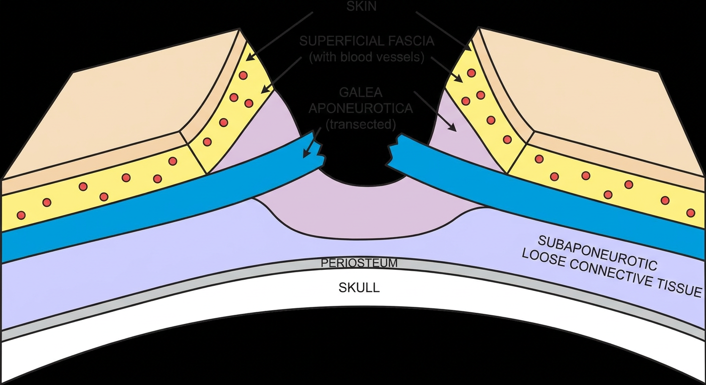
Schematic diagram of gaping wound on scalp
Reading File
This is a visual/anatomy query. Let me search the medical library for scalp wound diagrams.
Finding Sources
Finding Sources
Good results. Let me read the Roberts and Hedges section on scalp lacerations and the Rosen's section for diagrams/images.
Reading File
Reading File
I found exactly what's needed. There's an anatomy diagram and a Figure 35.53 showing the gaping wound. Let me also look for the actual image URL for Figure 35.53.
Reading File
I see Figure 35.53 is referenced but its image URL isn't shown inline - it seems the image might be just before line 1628. Let me search nearby for the image URL, and also fetch the anatomy diagram already found.
Reading File
Loading Image
Excellent - I have the scalp anatomy diagram. Now let me look for Figure 35.53 itself (the gaping wound image) - it seems the image tag may be just before line 1628. Let me check earlier lines.
Finding Sources
Reading File
The Figure 35.53 image tag appears to be missing/embedded differently. Let me generate a schematic diagram of a gaping scalp wound to complement the textbook anatomy diagram, and provide a comprehensive explanation.
Generating Image
Schematic cross-section diagram of a gaping scalp wound showing the five anatomical layers: skin, superficial fascia (with blood vessels), galea aponeurotica (transected/cut), subaponeurotic loose connective tissue, periosteum, and skull. The wound is gaping open wide because the galea aponeurotica has been transected. Labels point to each layer. The wound edges are pulled apart showing the depth of injury. Medical educational illustration style, clean line art with color coding of each layer.
Here is a complete schematic overview of a gaping scalp wound, drawing from Roberts and Hedges' Clinical Procedures in Emergency Medicine.
Gaping Wound of the Scalp
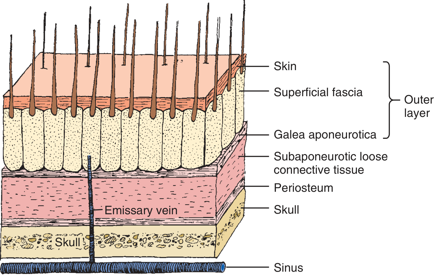
Scalp Anatomy (Fig. 35.52)
Schematic of a Gaping Scalp Wound
The 5 Layers of the Scalp (SCALP mnemonic)
| Layer | Description |
|---|---|
| S - Skin | Thick, hair-bearing outer covering |
| C - Connective tissue (superficial fascia) | Contains blood vessels - major source of bleeding in lacerations |
| A - Aponeurosis (Galea aponeurotica) | Tough fibrous sheet connecting frontalis and occipitalis muscles |
| L - Loose areolar (subaponeurotic) tissue | Contains emissary veins draining to intracranial sinuses |
| P - Periosteum | Thin, adhered to skull; cannot be sutured |
Why Does a Scalp Wound Gape?
This is the key clinical point from Roberts and Hedges' Clinical Procedures in Emergency Medicine:
- The outer layer (skin + superficial fascia + galea) is maintained by inelastic, tough fibrous septa
- These septa keep wounds from gaping unless all three portions have been traversed
- A wound that gapes open = the laceration has extended beneath the galea layer
- The galea is firmly attached to the underside of the subcutaneous fascia and acts like a retractor - when cut, it pulls the wound edges wide apart
Clinical Significance of Gaping
1. Hemostasis
- Blood vessels in the superficial fascia remain patent because fibrous SQ septa prevent normal vessel retraction - causing profuse, persistent hemorrhage
- A gaping wound with galeal transection requires suturing of the galea to achieve hemostasis
2. Infection risk
- The subaponeurotic loose tissue contains emissary veins connecting to intracranial venous sinuses
- A gaping wound breaching this layer can allow bacterial spread, potentially causing osteomyelitis, meningitis, or brain abscess
3. Wound closure
- Gaping (galeal) wounds: require single-layer sutures incorporating skin + SQ fascia + galea, or separate galeal closure with absorbable sutures (3-0 or 4-0, inverted/buried knot)
- Non-gaping wounds (galea intact): can be closed with superficial sutures only
- Horizontal mattress sutures are particularly useful for gaping scalp lacerations due to minimal skin mobility
Source: Roberts and Hedges' Clinical Procedures in Emergency Medicine, Figures 35.52-35.53 (pp. 813-814)
Which layer is called dangerous area.of scalp and why
The subaponeurotic loose areolar tissue (Layer 4 - the "L" in SCALP) is called the dangerous area of the scalp. It is dangerous because it contains emissary veins that communicate directly with the intracranial venous sinuses, allowing infections to spread into the skull.
Searching sources...
Finding Sources
Finding Sources
Finding Sources
Finding Sources
Finding Sources
Finding Sources
The FTS isn't matching - likely the content is in the block we already read. Let me use the Roberts and Hedges text from the previous session (which we already read) and also check the anatomy textbooks directly.
Finding Sources
Finding Sources
Found it - the Forensic Medicine textbook (P C Dikshit) and Parikh's likely have the "dangerous area" description. Let me read those sections, plus Gray's Anatomy and the Scott-Brown's sections.
Reading File
Reading File
Reading File
Excellent - rich content from multiple authoritative sources. Let me also fetch the coronal section diagram from Dikshit's Forensic Medicine.
Loading Image
This is an outstanding diagram that perfectly illustrates the answer. Now I have everything needed for a comprehensive, well-illustrated response.
The "Dangerous Area" of the Scalp
Layer 4 - Subaponeurotic Loose Areolar Tissue
The 4th layer of the scalp - the subaponeurotic loose areolar tissue (also called the subgaleal space) - is classically called the "dangerous area" of the scalp.
Coronal Section: Scalp to Brain (Fig. 15.1)
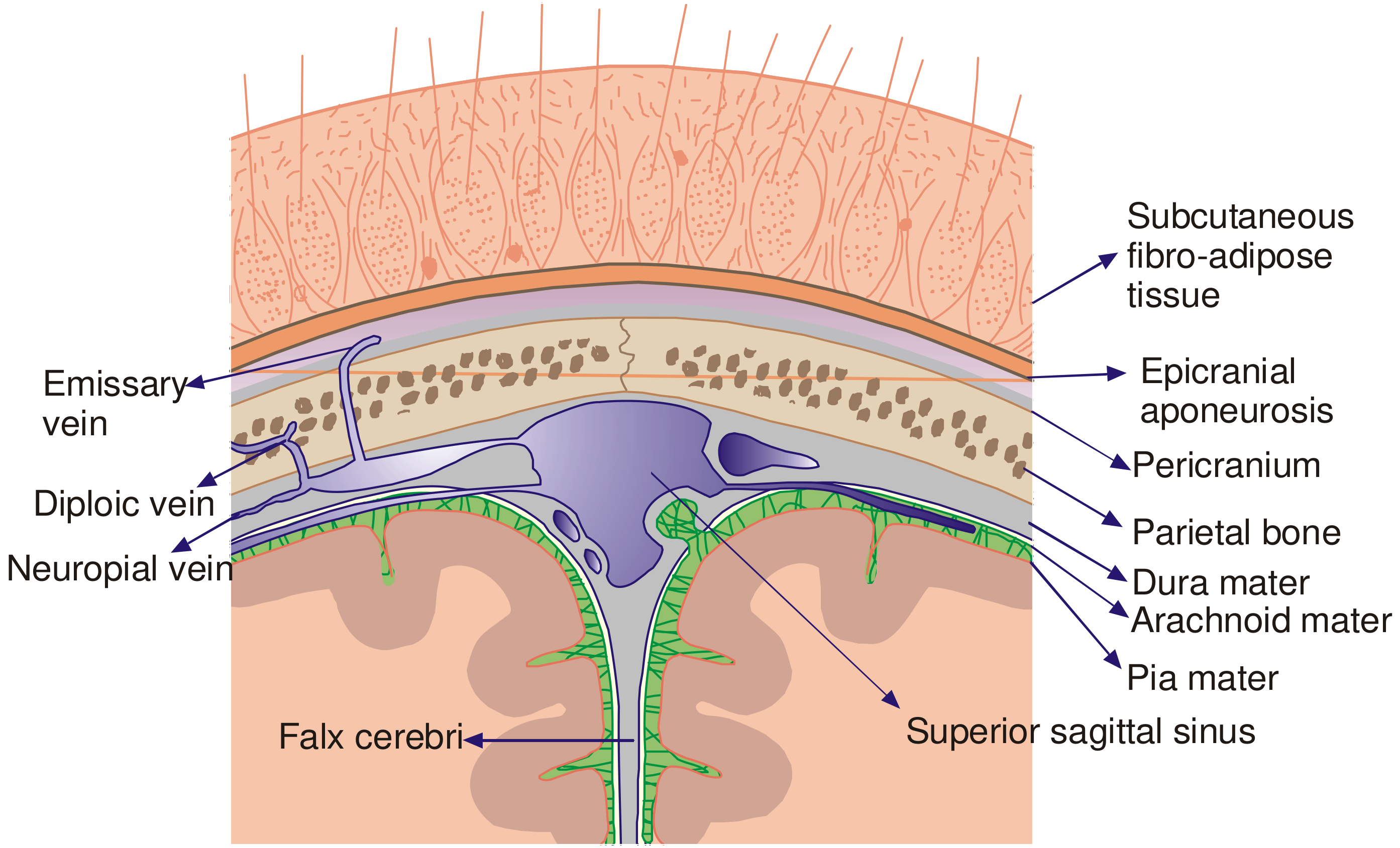
This diagram clearly shows the emissary veins passing through the skull connecting the scalp to the intracranial sinuses - the anatomical basis for why this layer is so dangerous.
The Five Scalp Layers (SCALP)
| # | Layer | Clinical Notes |
|---|---|---|
| 1 | S - Skin | Thickest skin on the body (3-8 mm) |
| 2 | C - (Sub)Cutaneous fibro-adipose tissue | Contains blood vessels; bleeds profusely when cut |
| 3 | A - Aponeurosis (Galea aponeurotica) | When transected → wound gapes open |
| 4 | L - Loose areolar tissue (subaponeurotic) | ⚠️ THE DANGEROUS AREA |
| 5 | P - Pericranium (Periosteum) | Adhered to skull; cannot be sutured |
Why Is It Called the "Dangerous Area"?
This layer is dangerous for four interconnected reasons:
1. Direct Vascular Communication with Intracranial Sinuses
The loose areolar tissue of this layer is traversed by emissary veins - valveless veins that pass through foramina in the skull connecting:
- Scalp/face veins ↔ Diploic veins (in the spongy bone between inner and outer skull tables)
- Diploic veins ↔ Meningeal veins and intracranial venous sinuses (parasagittal, lateral, and cavernous sinuses)
Because these veins have no valves, blood and, critically, infection can flow in either direction.
2. Rapid Spread of Infection
A septic wound or abscess in this layer can spread infection to:
- Meninges → Meningitis
- Skull bone → Osteomyelitis
- Intracranial sinuses → Cavernous sinus thrombosis / thrombophlebitis of cranial sinuses
- Brain → Brain abscess
As stated in Roberts and Hedges': "In scalp wounds that penetrate this layer, bacteria may be carried by these vessels to the meninges and the intracranial sinuses."
3. Pus / Blood Can Spread Widely in Any Direction
The loose areolar nature of the tissue offers almost no resistance to spread. A collection (blood, pus, oedema) in this layer can:
- Spread circumferentially all around the skull under the galea
- Track anteriorly and gravitate into the orbital region (causing "black eyes" / supraorbital haematoma) from a blow to the top or back of the head - a phenomenon sometimes misleadingly suggesting a basal skull fracture
- Spread behind the ear from a temporal injury
4. Subgaleal Haematoma
Bleeding into this layer is not limited by fibrous septa (unlike the subcutaneous layer above). A subgaleal haematoma can therefore accumulate a very large volume of blood, which is particularly dangerous in neonates (caput succedaneum exceeds this space) and can cause haemorrhagic shock.
Summary Table: Why "Dangerous"
| Feature | Consequence |
|---|---|
| Valveless emissary veins → intracranial sinuses | Infection spreads to brain/meninges |
| Loose, non-compartmentalised tissue | Rapid circumferential spread of pus/blood |
| Communication with diploic veins | Osteomyelitis of skull |
| Large potential space | Massive subgaleal haematoma → shock (especially in neonates) |
Sources:
- P C Dikshit Textbook of Forensic Medicine and Toxicology, p. 192 (Fig. 15.1)
- Parikh's Textbook of Medical Jurisprudence Forensic Medicine and Toxicology, p. 323
- Roberts and Hedges' Clinical Procedures in Emergency Medicine, p. 813-814
- Scott-Brown's Otorhinolaryngology Head & Neck Surgery, p. 1314
This is a shared conversation. Sign in to Orris to start your own chat.