
How to know the difference between between true and psyedoaneurysm in ct and radiology
Reading File
Quick answer: On CT/MRI, a true aneurysm has all three vessel wall layers intact and tapers smoothly from the parent artery, while a pseudoaneurysm (false aneurysm) lacks true vessel wall layers - it is contained only by surrounding soft tissue or clot. The key CT sign is that a pseudoaneurysm has a "neck" connecting it to the artery with blood swirling inside a contained hematoma, often with irregular margins and surrounding soft tissue changes.
Searching sources...
Finding Sources
Finding Sources
Searching Images
pseudoaneurysm vs true aneurysm CT scan radiology
Good results. Let me now read the key sections from multiple books simultaneously.
Reading File
Reading File
Reading File
Finding Sources
I have a great diagram URL from Robbins. Let me fetch it and search for more imaging-specific content.
Loading Image
Finding Sources
Finding Sources
Excellent - now I have the yin-yang sign mentioned. Let me read those key sections on imaging findings.
Reading File
Reading File
Reading File
I now have rich content plus image URLs. Let me fetch the key ultrasound images and also look for CT-specific features.
Loading Image
Loading Image
Finding Sources
I now have comprehensive textbook content and imaging. Let me compose the full answer.
True Aneurysm vs. Pseudoaneurysm: Radiological Differentiation
Fundamental Structural Difference
This is the foundation of every imaging distinction:
| Feature | True Aneurysm | Pseudoaneurysm (False Aneurysm) |
|---|---|---|
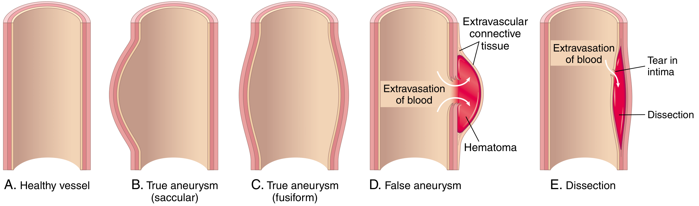
| Wall structure | All 3 layers intact (intima, media, adventitia) - just dilated/thinned | No true vessel wall - wall is only surrounding fibrous tissue/organized clot |
| Mechanism | Weakening/dilation of existing wall | Breach/rupture of vessel wall with contained hemorrhage |
| Communication | Direct - the sac IS the vessel | Indirect - blood leaks out via a "neck" into extravascular space |
| Rupture risk | Increases with size (Laplace's law) | High - relatively thin-walled sac under arterial pressure |
As Robbins Pathologic Basis of Disease illustrates:
1. CT Angiography (CTA) - Key Differentiating Features
True Aneurysm on CTA:
- Smooth, well-defined dilation of the vessel that is in continuity with the artery (the wall is the vessel wall)
- Fusiform or saccular shape, continuous with the vessel lumen
- Wall may show calcification (especially atherosclerotic aneurysms)
- Mural thrombus can be seen as a non-enhancing crescent lining the wall
- No surrounding soft tissue hematoma in uncomplicated cases
- The lumen enhances uniformly and brightly with contrast
Pseudoaneurysm on CTA:
- A rounded sac adjacent to (not truly part of) the vessel, connected via a neck - this neck is the breach in the vessel wall
- The sac shows contrast enhancement (blood actively swirling in) but is surrounded by non-enhancing hematoma or soft tissue
- Irregular, non-calcified "wall" (it's fibrous tissue, not true vessel wall)
- Surrounding soft tissue changes, edema, or hematoma are common
- On late phases, the sac may show "swirling" enhancement
- History of trauma, surgery, catheterization, or infection is a critical clinical clue
Practical CTA clue: If you see a contrast-enhancing sac that is NOT smoothly contiguous with the vessel lumen but connects via a narrow channel, surrounded by hematoma - that is a pseudoaneurysm. If the sac is a smooth outpouching with calcified walls and no surrounding hematoma, favor a true aneurysm.
2. Doppler Ultrasound - The Most Specific Bedside Tool
Duplex ultrasound with color Doppler is the diagnostic test of choice (sensitivity 94%, specificity 97%) and most clearly separates the two - Current Surgical Therapy 14e.
True Aneurysm on Duplex:
- Fusiform or saccular dilation of the vessel with smooth, continuous walls
- Laminar or slow turbulent flow on color Doppler
- B-mode shows calcified, echogenic walls
- No discrete neck; the vessel simply widens
Pseudoaneurysm on Duplex - Pathognomonic Signs:
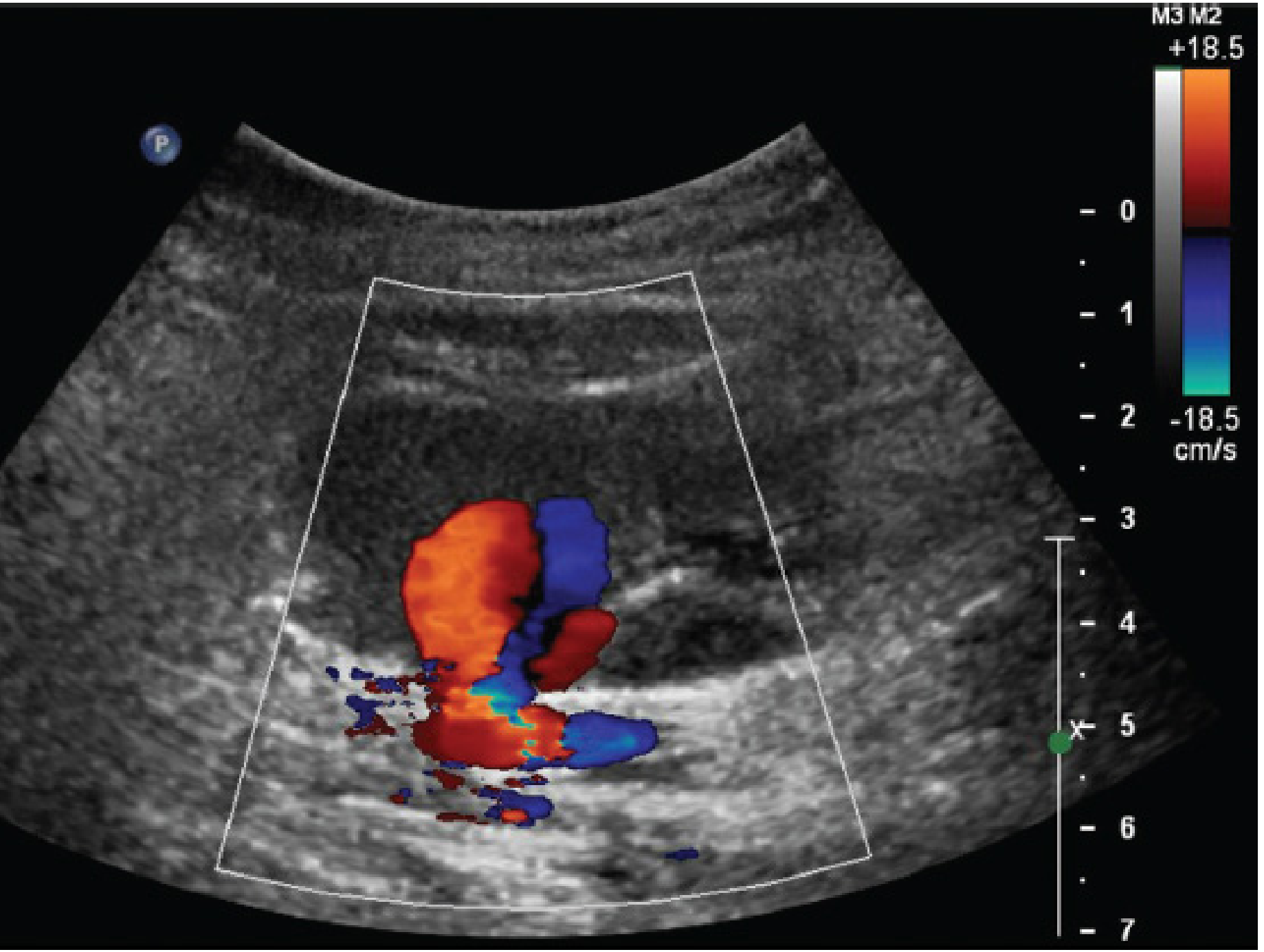
A. The Yin-Yang Sign (color Doppler)
During systole, blood jets into the sac (one color); during diastole, it flows back out (opposite color). This creates the characteristic red-blue swirling pattern within the sac, resembling a yin-yang symbol. This is the hallmark finding.
B. To-and-Fro Waveform (spectral Doppler at the neck)
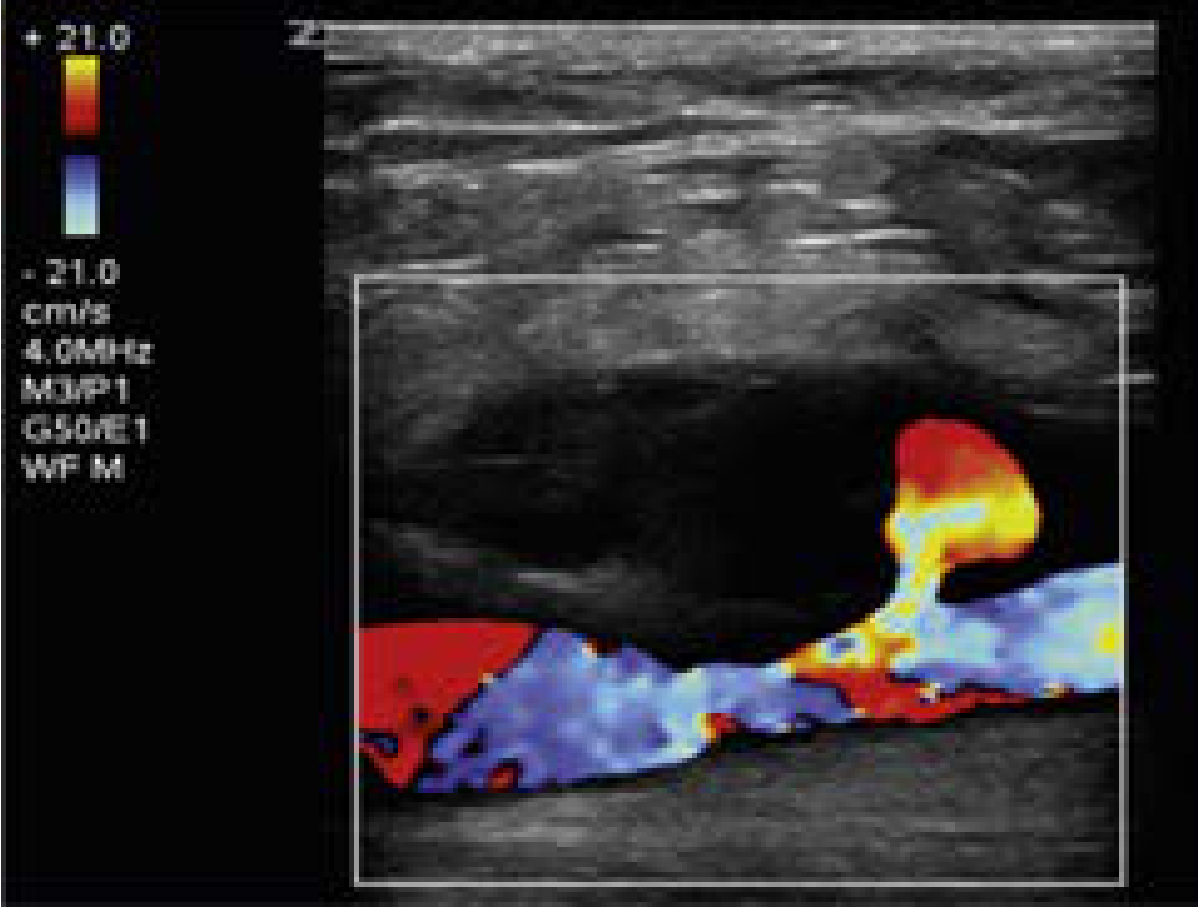
Spectral Doppler placed at the narrow neck of the pseudoaneurysm shows a biphasic signal: flow going INTO the sac in systole, flow returning OUT in diastole. This "to-and-fro" pattern is diagnostic of a pseudoaneurysm. A true aneurysm has no neck and no reversal of flow.
C. Identifiable Neck
The narrow channel connecting the parent artery to the sac is visible on B-mode and color Doppler. True aneurysms have no such neck.
D. B-mode alone is insufficient - a pseudoaneurysm sac can look just like a hematoma on grayscale. Color Doppler is mandatory to confirm active flow within the sac.
3. Summary Comparison Table
| Imaging Feature | True Aneurysm | Pseudoaneurysm |
|---|---|---|
| Wall | Calcified, smooth, 3-layer | Thin fibrous, no calcification |
| Shape | Fusiform or saccular, continuous with vessel | Rounded sac, OFF the vessel, connected via neck |
| Neck | Absent | Present (key feature) |
| Surrounding tissue | Normal | Hematoma, edema, soft tissue changes |
| CT enhancement | Uniform, same phase as vessel | Sac enhances; surrounding hematoma does not |
| Color Doppler | Laminar/slow flow, no directional reversal | Yin-yang sign (swirling) |
| Spectral Doppler at neck | N/A | To-and-fro waveform (pathognomonic) |
| B-mode alone | Usually diagnostic | Cannot distinguish from hematoma - need Doppler |
| Cause | Atherosclerosis, connective tissue, congenital | Trauma, catheterization, iatrogenic, infection |
| Spontaneous thrombosis | Does not occur | Can occur (especially <2 cm) |
4. Cardiac (Ventricular) Pseudoaneurysm - Special Case
On echocardiography/cardiac MRI after MI, a ventricular pseudoaneurysm has narrow neck : sac ratio < 0.5 (the neck is much narrower than the sac), while a true ventricular aneurysm has a wide mouth (neck : sac ratio ≥ 0.5). Pseudoaneurysms post-MI are contained ruptures - the walls are pericardium only, not myocardium - Textbook of Clinical Echocardiography.
Key Clinical Take-Away
The CT angiography triad for pseudoaneurysm:
- Contrast-filled sac adjacent to (not continuous with) a vessel
- A visible neck connecting them
- Surrounding hematoma or soft tissue changes
The ultrasound triad for pseudoaneurysm:
- Anechoic/hypoechoic sac next to an artery
- Yin-yang color pattern within the sac
- To-and-fro spectral waveform at the neck
If all three Doppler signs are present, the diagnosis is virtually certain and no further imaging is needed before treatment.
This is a shared conversation. Sign in to Orris to start your own chat.