
Nabothian cyst
nabothian cyst cervix
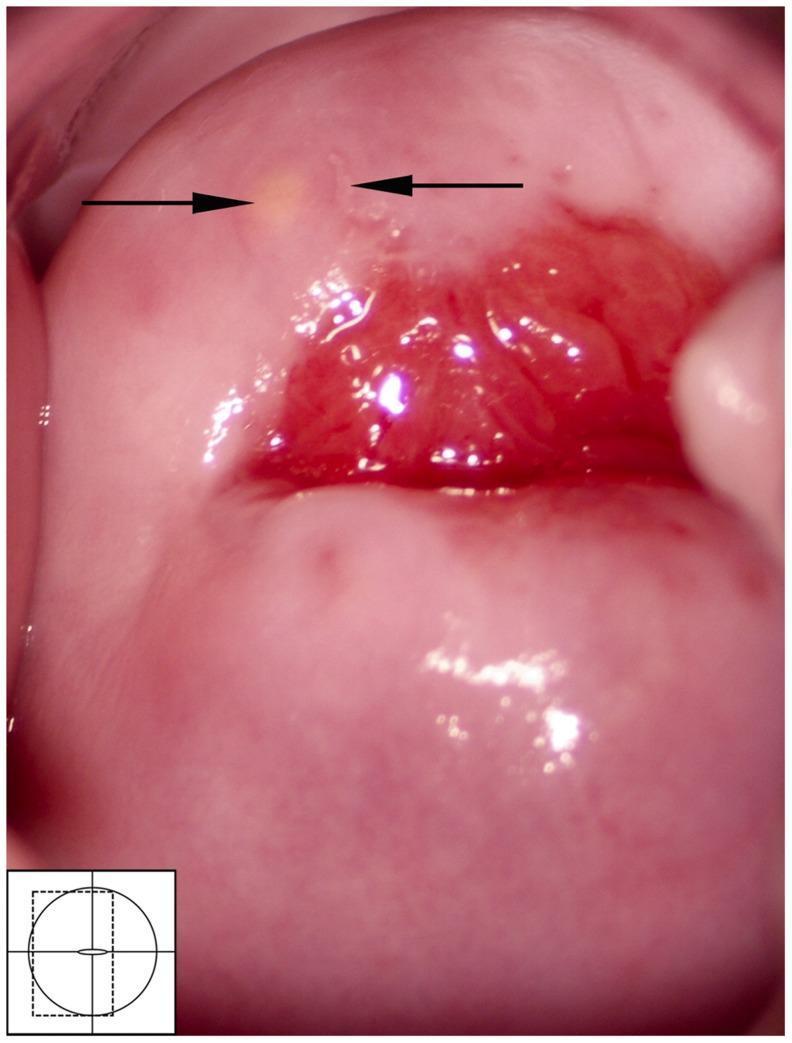
This colposcopic image provides a detailed view of the human cervix, centered on the external os and the transformation zone. On the anterior lip of the cervix, approximately at the 11 o'clock position (indicated by the left arrow), a small, circular, yellow-colored Nabothian cyst is visible. The cyst appears as a smooth, localized elevation representing mucus retention beneath metaplastic squamous epithelium. Adjacent to the cyst (indicated by the right arrow), there is a small, irregular area of off-white leukoplakia, which represents a mucosal abnormality. The central region surrounding the cervical os shows physiological erythema typical of the columnar epithelium. This clinical photograph is used in medical education to differentiate benign findings like Nabothian cysts from other cervical pathologies, such as those associated with female genital schistosomiasis, herpes simplex virus, or cervical intraepithelial neoplasia (CIN).
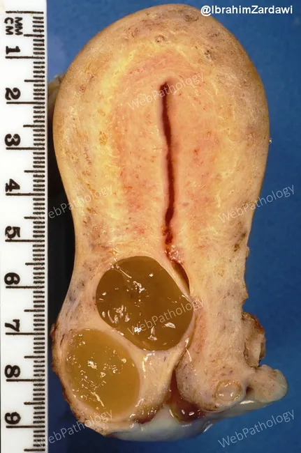
Gross pathology photograph of a grossly opened cervix displaying Nabothian cysts. Specimen obtained from a multiparous patient and opened in longitudinal fashion to expose the endocervical mucosa. Multiple translucent to yellow-brown cystic dilatations measure ranging from 0.2 to 1.0 cm; in this case several cysts are larger. The cysts are situated at the surface and within the cervical wall, with some approaching the outer surface and extending into paracervical soft tissue. The cyst contents are gelatinous, tenacious mucin. The cyst walls are smooth to slightly irregular, with a single layer of mucinous columnar or flat epithelium lining; no gross mass lesion is evident. Background stroma appears unremarkable; there is a chronic inflammatory background compatible with cervicitis in some glands. The differential diagnosis at gross inspection includes minimal deviation adenocarcinoma; however, there is no solid mass, no desmoplastic response, and no obvious invasion. The findings are typical of benign Nabothian cysts arising from blocked endocervical glands. This image highlights endocervical gland dilation and mucus retention as common incidental gynecologic pathology. Clinically, Nabothian cysts are usually asymptomatic, may be associated with chronic cervicitis, and are important to distinguish from malignant glandular tumors to avoid overtreatment. Useful for medical education and radiologic correlation.
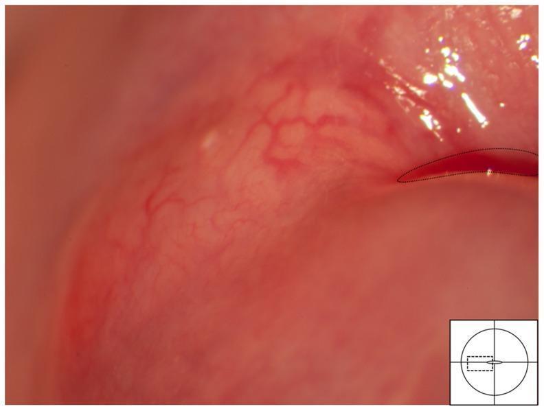
This colposcopic image displays a clinical photograph of the uterine cervix showing a Nabothian cyst. The cyst is visible as a pale yellow, spheroid structure localized within the transformation zone, adjacent to the squamo-columnar junction (demarcated by a dashed line). It exhibits a characteristic elevated surface caused by the retention of mucus beneath the metaplastic squamous epithelium. A distinct vascular network is draped over the cyst's surface; these mucosal blood vessels show regular branching patterns and appear to be pushed aside or stretched by the underlying cystic mass. This benign finding serves as an important differential diagnosis for other cervical mucosal lesions, such as the homogenous sandy patches and rubbery papules associated with Female Genital Schistosomiasis (FGS). The image illustrates the typical appearance of a retention cyst in a gynecological context, emphasizing the relationship between smooth surface morphology and regular vascular distribution.
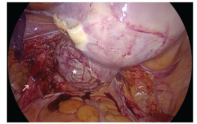
This intraoperative laparoscopic photograph provides a view of the pelvic cavity during a gynecological surgical procedure. The primary focus is a large, well-defined cervical cyst, marked with a white star, located in the posterior left lateral wall of the cervix. The cyst appears as a rounded, subserosal mass with a slightly irregular, lobulated surface and prominent superficial vascular markings. The sheer size of the cyst causes visible displacement of the cervix to the right lateral anterior side. An arrow points to the left ureter, which is identified in the retroperitoneal space following the dissection of the posterior leaf of the broad ligament and the uterosacral ligament. The ureter appears as a thin, tubular structure in close proximity to the cyst wall, emphasizing the risk of secondary ureteral compression and hydroureter in cases of large cervical masses like Nabothian cysts or cervical adenomyosis. The surrounding surgical field shows the uterus, pelvic peritoneum, and initial stages of tissue dissection required for a laparoscopic hysterectomy.
nabothian cyst cervix
Nabothian Cyst
Definition & Synonyms
Pathogenesis
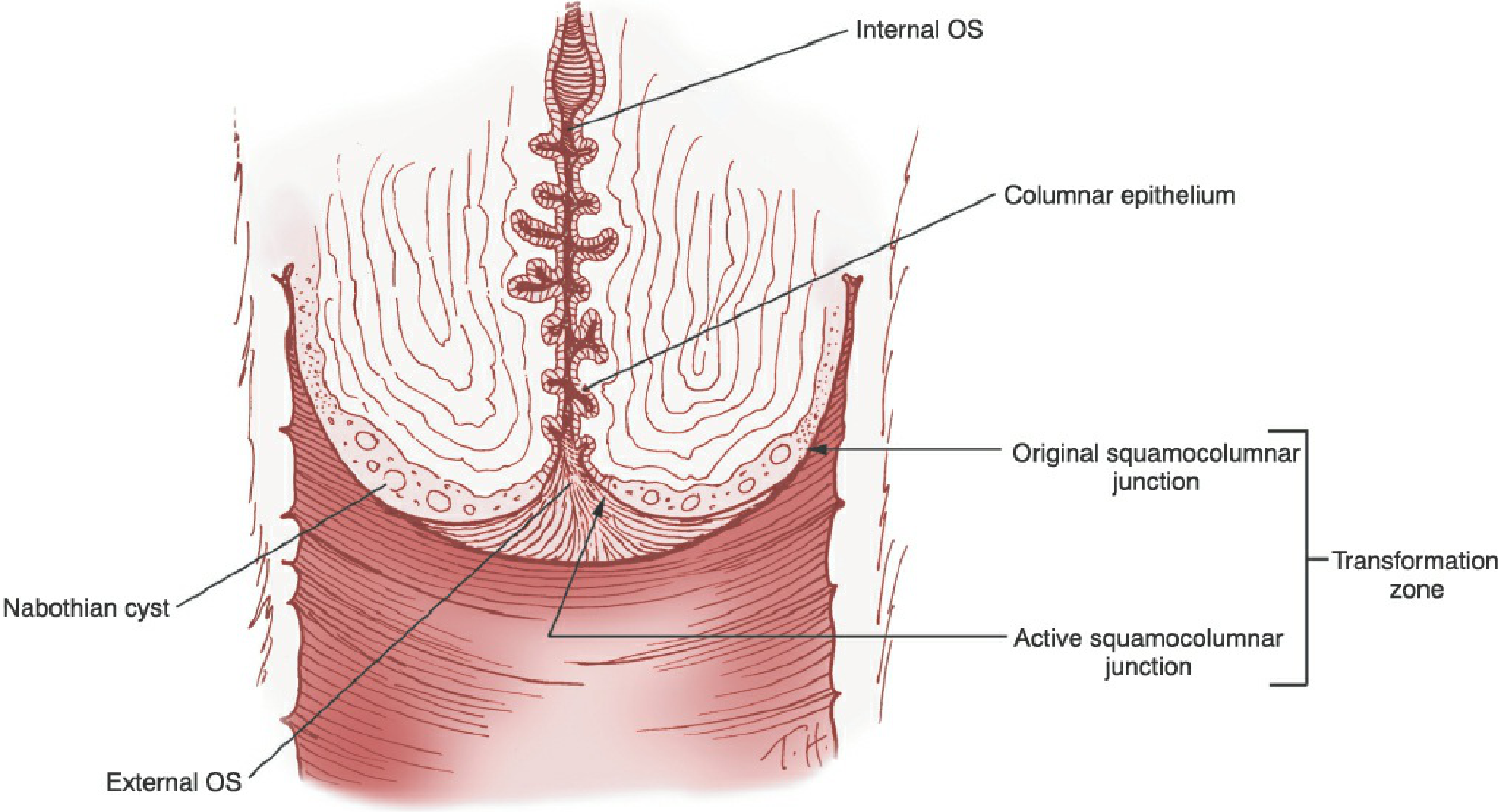
- Columnar (mucus-secreting) epithelium originally covers the ectocervix. Under the influence of the acidic vaginal environment, squamous metaplasia begins - reserve cells proliferate and gradually replace the columnar epithelium.
- As squamous metaplasia advances into the cervical clefts, it can trap islands of mucus-secreting columnar epithelium beneath the new squamous layer.
- Some of these trapped glands retain surface openings (visible as gland openings on colposcopy); others become completely encased, with mucus accumulating inside - these form Nabothian cysts.
- Gland openings and Nabothian cysts therefore mark the original squamocolumnar junction (SCJ) and the outer edge of the original transformation zone.
- Berek & Novak's Gynecology
Clinical Features
| Feature | Detail |
|---|---|
| Appearance | Smooth, pale-yellow or whitish/translucent dome-shaped elevations on the ectocervix |
| Size | Usually a few millimeters; rarely up to 1-4 cm (giant Nabothian cysts) |
| Number | Single or multiple |
| Symptoms | Almost always asymptomatic - an incidental finding on speculum exam |
| Vascularity | Regular branching vessels draped over the surface (important colposcopic feature distinguishing them from malignancy) |
Colposcopic Appearance
- Pale yellow, spheroid structures within the transformation zone, adjacent to the SCJ
- A smooth, raised surface with a regular vascular network stretched over the dome
- The active TZ contains gland openings, Nabothian cysts, and islands of columnar epithelium surrounded by metaplastic squamous epithelium - Pfenninger and Fowler's Procedures for Primary Care, 3rd ed.
Differential Diagnosis of Cervical Lesions
- Cervical polyp
- Condyloma
- Cervical/endometrial malignancy
- Prolapsed myoma
- Squamous papilloma
- Sarcoma
- Retained products of conception
- Smooth surface, regular vessels (no atypical vascularity)
- No friability, no acetowhite change (or only mild)
- Clearly cystic - translucent when illuminated
- No stalk (unlike polyps)
Management
- Giant Nabothian cysts (rare) can occasionally cause symptoms like pelvic pressure, dyspareunia, or - as seen in the laparoscopic image above - can compress adjacent structures like the ureter, potentially causing hydroureter. These may require surgical drainage or excision.
- If a cyst has an atypical appearance (irregular surface, atypical vessels, rapid growth), biopsy should be considered to exclude minimal deviation adenocarcinoma (adenoma malignum), which can grossly mimic large Nabothian cysts.