
Osteoarthritis
Reading File
Osteoarthritis (OA) is the most common joint disorder, characterized by progressive cartilage degradation, subchondral bone changes, osteophyte formation, and synovial inflammation. Now searching the medical library for comprehensive coverage.
Searching sources...
Finding Sources
Searching PubMed
"osteoarthritis"[MeSH Terms] AND "management"[MeSH Terms]
Good. I have strong hits from Robbins (pathology), Goldman-Cecil (clinical medicine), Firestein & Kelley's Rheumatology, and Rosen's Emergency Medicine. Let me read the most comprehensive sections in parallel.
Reading File
Reading File
Reading File
Good - I have Robbins pathology and some Goldman-Cecil content (but the block was adjacent content). Let me read more of the Goldman-Cecil OA block, plus the Robbins pathology continuation for morphology and clinical features, and fetch the OA diagram.
Reading File
Reading File
Loading Image
Excellent. Now let me get the Goldman-Cecil OA clinical chapter (block40 OA section) and Rosen's EM for management details, plus the histology/morphology image.
Reading File
Reading File
Loading Image
I now have rich content from Goldman-Cecil and Rosen's EM. Let me read more of the Goldman-Cecil pathobiology and management sections to get the full clinical picture.
Reading File
Reading File
Now let me get the Goldman-Cecil diagnosis and treatment sections, which are further in that block.
Reading File
I now have excellent, comprehensive content to write a thorough answer. Let me compile everything.
Osteoarthritis (OA)
Definition
Osteoarthritis is characterized by failed repair of synovial joint tissues after intra-articular stress. The resulting breakdown of cartilage and bone leads to symptoms of pain, stiffness, and functional disability. Although the name implies inflammation, it is primarily a degenerative disorder, with inflammation acting as a secondary contributor. - Goldman-Cecil Medicine, Ch. 241
Epidemiology
- Affects >500 million people worldwide; women are disproportionately affected
- The incidence increases rapidly between ages 50-75; ~40% of people >70 are affected
- An estimated 25% of the U.S. adult population will have physician-diagnosed OA by 2040
- OA is the 15th highest cause of years lived with disability worldwide
- Lifetime risk of symptomatic knee OA: ~40% in men, ~47% in women; rises to ~60% in persons above a healthy weight
- Knee OA accounts for ~85% of the total burden of OA
Goldman-Cecil Medicine, Ch. 241
Classification
| Type | Features |
|---|---|
| Primary (idiopathic) | Appears without identifiable cause; associated with aging; oligoarticular; affects weight-bearing joints |
| Secondary | Occurs in younger individuals; predisposing causes include prior joint injury, deformity, diabetes, or obesity (~5% of cases) |
Pathogenesis
Stages of Joint Damage
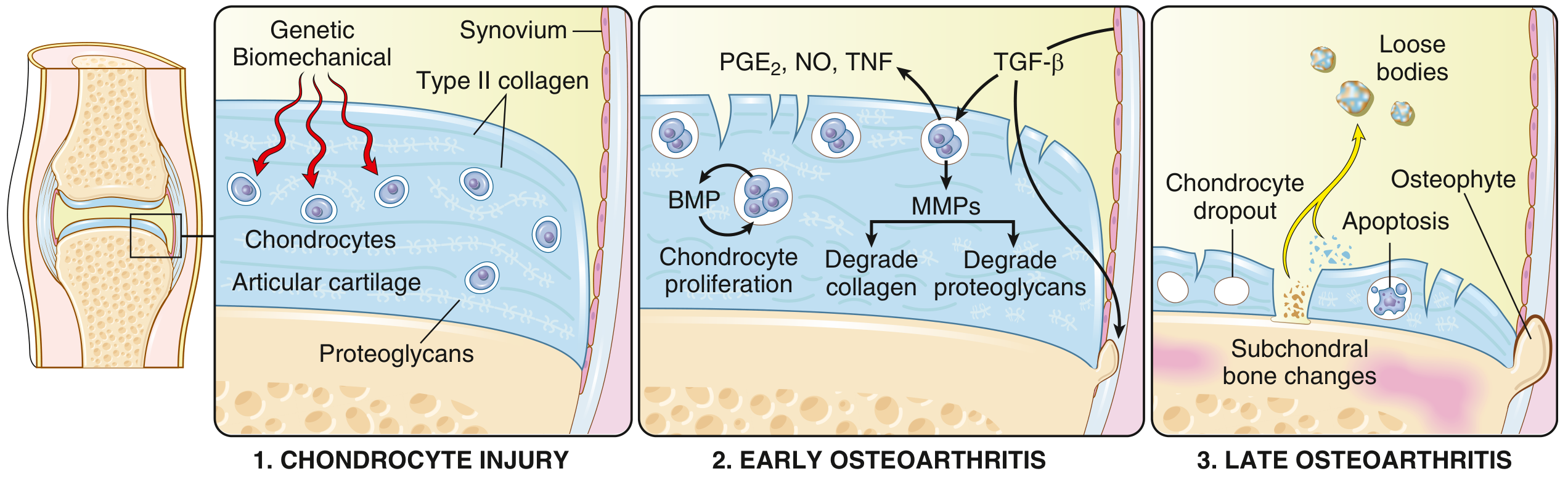
Fig. 19.31 - Robbins & Kumar Basic Pathology: Progression from chondrocyte injury (stage 1) to early OA (stage 2) to late OA (stage 3)
1. Chondrocyte injury - Genetic and biomechanical stresses injure chondrocytes, triggering changes in the extracellular matrix (Type II collagen and proteoglycans disrupted)
2. Early OA - Hypertrophic chondrocytes proliferate and attempt repair, but simultaneously release:
- Matrix metalloproteinases (MMPs) - degrade collagen and proteoglycans
- Proinflammatory mediators: PGE₂, NO, TNF
- TGF-β drives some compensatory repair (BMP signaling)
- Degradation eventually exceeds repair
3. Late OA - Chondrocyte dropout via apoptosis, full-thickness cartilage loss, subchondral bone changes, osteophyte formation, and loose bodies
Robbins & Kumar Basic Pathology, p. 790
Mechanics vs. Inflammation
Mechanical load is the primary driver when:
- Excessive strain is placed on a normal joint (obesity, occupational overuse, malalignment)
- Normal load traverses a joint that has lost mechanical protection (ligament rupture, meniscal damage, muscle weakness)
This altered mechanical load then activates inflammatory signaling pathways, which drive cartilage breakdown. Systemic obesity-related inflammation further amplifies the process. - Goldman-Cecil Medicine, Ch. 241
Morphology (Gross and Histologic)
Histology (early): Fibrillation (fraying) of the articular cartilage surface is the earliest change
Advanced disease:
- Full-thickness cartilage sloughing
- Loose bodies ("joint mice") - dislodged cartilage and bone fragments
- Bone eburnation - exposed subchondral bone polished by friction (ivory appearance)
- Subchondral cysts - synovial fluid forced into bone via ball-valve mechanism through fracture gaps
- Osteophytes - bony outgrowths capped by fibrocartilage at joint margins
- Synovium: mildly congested, fibrotic, with scattered chronic inflammatory cells
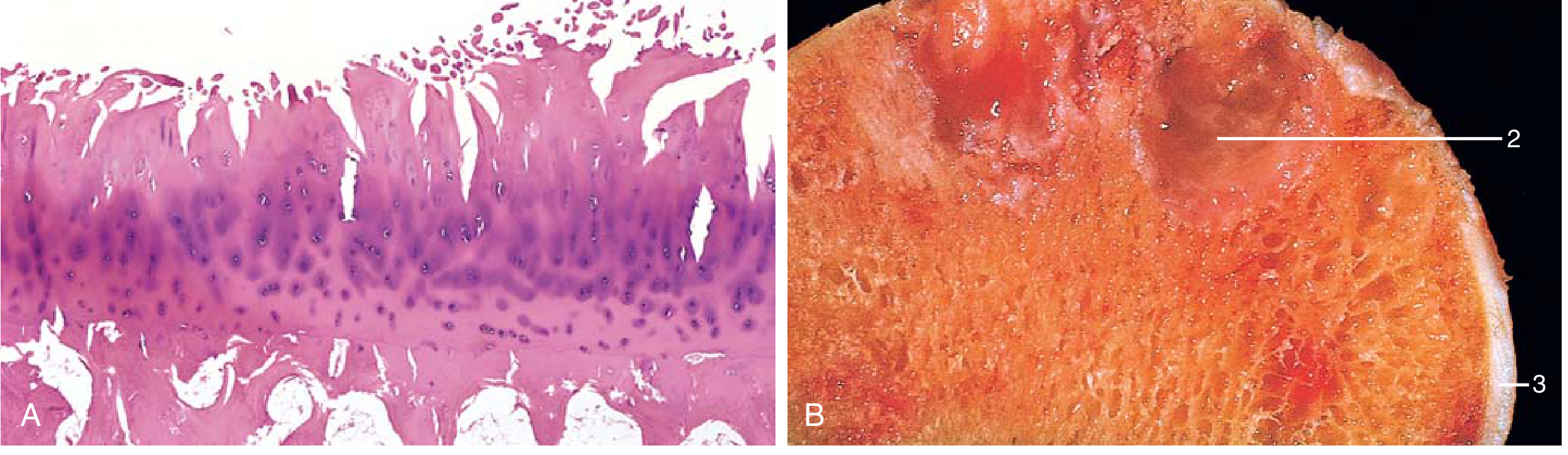
Fig. 19.32 - Robbins & Kumar: (A) Fibrillation of articular cartilage on histology; (B) Gross specimen showing eburnated surface (1), subchondral cyst (2), and residual cartilage (3)
Risk Factors
| Modifiable | Non-modifiable |
|---|---|
| Obesity (BMI - causative via Mendelian randomization) | Age |
| Joint malalignment | Sex (female > male) |
| Muscle weakness | Genetics (>80 OA loci identified in GWAS) |
| Sedentary lifestyle | Prior joint injury |
| Occupational exposure | Ethnicity |
| Athletic injuries |
Key genetic loci include GDF5 (chromosome 20), RUNX2, SMAD3, and PTHLH - genes important in skeletal and bone development. - Firestein & Kelley's Textbook of Rheumatology
OA vs. Rheumatoid Arthritis (Key Comparison)
| Feature | Osteoarthritis | Rheumatoid Arthritis |
|---|---|---|
| Primary mechanism | Mechanical injury to cartilage | Autoimmunity |
| Inflammation | Secondary; exacerbates damage | Primary cause of destruction |
| Joints involved | Weight-bearing (knees, hips); DIP/PIP/CMC | Small joints of fingers first; then multiple joints |
| Pathology | Cartilage degeneration, bone spurs, subchondral cysts; minimal inflammation | Inflammatory pannus; severe chronic inflammation; ankylosis |
| Serology | Negative (no ACPA, RF) | ACPA, rheumatoid factor positive |
| Extra-articular | None | Lungs, heart, other organs |
Robbins & Kumar Basic Pathology, Table 19.2
Clinical Features
Symptoms:
- Pain - mechanical in nature; worse with activity, better at rest; worse toward end of day; at rest in advanced disease
- Morning stiffness - localized, lasts <30 minutes (distinguishes from RA, which is >1 hour)
- Crepitus - audible and palpable during joint movement
- Functional limitation - difficulty with stairs/rising (knee), opening jars (hand), putting on shoes (hip)
- Catching or locking - especially knee (associated with falls)
Signs:
- Tenderness along joint line
- Bony swelling (osteophytes) + soft tissue swelling (effusion)
- Reduced range of motion
- Heberden nodes - osteophytes at DIP joints (more common in women)
- Bouchard nodes - osteophytes at PIP joints
- Knee: varus deformity, fixed flexion, ligamentous instability
- Hip: early loss of internal rotation
Joints most commonly affected: Hips, knees, lower lumbar and cervical vertebrae, PIP and DIP joints of fingers, first carpometacarpal joints, first tarsometatarsal joints
Spinal involvement: Osteophyte impingement on spinal foramina causes cervical/lumbar nerve root compression, radicular pain, muscle spasms, neurologic deficits
Diagnosis
Diagnosis is clinical based on:
- Symptoms: pain, brief morning stiffness, functional limitation
- Examination: crepitus, restricted/painful movement, joint tenderness, bony enlargement
Investigations:
- Plain radiographs: not required for typical presentations; useful for atypical cases
- Shows: asymmetric joint space narrowing, osteophytes, subchondral cysts, subchondral sclerosis
- Note: radiographic severity correlates poorly with symptoms
- Laboratory tests: limited role; mainly to exclude alternatives (infection, crystal arthropathy, RA)
- Synovial fluid: mildly inflammatory, low WBC (<2000 cells/µL), few PMNs
Red flags suggesting alternative diagnosis:
- Prolonged morning stiffness (>1 hr) → RA
- Hot, swollen joint → crystal disease (gout/pseudogout) or septic arthritis
- Recent trauma
Goldman-Cecil Medicine, Ch. 241; Rosen's Emergency Medicine
Management
Management should be individually tailored and focus on core treatments.
Non-Pharmacologic (First-Line)
- Exercise (land and water-based) - most evidence-based intervention; reduces pain and improves function
- Weight loss - BMI reduction relieves joint load; particularly effective for knee/hip OA
- Self-management education - activity modification, pacing
- Physical/occupational therapy - strengthening, gait aids, assistive devices
- Orthoses/bracing - appropriate footwear, splints, knee braces for malalignment
- TENS, acupuncture - limited but some evidence for pain relief
Pharmacologic
| Drug | Notes |
|---|---|
| Acetaminophen (paracetamol) | First-line; best safety profile; recent data suggest minimal benefit as monotherapy in knee OA |
| Topical NSAIDs (diclofenac gel) | Similar efficacy to oral NSAIDs; lower GI risk; preferred in elderly/high GI-risk patients |
| Oral NSAIDs | More effective than acetaminophen for pain; use when acetaminophen fails; risk: GI bleed, renal impairment, CV events |
| COX-2 inhibitors | Lower GI bleeding risk than non-selective NSAIDs; residual CV risk with long-term use |
| Duloxetine | Centrally-acting SNRI; effective for chronic musculoskeletal pain/knee OA; initiated in outpatient setting |
| Topical capsaicin | Useful for small hand joints; systemic toxicity low; significant local skin reaction limits use |
| Intra-articular corticosteroids | For acute flares; pain relief for weeks; unpredictable; no long-term benefit; may worsen arthritis with repeated use |
| Intra-articular hyaluronic acid | Best studied in knee; may improve function; no strong long-term benefit |
| Tramadol/Opioids | Not recommended; risks outweigh benefits for long-term OA management |
| Glucosamine/chondroitin | Supplements with limited demonstrated benefit |
Rosen's Emergency Medicine; Goldman-Cecil Medicine, Ch. 241
Surgical
- Joint replacement (arthroplasty) - indicated when:
- Functional status interferes with activities of daily living
- Supportive care and other measures have failed
- Pain is severe and persistent
- Knee replacement and hip replacement are among the most successful surgical procedures in medicine
Prognosis
- Natural history is highly variable; usually evolves over decades
- Progressive disease punctuated by periods of increased activity
- 80% of affected individuals have movement limitations
- 25% have difficulty with major activities of daily living
- No disease-modifying treatment currently exists - all current therapies are symptomatic
- Joint fusion (ankylosis) does NOT occur (distinguishing OA from RA)
Sources: Robbins & Kumar Basic Pathology, p. 789-791 | Goldman-Cecil Medicine, Ch. 241 | Rosen's Emergency Medicine, Ch. 102 | Firestein & Kelley's Textbook of Rheumatology
This is a shared conversation. Sign in to Orris to start your own chat.