
uterine inversion degrees classification diagram
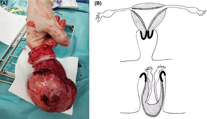
The image consists of two parts: (A) a clinical photograph of a surgically resected uterus and (B) an anatomical diagram illustrating uterine inversion. Panel A shows a gross pathological specimen of a total hysterectomy. The uterus appears as an enlarged, reddish, irregular mass with a bulbous uterine body and a distinct whitish, constricted cervical region. A surgical operator's finger is inserted into a central depression, representing the fundic invagination, which demonstrates the inverted state where the endometrial surface is on the exterior. Panel B provides a schematic comparison between normal uterine anatomy (top) and total uterine inversion (bottom). The diagram highlights the descent of the uterine fundus through the cervical canal and the subsequent 'cupping' or invagination of the adnexal structures (fallopian tubes and ovaries) into the resulting void. This visual provides clinical context for non-puerperal uterine inversion, often associated with heavy intrauterine masses like adenomyosis or myomas, emphasizing the structural transformation that occurs during this rare gynecological emergency.
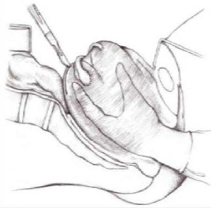
This medical illustration depicts Haultain’s procedure, a surgical technique used for the correction of chronic uterine inversion. The diagram shows a surgical field where a gloved hand stabilizes the inverted uterus. A second hand in the upper-left quadrant applies a scalpel to the posterior rim of the constriction ring, illustrating the longitudinal hysterotomy required to release the incarceration of the uterine fundus. To the right, another surgical assistant's hand is visible, potentially using a traction suture or loop to provide counter-tension. The illustration highlights the anatomical relationship between the cervical constriction ring and the inverted fundus. This surgical diagram serves as an educational resource for obstetrics and gynecology, demonstrating the operative management of life-threatening postpartum complications when manual reduction (Johnson maneuver) has failed.
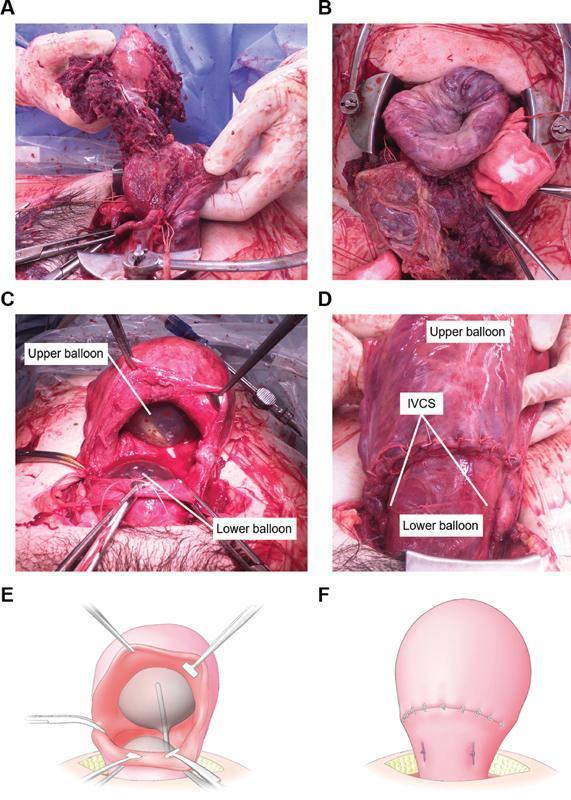
A composite image containing clinical photographs and schematic diagrams (A-F) documenting the management of postpartum hemorrhage (PPH) caused by uterine inversion and severe uterine atony. Panels A and B show the surgical field during cesarean section with an inverted, dark red, congested uterus and adherent placental tissue. Panels C and E (photograph and diagram) demonstrate the intraoperative placement of a double-balloon tamponade (UBT) system through a uterine incision, with an 'upper balloon' positioned in the uterine corpus and a 'lower balloon' in the isthmus. Panels D and F (photograph and diagram) illustrate the postoperative state where Isthmus Vertical Compression Sutures (IVCS) have been applied. These white, vertical sutures compress the lower uterine segment between the two inflated balloons to achieve hemostasis. The sequence highlights a step-by-step minimally invasive technique combining mechanical pressure and surgical compression to treat life-threatening obstetric hemorrhage.
Clinical Case: Uterine Inversion - Complete Analysis
Case Summary
Q1. Spot Diagnosis
- Reddish mass protruding from the vagina/introitus after delivery (inverted uterine fundus)
- Absent uterine fundus on abdominal palpation
- Profound hemodynamic collapse disproportionate to visible blood loss
Q2. Type of Shock
-
Hypovolemic/hemorrhagic shock - from postpartum hemorrhage due to uterine atony (inverted uterus cannot contract normally)
-
Neurogenic (vasovagal/distributive) shock - the peritoneum and broad ligament are stretched and inverted with the uterus, stimulating intense vagal discharge via afferent pelvic nerve traction. This causes profound bradycardia and vasodilation.
Q3. Why is Shock Out of Proportion to Blood Loss?
- The round ligaments, fallopian tubes, ovaries, and broad ligament are dragged inward
- This traction stimulates intense parasympathetic (vagal) afferent signals
- Result: vasodilation + bradycardia independent of blood volume loss
- The patient can be in severe shock even before significant external hemorrhage occurs
Q4. Classification by Degrees and Timing
By Degree (Anatomical):
| Grade | Description |
|---|---|
| 1st degree (Incomplete) | Fundus inverts but remains within the uterine cavity; does not reach the cervical os |
| 2nd degree (Complete) | Inverted fundus passes through the cervix, lies in the vagina |
| 3rd degree (Prolapsed/Total) | Inverted uterus protrudes through the vaginal introitus |
| 4th degree (Total prolapse) | Uterus and vaginal walls both inverted and visible externally |
By Timing:
| Type | Definition |
|---|---|
| Acute | Occurs within 24 hours of delivery (most common; best prognosis for manual reduction) |
| Subacute | Occurs between 24 hours and 4 weeks postpartum |
| Chronic | Occurs >4 weeks postpartum (tissue edema, constriction ring, harder to reduce) |
Q5. Investigations to Send
Immediate (resuscitation phase):
- CBC/FBC - hemoglobin, hematocrit, platelet count
- Coagulation profile - PT, aPTT, INR, fibrinogen (DIC screen - critical given massive PPH)
- Blood group and cross-match - for urgent transfusion; order at least 4 units packed RBCs
- Serum electrolytes, urea, creatinine - renal function (risk of acute tubular necrosis)
- Blood glucose
- Arterial blood gas (ABG) - assess acidosis, oxygenation (SpO2 94% is concerning)
- Serum lactate - marker of tissue hypoperfusion
- Liver function tests (baseline)
Imaging:
- Bedside ultrasound (TVS/TAS) - confirms inversion if diagnosis uncertain, identifies placental position
- Chest X-ray - baseline before any surgical intervention
Q6. Management
Immediate Resuscitation (simultaneous with diagnosis):
- 2 large-bore IV cannulas (16G or larger)
- Aggressive IV fluid resuscitation (crystalloids initially; blood products urgently)
- Oxygen supplementation (SpO2 94% - aim >95%)
- Foley catheter (monitor urine output)
- Warm the patient (prevent hypothermia)
- Call anesthesiologist immediately
A. Shock Management (Damage Control Resuscitation):
- Massive transfusion protocol: pRBC : FFP : Platelets in 1:1:1 ratio
- Tranexamic acid 1g IV STAT (within 3 hours of delivery for PPH) - WHO PPH guidelines support its use
- Correct hypothermia, acidosis, coagulopathy (the "lethal triad")
- Vasopressors (norepinephrine) if fluid-refractory shock
B. Uterine Reinversion (MOST CRITICAL STEP - do not delay):
- Do NOT remove the placenta first if still attached (causes catastrophic bleeding)
- Place the inverted fundus in the palm of the hand, with fingers directed toward the posterior fornix
- Apply upward pressure along the axis of the uterus, pushing fundus back through the cervix
- Maintain pressure until the cervical ring contracts around the reinverted uterus
- Terbutaline 0.25 mg IV/SC
- Magnesium sulfate 4-6 g IV over 15-20 min
- IV nitroglycerin (50-100 mcg bolus) - fastest acting
- General anesthesia with halogenated agents (sevoflurane/halothane) - causes uterine relaxation
- Restart uterotonics (oxytocin infusion)
- Now remove the placenta manually
- Bimanual compression if atony persists
- Huntington procedure (laparotomy, traction on round ligaments)
- Haultain procedure (posterior longitudinal incision through cervical ring to allow reinversion)
- Hysterectomy as last resort
C. Coagulopathy Management:
- Replace clotting factors: FFP, cryoprecipitate (if fibrinogen < 2 g/L), platelets
- Monitor with TEG/ROTEM if available
- Target: fibrinogen >2 g/L, platelets >50 x10⁹/L, INR <1.5
D. Infection Prevention:
- Broad-spectrum IV antibiotics (e.g., co-amoxiclav or cefazolin + metronidazole)
- Especially important given delivery by nursing staff at PHC without full sterile technique
E. Acidosis Management:
- Primarily correct by restoring perfusion (transfusion, vasopressors)
- ABG-guided bicarbonate if severe metabolic acidosis (pH < 7.1)
F. Hypothermia Prevention:
- Warm IV fluids
- Warm blankets
- Warm operating room if surgery needed
- Target core temperature > 36°C
Q7. Risk Factors for Uterine Inversion
| Risk Factor | Mechanism |
|---|---|
| Grand multiparity (P6L6) | Uterine atony, lax myometrium |
| Fundal placentation | Found in virtually ALL reported cases; thins the fundal wall |
| Excessive cord traction | Especially dangerous with fundal placenta |
| Fundal pressure during delivery | Pushes fundus inward |
| Placenta accreta spectrum | Abnormal adherence prevents normal separation |
| Short umbilical cord | Traction pulls fundus down |
| Uterine anomalies | Congenital structural weakness |
| Magnesium sulfate use | Uterine relaxation |
| Primiparity | Also listed (primiparas have less uterine "experience") |
| Connective tissue disorders | Uterine wall laxity (e.g., Marfan's, Ehlers-Danlos) |
| Mismanagement of 3rd stage | Premature cord traction, Crede's maneuver on an uncontracted uterus |
Sources: Creasy & Resnik's Maternal-Fetal Medicine, p. 948-949; Rosen's Emergency Medicine, p. 3421; Tintinalli's Emergency Medicine, p. 686
Questions 8-13: Uterine Inversion (Continued)
Q8. Signs of Placental Separation
| Sign | Description |
|---|---|
| Uterus becomes globular and rises (Calkin's sign) | As the placenta descends into the lower segment, the fundus rises up and becomes firm, round, and hard |
| Cord lengthening | The umbilical cord advances 5-10 cm out of the vagina as the placenta descends |
| Sudden gush of blood | A small gush of dark retroplacental blood appears at the vaginal introitus as the placenta separates |
- Schultze mechanism - placenta delivers fetal surface first (shiny), like an inverted umbrella; accounts for ~80% of separations; associated with the blood gush coming after placental delivery
- Matthews Duncan mechanism - placenta delivers edge first (maternal surface/dull side), slides out sideways; blood trickles throughout separation; associated with lower-segment implantation
Do NOT attempt to deliver the placenta before these signs appear - forced removal is a major cause of uterine inversion.
Q9. Steps of AMTSL (Active Management of the Third Stage of Labor)
Step 1 - Uterotonic Drug (within 1 minute of delivery)
- Oxytocin 10 IU IM (drug of choice; given into anterolateral thigh immediately after baby is born, before placental delivery)
- Alternatives: Misoprostol 600 mcg oral/sublingual (if oxytocin not available), ergometrine, oxytocin+ergometrine (Syntometrine)
- Given BEFORE placenta delivers
Step 2 - Controlled Cord Traction (CCT) - after signs of separation appear
- Clamp the cord close to the perineum
- Apply counter-pressure (Brandt-Andrews maneuver): place the edge of one hand above the symphysis pubis to stabilize the uterus (push upward) while applying gentle, steady downward traction on the cord with the other hand
- Never apply cord traction without counter-pressure - this is how uterine inversion is caused
- Ask the mother to push/bear down with contractions
Step 3 - Uterine Massage (after placental delivery)
- Immediately after placental delivery, feel for the fundus through the abdominal wall
- Apply gentle, sustained, circular massage until the uterus is firm and well-contracted
- Check the placenta for completeness (membranes and cotyledons)
- Monitor for bleeding for at least 1 hour
- Early cord clamping (1-3 minutes after birth, or after cord stops pulsating)
- Examine placenta and membranes for completeness
- Examine birth canal for lacerations
- Monitor closely for 2 hours postpartum
Q10. Methods of Replacement of the Uterus
Non-surgical (Manual) Methods:
- Apply the inverted fundus in the palm of the hand with fingers pointing toward the posterior fornix
- Elevate the uterus progressively through the vagina, cervix, and into the abdominal cavity
- The direction of pressure is upward toward the umbilicus
- Maintain pressure until cervical ring contracts
- If placenta still attached: do NOT remove it first - replace uterus with placenta in situ
- Success rate is highest immediately after inversion (before cervical ring forms)
- Used when manual reduction fails and cervical ring is formed
- Patient in Trendelenburg position
- Warm saline (3-5 litres) infused into the vagina under pressure via a rubber tube/douche nozzle placed in the vaginal vault; the operator's hand seals the introitus
- Hydraulic pressure distends the vagina and pushes the fundus back into place
- Effective because it uses fluid pressure to gradually dilate the cervical ring
Pharmacological Adjuncts (Tocolysis to relax the cervical ring):
- Terbutaline 0.25 mg IV or SC (beta-2 agonist)
- Magnesium sulfate 4-6 g IV over 15-20 min (preferred)
- IV Nitroglycerin 50-100 mcg IV bolus (fastest acting; very brief effect)
- Halogenated inhalational anesthetics (sevoflurane, halothane) - via anesthesiologist
Surgical Methods (when all else fails):
| Procedure | Details |
|---|---|
| Huntington procedure (laparotomy) | Clamps placed on round ligaments ~1 inch inside the inverted uterus; upward traction applied while an assistant pushes from below vaginally |
| Haultain procedure | A longitudinal posterior incision is made through the cervical constriction ring to enlarge it, allowing manual reinversion; incision is then closed (like a classical cesarean scar) |
| Hysterectomy | Last resort if uterus cannot be replaced or hemorrhage is uncontrollable |
- Restart uterotonics immediately
- Now perform manual removal of placenta if it was left in situ
- Assess for perforation, lacerations, and coagulopathy
Q11. When Should the Placenta Be Removed? How?
In the context of uterine inversion:
- The placental bed lacks tone to contract and control bleeding when the uterus is inside out
- Premature separation tears open venous sinuses in an uncontracted, inverted myometrium
- The placenta can then be manually removed with the uterus now in proper position
- Ensure uterus is well contracted (oxytocin infusion running)
- Perform manual exploration to confirm no retained fragments
In general obstetric practice (retained placenta):
- Wait up to 30 minutes for spontaneous delivery
- If retained beyond 30 min: attempt manual removal
- Manual removal technique:
- General or regional anesthesia
- Empty the bladder
- Introduce the dominant hand into the uterine cavity along the cord
- Find the placental edge; use a sideways, "peeling" motion (like opening a book) along the cleavage plane between placenta and uterine wall
- Grasp placenta with the other hand at the introitus and extract
- Inspect for completeness
- Administer uterotonics + broad-spectrum antibiotics
- If placenta is abnormally adherent (accreta/increta/percreta): do NOT attempt forcible removal - prepare for hysterectomy
Q12. How to Prevent Recurrence
In Future Pregnancies:
- Elective caesarean section is strongly advised in any subsequent pregnancy, especially if the inversion was related to placenta accreta or if reinversion was difficult
- Deliver in a tertiary hospital with full surgical backup
- Pre-operative cross-match and consent for hysterectomy
During Future Deliveries (if vaginal birth attempted):
- Management of third stage ONLY by experienced obstetrician
- Strict AMTSL with proper controlled cord traction (never forceful)
- No fundal pressure
- Ultrasound in advance to confirm placental site (warn team if fundal)
- Have uterotonics ready; avoid magnesium sulfate in labour if possible
- Careful watch during third stage - at first sign of inversion, immediate intervention
Education & System Level:
- Train birth attendants (nurses, midwives at PHC) in proper 3rd stage management
- Never apply aggressive fundal pressure
- Never apply traction before signs of placental separation
- As in this case, PHC delivery by nursing staff without training in AMTSL directly caused this complication
Q13. Short-Term and Long-Term Complications
Short-Term (Acute) Complications:
| Complication | Mechanism |
|---|---|
| Hemorrhagic shock | Atonic PPH from inverted, non-contracting uterus |
| Neurogenic shock | Vagal stimulation from peritoneal/broad ligament traction |
| DIC (Disseminated Intravascular Coagulation) | Massive hemorrhage depletes clotting factors; hypoperfusion triggers coagulation cascade |
| Acute Tubular Necrosis (ATN) | Renal hypoperfusion from prolonged shock |
| Hypoxic encephalopathy | Cerebral hypoperfusion |
| Hypothermia | From massive transfusion, prolonged surgery |
| Cardiac arrest | From severe vasovagal response or uncontrolled hemorrhage |
| Sepsis/endometritis | Especially if delivery was at PHC without aseptic technique, with prolonged manipulation |
| Uterine necrosis | If reinversion is delayed - tissue edema, ischemia |
| Bladder/urethral injury | During surgical reinversion (Haultain) |
| Adult Respiratory Distress Syndrome (ARDS) | From massive transfusion, shock |
Long-Term Complications:
| Complication | Details |
|---|---|
| Sheehan's syndrome (pituitary necrosis) | Anterior pituitary infarction due to prolonged hypotension during PPH; presents as failure of lactation, amenorrhea, adrenal/thyroid insufficiency |
| Asherman's syndrome | Intrauterine adhesions from uterine manipulation/infection |
| Infertility | Secondary to Asherman's, or if hysterectomy was required |
| Chronic pelvic pain | From pelvic adhesions post-surgery |
| Recurrent inversion | In future pregnancies (as discussed in Q12) |
| Psychological trauma | Post-traumatic stress, tokophobia (fear of future pregnancy) |
| Hypothyroidism / adrenal insufficiency | If Sheehan's syndrome is present |
| Hysterectomy | Loss of reproductive potential if surgery was unavoidable |
Sources: Rosen's Emergency Medicine, p. 3412, 3421; Pfenninger and Fowler's Procedures for Primary Care, p. 7160; Creasy & Resnik's Maternal-Fetal Medicine, p. 948-949