
Paraquat poisning detailed explanation
paraquat poisoning
paraquat poisoning
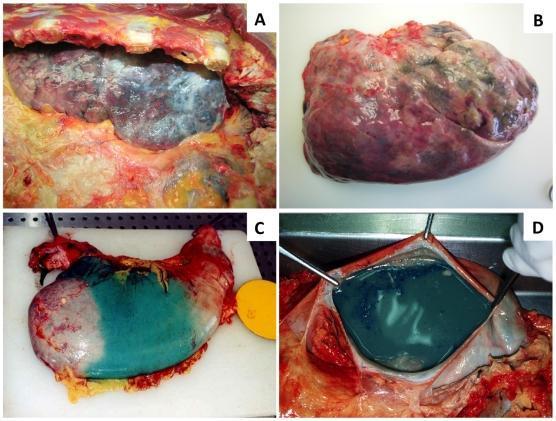
This composite of four clinical photographs displays macroscopic findings from a human autopsy following fatal paraquat poisoning. Panels A and B focus on the lungs. Image A shows a lung in situ exhibiting significant fibrosis, characterized by a mottled, pale-white to grayish appearance interspersed with dark hemorrhagic patches and a firm, contracted texture. Image B depicts an excised lung with severe pulmonary edema and vascular congestion, appearing enlarged, heavy, and dark reddish-purple. Panels C and D illustrate pathognomonic findings in the gastrointestinal tract. Image C shows an external view of the stomach with a striking, localized greenish-blue discoloration, a characteristic artifact of the blue dye added to paraquat formulations to prevent accidental ingestion. Image D shows the gastric lumen opened, revealing a large volume of matching greenish-blue fluid contents. These images serve as educational materials for forensic pathology and toxicology, demonstrating the acute internal manifestations of herbicide ingestion.
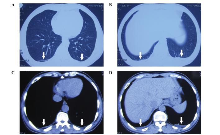
Diagnostic Image: Four axial chest computed tomography (CT) scans (labeled A, B, C, and D) obtained three days following acute paraquat poisoning. Images A and B are displayed in lung window settings, while C and D are in mediastinal/soft tissue window settings. The scans demonstrate bilateral pathology in the dependent portions of the lower lobes. White arrows indicate the presence of small pleural effusions and subpleural 'effusion shadows' or early consolidative changes characterized by increased opacity and poorly defined margins. Anatomical structures visible across the sections include the pulmonary parenchyma, heart, descending aorta, liver, and vertebral bodies. The clinical significance of these findings relates to the early pulmonary manifestations of paraquat toxicity, which typically progresses from initial edema and inflammation to subsequent pulmonary fibrosis. This content is intended for intermediate to advanced medical education in emergency medicine, toxicology, and radiology, highlighting the characteristic early imaging features of chemical-induced lung injury.
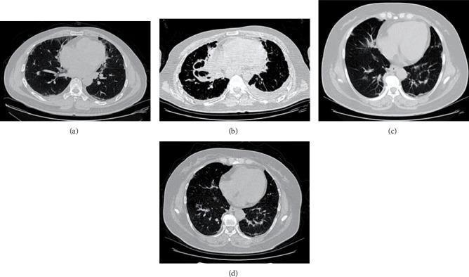
This composite diagnostic image features four axial non-contrast chest CT scans (a-d) documenting the progression of lung injury in two different patients following paraquat poisoning. Patient 1: Image (a) represents the initial scan showing mild peripheral ground-glass opacities (GGO) and focal consolidation. Image (b) shows follow-up at 2 months, demonstrating severe disease progression characterized by dense bilateral consolidation, architectural distortion, and the development of cystic airspaces or honeycombing, indicative of progressive pulmonary fibrosis. Patient 2: Image (c) shows the baseline scan with subtle, patchy ground-glass opacities. Image (d) shows follow-up at 2 months, revealing an increase in reticular markings and persistent GGO, though with significantly less consolidation and cystic change compared to Patient 1. The series illustrates the spectrum of acute lung injury evolving into chronic interstitial fibrosis, a hallmark of paraquat toxicity, and serves as an educational tool for comparing variable prognostic outcomes in toxic inhalation or ingestion cases.
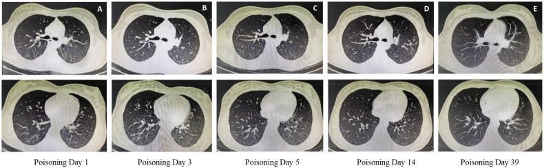
This diagnostic image series displays a sequence of axial chest CT scans (lung window) demonstrating the progression and resolution of pulmonary abnormalities in a patient with paraquat poisoning undergoing FPSA-CVVH treatment. The series is labeled A through E, representing Poisoning Day 1, 3, 5, 14, and 39. Image A shows bilateral, scattered ground-glass opacities (GGOs) with ill-defined boundaries, primarily located in the right lung and left lower lobe. Image B illustrates a progression of these inflammatory shadows. From Day 5 (Image C) to Day 14 (Image D), there is a gradual absorption and reduction of the lung opacities. The final scan (Image E) shows complete resolution of the pulmonary abnormalities, with clear lung parenchyma and no residual GGOs or fibrosis. This longitudinal comparison illustrates the typical imaging manifestation of acute chemical pneumonitis and its potential for resolution following intensive detoxification therapy. It serves as an educational reference for radiological monitoring in emergency toxicology and critical care.
paraquat poisoning management cyclophosphamide methylprednisolone hemoperfusion evidence review
Paraquat Poisoning
1. What it is
2. Toxicokinetics
- Oral bioavailability is under 30% (the forensic text says only 5-10% is absorbed). Protein binding is about 5%. Vd is about 1 L/kg.
- Plasma levels peak at about 2 hours, and tissue distribution is maximal over the next 6 hours.
- Paraquat concentrates in the lungs and kidneys, and also reaches the liver and muscle. Muscle can release it back into the circulation later.
- The kidneys excrete it, mostly unchanged in urine within 24 hours. Half-life is about 12 hours with normal renal function and over 48 hours in renal failure. - Brenner and Rector's The Kidney, p. 2868; Essentials of Forensic Medicine and Toxicology, p. 496
- Lethal dose of 20% concentrate is about 10-20 mL in an adult and 4-5 mL in a child. - Tintinalli's Emergency Medicine, p. 1347
3. Mechanism of toxicity
- Redox cycling. Paraquat accepts an electron from NADPH-dependent reduction. The radical then reacts with O2 to regenerate paraquat and produce superoxide and hydroxyl radicals. Each cycle consumes NADPH and generates more reactive oxygen species.
- Lung accumulation. Type I and II alveolar cells actively take paraquat up through an energy-dependent polyamine transporter. The lung has plenty of O2 and electrons, so the cycle runs continuously.
- Cell injury. Lipid peroxidation, membrane damage and cell death follow.
- Two lung phases. The first is a destructive phase with loss of type I and II cells, inflammation and hemorrhage. The second is a proliferative phase with interstitial and alveolar fibrosis.
- Oxygen worsens it. Oxygen sustains the redox cycle, so supplemental O2 increases toxicity.
- Local corrosion. Concentrated solutions directly burn mucosa.
4. Clinical features by dose
| Severity | Dose | Course |
|---|---|---|
| Mild | <20 mg/kg (<7.5 mL of 20% solution) | Asymptomatic, or vomiting, diarrhea and oral ulcers. Complete recovery. |
| Moderate-severe | 20-40 mg/kg | Delayed multi-organ failure. Renal and hepatic injury at days 1-4, then cough, hemoptysis and fibrosis over 1-2 weeks. Most die in 2-3 weeks from respiratory failure. |
| Fulminant | >40-50 mg/kg | Death within 1-4 days from multi-organ failure, cardiogenic shock and corrosive effects. |
- GI. A burning mouth and throat develops, with ulceration at 1-2 days. Other features include dysphagia, aphonia and hematemesis. Esophageal perforation can cause mediastinitis and pneumothorax. Pancreatitis can occur.
- Kidney. Acute tubular necrosis with proximal tubular dysfunction. It is usually reversible if the patient survives.
- Liver. Centrilobular necrosis and cholestasis.
- Lung. Progresses to ARDS-like illness, then irreversible fibrosis and refractory hypoxemia. This is the main cause of death.
- Heart and adrenals. Toxic myocarditis, shock, and adrenal necrosis.
- CNS. Paraquat crosses the blood-brain barrier poorly, so CNS signs are uncommon. Late coma or seizures are reported.
- Metabolic. Lactic acidosis.
5. Diagnosis and prognosis
- History. Establish the product, concentration, amount and timing.
- Urine sodium dithionite test. Add dithionite to alkalinized urine. A blue color confirms recent exposure, and a deeper blue means a worse prognosis. A clear result at 6-24 hours favors survival. Patients observed for at least 12 hours with a negative test are unlikely to develop toxicity.
- Plasma paraquat level. Use with a time-since-ingestion nomogram (Hart, Proudfoot). It is the best mortality predictor but is rarely available.
- Other markers. Lactate above about 3.4-4.4 mmol/L is associated with death. Rapid creatinine or cystatin C rise (creatinine >0.049 mg/dL/h over 6 h) predicts death. Serum amylase also has prognostic value - 2026 meta-analysis.
- Imaging. CXR may show infiltrates, pneumomediastinum or pneumothorax. CT shows ground-glass opacities and consolidation early, then fibrosis and honeycombing.
- Monitoring. Serial ABG, renal and liver tests, CXR and pulmonary function tests.
6. Management
- Replace fluids and electrolytes and maintain urine output. Forced diuresis does not speed paraquat clearance.
- Avoid supplemental O2 unless the patient is severely hypoxic. Oxygen accelerates injury.
- Treat pain with opioids. Contact with caustics is very painful.
- Remove contaminated clothing, wash skin with soap and water, and irrigate the eyes.
- Gastric lavage is contraindicated because of corrosive risk. Emetics are also contraindicated.
- If the patient presents within 1-2 hours, give an adsorbent orally or by nasogastric tube. Options are activated charcoal 1-2 g/kg, Fuller's earth 1-2 g/kg as a 15% suspension, or 7% bentonite. Indian textbooks add a cathartic (20% mannitol) afterward.
- Consider endoscopy for the extent of mucosal injury.
- Paraquat is small and unbound, so hemoperfusion or hemodialysis can clear it from plasma. But it moves rapidly into the lungs and becomes trapped there. Benefit is limited to very early use, ideally within 4 hours. The forensic text allows up to 12 hours. Rebound after treatment is common.
- Hemoperfusion is preferred. Intermittent hemodialysis is an alternative. CVVH and peritoneal dialysis are not useful for removal, though CVVH can support AKI. - Brenner and Rector's The Kidney, p. 2869; Tintinalli, p. 1348
- Pulse cyclophosphamide plus methylprednisolone followed by dexamethasone is widely used (Lin protocol). Small studies suggested benefit. However, a large randomized trial found none, and Tintinalli and Brenner's The Kidney say no regimen has clearly changed outcome.
- Tintinalli suggests dexamethasone 8 mg IV every 8 hours for 72 hours and N-acetylcysteine, since both have low toxicity.
- Vitamins C and E, deferoxamine, selenium, salicylate, superoxide dismutase and radiotherapy have not been proven useful.
- A 2025 review in BMC Public Health discusses regulation of paraquat. No treatment has been shown to reliably change the outcome.
- Fibrosis can lead to refractory respiratory failure. Lung transplantation has been reported in rare cases, but I could not confirm this from the sources I retrieved.
- Involve palliative care early. In severe poisoning the most humane approach may be analgesia and comfort care.
7. Autopsy findings
- Oral and pharyngeal ulceration, and esophageal epithelial casts.
- Gastric erosions, or normal stomach.
- Kidney: cortical pallor and tubular damage. Liver: centrilobular necrosis and fatty change.
- Lungs: early edema and hemorrhage, with hyaline membranes. Over days to weeks they become fibrotic and stiff, which can be mistaken for diffuse pneumonia.
8. Key points
- Ingested paraquat causes delayed, progressive lung fibrosis, and death is usually from respiratory failure.
- Paraquat is taken up into lungs via the polyamine transporter, and oxygen worsens injury.
- Dithionite urine test: blue means positive.
- Avoid lavage, emetics and routine oxygen.
- Adsorbent early, hemoperfusion within hours, and supportive care.
- No proven antidote, and immunosuppression remains controversial.
- Prevention matters most. Restricting access and banning the herbicide has reduced suicide deaths in several countries.