
Shortgut syndrome
Reading File
Now I'll search the medical library and PubMed in parallel for short gut syndrome.
Finding Sources
Finding Sources
Searching PubMed
"short bowel syndrome" management
Good results. Now let me read the most comprehensive sections from Sabiston, Schwartz's, Harrison's 22E, and Goldman-Cecil in parallel.
Reading File
Reading File
Reading File
Reading File
Let me read the rest of Harrison's treatment section and Schwartz's remaining content on management/surgical options.
Reading File
Reading File
Reading File
Excellent - I have very rich content. Let me fetch the surgical procedure diagram and quickly check the teduglutide (GLP-2 agonist) pharmacology from Goodman & Gilman and Katzung.
Loading Image
Reading File
I now have comprehensive information from multiple authoritative textbooks. Let me compose the full answer.
Short Bowel Syndrome (SBS)
Definition
Short bowel syndrome (SBS) is a state of significant malabsorption - of macronutrients, micronutrients, fluid, and electrolytes - resulting from extensive small intestinal resection or, rarely, congenital bowel anomaly. The most widely used anatomical threshold is <200 cm of residual small bowel in adults. Intestinal failure is the functional endpoint: persistent parenteral nutrition (PN) dependence, generally seen when <100 cm of small bowel remains with no colon in continuity. - Harrison's Principles of Internal Medicine 22E, p. 2591; Schwartz's Principles of Surgery, p. 1281
Causes
Adults:
- Acute mesenteric ischemia (arterial or venous thrombosis) - single largest event cause (75% of SBS results from a single massive resection)
- Crohn's disease - most common cumulative cause (25%, repeated resections)
- Malignancy, volvulus, radiation enteritis, trauma, internal hernia
Children/Neonates:
-
Necrotizing enterocolitis (NEC) - most common (~35%)
-
Intestinal atresia (~25%)
-
Gastroschisis (~18%)
-
Midgut volvulus, malrotation (~14%)
-
Sabiston Textbook of Surgery, p. 1876; Sabiston (pediatric section)
Pathophysiology
Determinants of severity
The degree of malabsorption depends on several anatomical factors beyond just bowel length:
| Factor | Impact |
|---|---|
| Length of residual bowel | <100 cm (no colon) or <60 cm (intact colon) → likely lifelong TPN dependence in adults |
| Intact colon | Absorbs large fluid/electrolyte loads; bacteria ferment malabsorbed carbohydrates to short-chain fatty acids (SCFAs), adding several hundred kcal/day |
| Ileocecal valve (ICV) | Slows transit, prolongs contact time. The ICV may serve as a surrogate for an intact terminal ileum (the segment with greatest adaptive capacity) |
| Segment resected | Ileal loss is worse than jejunal loss - ileum is sole site of B12 absorption and active bile salt reabsorption |
| Residual bowel health | Diseased bowel (Crohn's) adapts poorly vs. healthy bowel |
- Schwartz's Principles of Surgery, p. 1282
Site-specific consequences of resection
- Jejunal resection: Better tolerated; ileum compensates; bile salt and B12 absorption preserved
- Ileal resection (<100 cm): Bile salt malabsorption → colonic secretory diarrhea (cholerheic diarrhea)
- Ileal resection (>100 cm): Hepatic bile salt synthesis cannot compensate → fat malabsorption → steatorrhea and fat-soluble vitamin (A, D, E, K) deficiency
- Loss of the ileal brake: The GLP-1-mediated suppression of gastric emptying is lost → unabated proximal secretions, maldigestion, lack of satiety
Gastric acid hypersecretion
After massive resection, hypergastrinemia causes gastric acid hypersecretion persisting for 1-2 years postoperatively. The acid load overwhelms duodenal buffering, inactivates digestive enzymes, and worsens malabsorption. - Schwartz's Principles of Surgery, p. 1282
Intestinal adaptation
-
Begins within 48 hours of resection
-
Residual bowel undergoes villous hyperplasia (increased crypt cell proliferation, villous lengthening, bowel dilation and elongation)
-
Continues for up to 2 years, and improvements can occur even at 3-5 years
-
Driven by luminal nutrients, pancreatico-biliary secretions, and gut hormones especially GLP-2 (secreted by L cells of terminal ileum/colon)
-
Other trophic factors: EGF, IGF-1, TGF-α, trefoil peptides, IL-11, growth hormone
-
Goldman-Cecil Medicine, p. 994; Harrison's 22E, p. 2590
Clinical Features
- Severe, high-volume diarrhea (worst in first 3 postoperative months)
- Weight loss, malnutrition
- Dehydration and electrolyte disturbances (Na, Cl, Mg, Zn losses)
- Malabsorption: fat, protein, carbohydrate, fat-soluble vitamins
- B12 deficiency (megaloblastic anemia, neuropathy) if ileum resected
- Specific to jejunostomy patients: high-output losses of Na, Cl, HCO3, Mg, Zn, water
Long-Term Complications
-
Metabolic bone disease / osteoporosis - Ca and vitamin D malabsorption; difficult to treat even with high-dose oral vitamin D
-
Renal calcium oxalate stones - in patients with shortened small bowel and intact colon: Ca is saponified by malabsorbed fatty acids, freeing oxalate to be absorbed in the colon → hyperoxaluria
-
Small bowel bacterial overgrowth (SIBO) - due to adhesions, abnormal motility, strictures
-
TPN-associated complications - catheter sepsis, venous thrombosis, hepatic failure (leading to combined liver-small bowel transplant), renal failure, osteoporosis
-
Fat-soluble vitamin deficiencies (A, D, E, K), copper, selenium, chromium deficiency
-
Harrison's 22E, p. 2591
Treatment
Phase 1: Acute (immediately post-resection)
- Aggressive IV fluid/electrolyte resuscitation
- Total parenteral nutrition (TPN) - nearly always required initially
- High-dose H2 blockers or proton pump inhibitors (e.g., omeprazole 40 mg/day, lansoprazole 30 mg/day) to counter gastric hypersecretion
- Treat underlying condition
Phase 2: Adaptation (months to ~2 years)
- Gradual introduction of enteral feeding - even while on TPN, luminal nutrients are required to drive intestinal adaptation
- Anti-motility agents: loperamide (2-8 mg up to 4x/day), diphenoxylate-atropine, codeine (15-60 mg up to 4x/day), or potent opiates (tincture of opium, liquid morphine) to slow transit and maximize absorption
- Octreotide (100-250 mcg SC TID, converted to long-acting monthly if effective) for high-volume secretion (>3 L/day output) - caution: may impair intestinal adaptation in animal models
- Dietary modification:
- With colon: low-fat, high complex carbohydrate diet; small frequent meals
- Without colon (high jejunostomy): high-salt, nutrient-rich diet; oral rehydration solution with sodium 75-90 mmol/L
- Vitamin B12 parenterally: 500-1000 mcg IM/SC monthly
- Oral vitamin/mineral supplements at doses above standard RDA; high-potency multivitamin daily
- Magnesium supplementation: liquid preparation added to ORS, sipped throughout the day
Pharmacological: Teduglutide (GLP-2 analogue)
The only drug specifically approved for SBS. Mechanism: Teduglutide is a 33-amino acid GLP-2 analogue (DPP-4 resistant due to amino acid substitution). GLP-2 is secreted by L cells of the ileum and colon and is the only known intestinotropic gut peptide. It:
- Enhances intestinal mucosal growth (via IGF-1 release and direct action)
- Improves fluid absorption and intestinal/portal blood flow
- Reduces gastric secretion
- Improves intestinal barrier function
Dose: 0.05 mg/kg SC once daily. t1/2: 1-2 h; excreted renally; catabolized by DPP-4 (slower than native GLP-2). Side effects: abdominal pain, nausea, injection-site reactions; monitor for polyp formation (requires colonoscopy before starting).
Growth hormone + glutamine: explored in clinical trials to reduce TPN requirements; short-term results promising but patients often revert at cessation. Evidence remains inconclusive. - Goodman & Gilman's, p. 1125; Sabiston, p. 1880
Enteral autonomy outcomes
-
50-75% of SBS patients who initially require TPN eventually achieve TPN independence
-
Infants with as little as 10 cm of residual bowel have been weaned from TPN
-
Adults with 35-40 cm of remaining bowel have ~50% probability of weaning from TPN; each additional centimeter increases enteral autonomy rate by ~4%
-
Schwartz's Principles of Surgery, p. 1283
Surgical Options (Non-Transplant)
Used when conservative management fails and there is a dilated residual segment:
1. Bianchi Procedure (LILT - Longitudinal Intestinal Lengthening and Tailoring)
The mesenteric vascular bed is separated into two systems; the dilated bowel is split longitudinally into two parallel segments each with its own mesenteric blood supply, then anastomosed isoperistaltically. Result: doubles bowel length, halves diameter. Technically demanding; risk of vascular injury. Primarily used in children.
2. STEP (Serial Transverse Enteroplasty)
Serial transverse stapling of dilated bowel from alternating sides creates a narrower, longer lumen without separating the dual mesenteric vasculature. Less technically demanding. In an international registry of 111 patients, 47% achieved enteral autonomy at median 21-month follow-up.
Older procedures (reversed intestinal segment, colon interposition, intestinal valves, electrical pacing) have largely fallen out of favor due to poor efficacy and risk of obstruction.
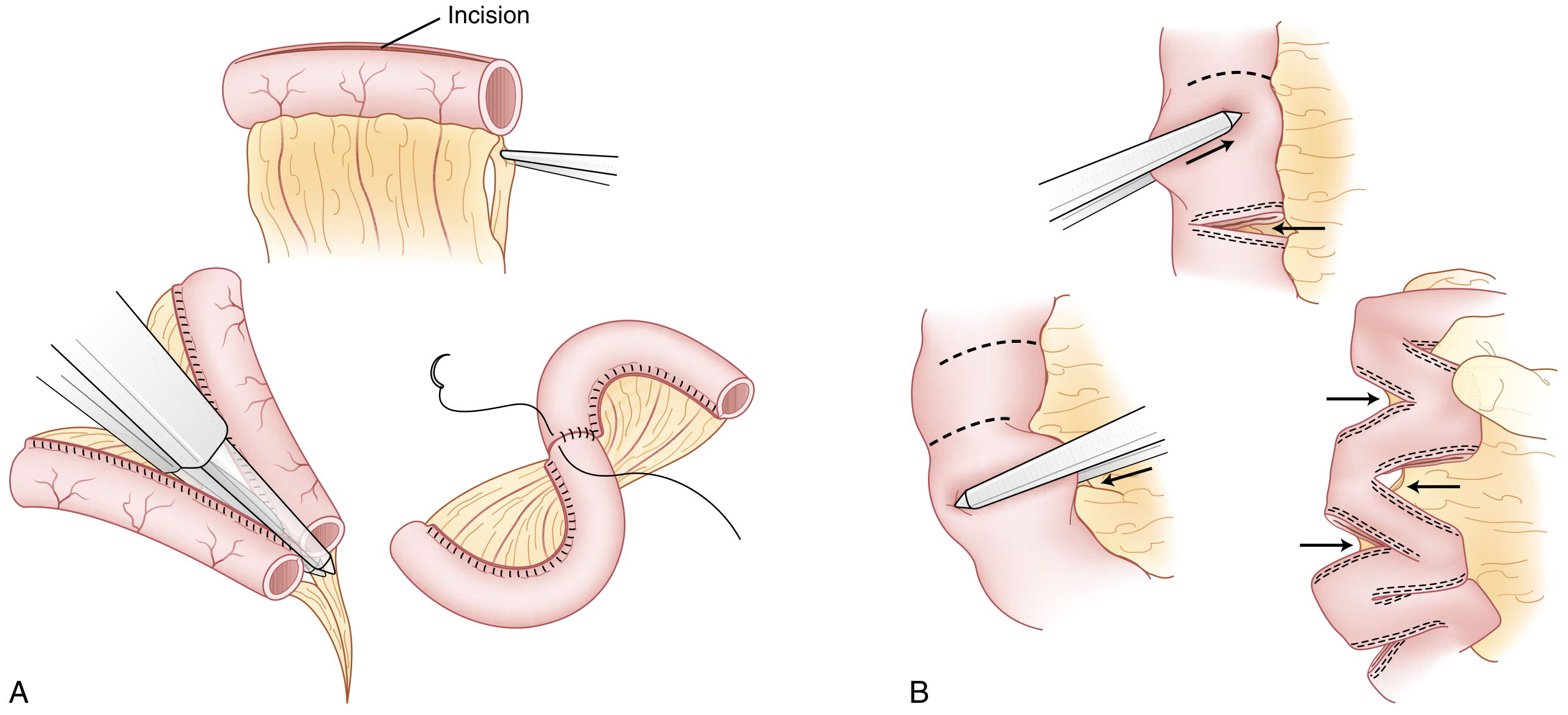
Fischer's Mastery of Surgery, Sabiston Textbook of Surgery
Intestinal Transplantation
Indications (life-threatening complications of intestinal failure / long-term TPN):
- Impending or overt liver failure
- Thrombosis of major central veins (loss of central venous access)
- Frequent catheter-related sepsis
- Frequent episodes of severe dehydration
Types: intestine alone (37%), intestine + liver + pancreas (30%), intestine + liver (24%).
5-year survival on long-term TPN is 50-75%; transplantation is considered when this window is threatened by the above complications. - Schwartz's Principles of Surgery, p. 1283
Monitoring
Patients require lifelong monitoring for:
-
Bone density (DEXA scans) - vitamin D, calcium
-
Fat-soluble vitamins (A, D, E, K), B12, folate, iron, Mg, Zn
-
Trace elements in TPN-dependent patients: copper, selenium, chromium
-
Clinical signs: hair loss, skin/nail changes, peripheral neuropathy
-
Harrison's 22E, p. 2591
Summary table:
| Residual bowel anatomy | Expected clinical course |
|---|---|
| >200 cm, intact colon | Usually manageable without long-term TPN |
| 60-100 cm + intact colon | May achieve enteral autonomy with adaptation |
| <60 cm + intact colon | Likely TPN dependent |
| <100 cm, no colon | Almost certainly TPN dependent; high risk of intestinal failure |
| <10 cm (neonatal) | TPN dependent initially; some achieve autonomy |
VHL syndrome
Von Hippel-Lindau (VHL) syndrome is an autosomal dominant hereditary cancer syndrome caused by mutations in the VHL tumor suppressor gene on chromosome 3p25. It predisposes to hemangioblastomas of the CNS/retina, clear cell renal cell carcinoma, pheochromocytoma, and pancreatic cysts/tumors. The VHL protein normally targets HIF-1α for degradation; when lost, HIF-1α accumulates and drives VEGF/PDGF overexpression, promoting angiogenesis and tumor growth.
Searching sources...
Finding Sources
Finding Sources
Searching PubMed
"von Hippel-Lindau" syndrome management
Good sources found. Let me read the most content-rich sections in parallel - Bradley & Daroff's (neurology), Robbins (pathology/molecular), NKF Primer (renal/comprehensive), and Goldman-Cecil.
Reading File
Reading File
Reading File
Reading File
Excellent content so far. Let me read the rest of the NKF Primer section (renal/surveillance/management) and fetch the MRI image of cerebellar hemangioblastomas.
Reading File
Reading File
Loading Image
I now have comprehensive, multi-source data. Let me compile the full answer.
Von Hippel-Lindau (VHL) Syndrome
Overview
VHL syndrome is an autosomal dominant hereditary multisystem tumor syndrome caused by germline mutations in the VHL tumor suppressor gene on chromosome 3p25. It predisposes patients to a characteristic set of benign and malignant tumors across multiple organs. Prevalence is approximately 1 in 36,000 live births. Penetrance exceeds 90% in mutation carriers. About 20% of cases arise from de novo mutations (no family history). - NKF Primer on Kidney Diseases, 8e
The condition was named after Eugen von Hippel (who described retinal angiomas) and Arvid Lindau (who described spinal hemangioblastomas).
Molecular Genetics and Pathogenesis
The VHL gene and protein
The VHL gene encodes two protein isoforms:
- pVHL30 - 30 kDa, 213 amino acids (full-length)
- pVHL19 - 19 kDa, lacks 53 N-terminal amino acids
Both isoforms appear to have similar tumor-suppressor function.
Two-hit mechanism
In accordance with Knudson's two-hit hypothesis, VHL is a tumor suppressor. Patients inherit one mutant allele; tumor development requires somatic inactivation of the second allele. VHL mutations are also found in a significant portion of sporadic clear-cell RCC (the most common form of sporadic kidney cancer).
HIF pathway (core mechanism)
Under normal oxygen tension:
- HIF-α subunits are hydroxylated by prolyl hydroxylases
- Hydroxylated HIF-α is recognized by pVHL, which is part of an E3 ubiquitin ligase complex
- pVHL tags HIF-α for ubiquitin-mediated proteasomal degradation
When pVHL is lost (VHL mutation):
-
HIF-α subunits escape degradation, stabilize, and translocate to the nucleus
-
HIF-α/HIF-β heterodimers activate hypoxia response elements (HREs) driving transcription of:
- VEGF - angiogenesis (explains highly vascular tumors)
- PDGF-B - tumor growth
- TGF-α - cellular proliferation
- Erythropoietin (EPO) - can produce paraneoplastic polycythemia
- GLUT-1, carbonic anhydrase IX - metabolic adaptation
-
Cyst formation is also explained by pVHL's role in maintaining primary cilia integrity and mediating nephron progenitor cell differentiation
-
Robbins & Kumar Basic Pathology; Bradley & Daroff's Neurology; NKF Primer
VHL Disease Classification
| Type | Pheochromocytoma | RCC | Key Features |
|---|---|---|---|
| Type 1 | Absent | Present | Hemangioblastomas, RCC, pancreatic tumors; usually due to large deletions or truncating mutations |
| Type 2A | Present | Absent | PCC + hemangioblastomas; missense mutations |
| Type 2B | Present | Present | PCC + hemangioblastomas + RCC; high-risk missense mutations |
| Type 2C | Present only | Absent | PCC only ("pheochromocytoma only"); specific missense mutations |
Type 2C mutations (e.g., L188V, V84L) affect pVHL's interaction with certain binding partners but spare HIF regulation - explaining why type 2C lacks hemangioblastomas and RCC. - NKF Primer, 8e
Clinical Manifestations
1. CNS Hemangioblastomas (60-80% of patients)
Hemangioblastomas are benign, slow-growing, highly vascular tumors. They appear as a mural nodule within a large fluid-filled cyst on MRI. Histology: capillary-sized thin-walled vessels surrounded by stromal cells with vacuolated lipid-rich cytoplasm (PAS-positive); stromal cells express inhibin (diagnostic marker). Mast cells within these tumors may produce EPO.
Sites (in order of frequency):
- Cerebellum (~50%) - most common
- Spinal cord (especially conus medullaris, cervicomedullary junction)
- Brainstem (area postrema of medulla)
- Cerebral hemispheres (<5%)
- Retina (retinal hemangioblastoma/retinal angioma)
Symptoms:
- Cerebellar: headache (most common), ataxia, nausea/vomiting, nystagmus
- Spinal: focal back/neck pain, sensory loss, weakness; frequently cause syringomyelia (due to intramedullary location)
- Brainstem: may cause syringobulbia
- Up to 20% have acute onset after mild head trauma
By age 60, 84% of VHL patients will develop at least one cerebellar hemangioblastoma.
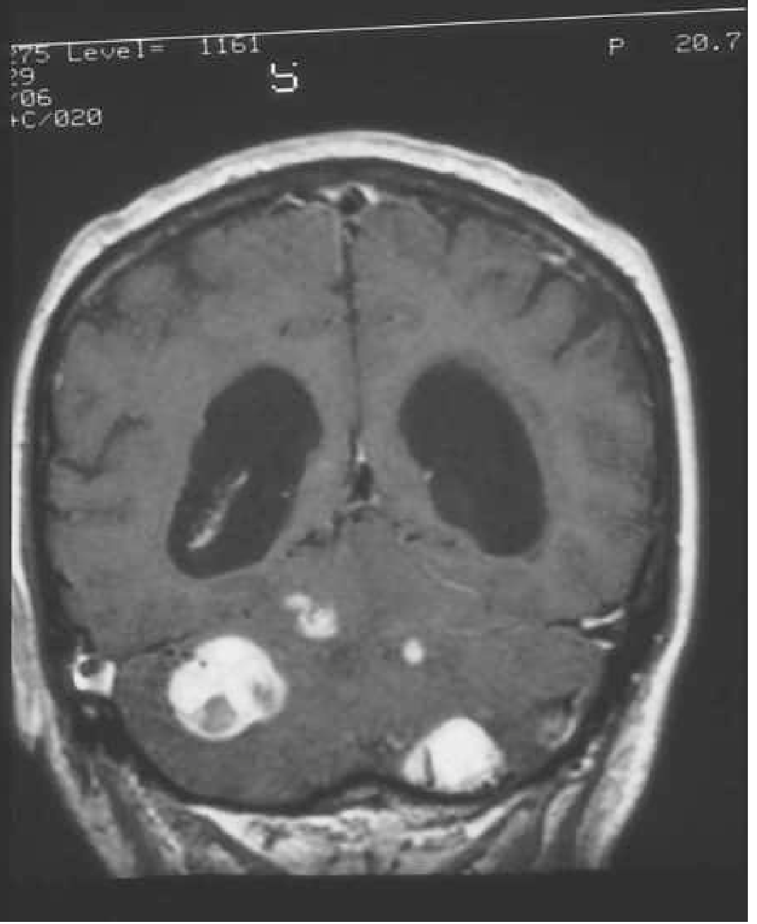
Contrast-enhanced MRI showing bilateral cerebellar hemangioblastomas (bright enhancing nodules) in a VHL patient - Bradley & Daroff's Neurology
2. Retinal Hemangioblastoma (Retinal Angioma)
- May occur as early as age 1
- Peripheral lesions can be asymptomatic; central or large lesions cause vision loss
- Arteriovenous shunting → fluid extravasation
- Complications: hemorrhage, retinal detachment, glaucoma, uveitis, macular edema, sympathetic ophthalmitis
3. Renal Cell Carcinoma (RCC)
- Most common malignant tumor in VHL and leading cause of death
- Lifetime risk up to 70% (varies by mutation type)
- Clear cell histology (same as most sporadic RCCs)
- Characteristically multifocal and bilateral
- Mean age at presentation: 40 years (younger than sporadic RCC)
- Arise from proximal tubular epithelium
- Renal cysts (from distal tubular epithelium) are present in >50% of patients
4. Pheochromocytoma (7-20%)
- Often bilateral and may be extraadrenal
- Mean age: 28 years (much younger than sporadic pheo)
- Risk varies by VHL mutation type (absent in Type 1; hallmark of Type 2)
- Symptoms: episodic/sustained hypertension, severe headache, diaphoresis, flushing, palpitations; risk of hypertensive crisis, MI, stroke, heart failure
- Biochemical diagnosis: elevated plasma/urine catecholamines and metanephrines
5. Endolymphatic Sac Tumors (ELSTs) (10-15%)
- Arise from the membranous labyrinth of the inner ear
- Bilateral ELSTs are pathognomonic of VHL
- Symptoms: sudden hearing loss (from hemorrhage), tinnitus, vertigo
6. Pancreatic Lesions (5-10% for tumors; cysts are common)
- Simple serous cysts - most common, rarely cause organ dysfunction
- Serous microcystic adenomas
- Non-secretory pancreatic neuroendocrine tumors (PNETs) - usually multiple; surgery indicated when >3 cm to prevent obstructive pancreatitis
- Adenocarcinoma (rare)
- Pancreatic cysts may obstruct the bile duct or cause exocrine insufficiency when numerous
7. Epididymal and Broad Ligament Lesions
- Papillary cystadenoma of the epididymis (males) - may be bilateral; often asymptomatic but palpable
- Papillary cystadenoma of the broad ligament (females)
- Bilateral epididymal cystadenomas are virtually pathognomonic of VHL
8. Hepatic Cysts
- Common but rarely significant clinically
Diagnosis
Clinical diagnostic criteria (either of):
- One characteristic VHL tumor + positive family history, OR
- Two or more characteristic VHL tumors (excluding epididymal/renal cysts alone) in a patient without family history
Genetic testing:
- Detection rate nearly 100% in patients with classic clinical features
- Recommended for: at-risk relatives with family history; sporadic cases of retinal/CNS hemangioblastoma, PCC, or ELST; clear-cell RCC diagnosed before age 50; bilateral or multifocal tumors
- (American College of Medical Genetics/National Society of Genetic Counselors guidelines)
Surveillance Protocol
| Manifestation | Screening modality | Starting age | Frequency |
|---|---|---|---|
| CNS hemangioblastomas | Contrast-enhanced MRI brain + spine (pre- and post-Gd T1, thin posterior fossa cuts) | 11-15 years | Every 2 years (or sooner if symptomatic) |
| Retinal hemangioblastoma | Ophthalmologic exam (fluorescein angiography) | 1 year | Annual |
| RCC / renal cysts | Abdominal MRI with/without contrast | 16 years | Every 2 years |
| Pheochromocytoma | Plasma/urine metanephrines; functional imaging | 5 years | Annual |
| Pancreatic lesions | Abdominal MRI | 16 years | Every 2 years |
| ELSTs | Audiometry; MRI | 11 years | Every 2-3 years |
- NKF Primer on Kidney Diseases, 8e
Management
CNS Hemangioblastomas
- Surgical resection is the standard treatment for symptomatic tumors
- Arteriography is not required for diagnosis but useful pre-operatively to identify feeding vessels
- Stereotactic radiosurgery is an alternative for small/surgically inaccessible lesions
Retinal Hemangioblastomas
- Laser photocoagulation or cryotherapy for small lesions
- Anti-VEGF therapy for macular involvement
- Early treatment before vision loss is key
Renal Cell Carcinoma
- Active surveillance until lesions reach 3 cm (threshold at which metastatic risk increases significantly)
- Nephron-sparing surgery (partial nephrectomy) or radiofrequency/cryoablation at the 3 cm threshold
- Total nephrectomy avoided as long as possible to preserve renal function (tumors are multifocal/bilateral)
- Tyrosine kinase inhibitors (TKIs) and anti-VEGF agents show promise in pilot studies for RCC and other lesions; further evidence needed
- A 2026 EAU Young Academic Urologists guideline (PMID: 42373875) specifically addresses kidney cancer management in VHL
Pheochromocytoma
- Alpha-blockade (phenoxybenzamine or selective alpha-1 blockers) followed by beta-blockade pre-operatively
- Laparoscopic adrenalectomy (cortical-sparing if bilateral to preserve adrenal cortex)
- Pre-operative screening for PCC is mandatory before any elective surgery in VHL patients (risk of intraoperative hypertensive crisis)
Pancreatic Tumors
- Surgery when PNETs reach >3 cm to prevent obstructive pancreatitis or malignant transformation
ELSTs
- Surgical resection when symptomatic; hearing rehabilitation as needed
Key Points Summary
- Gene: VHL on chromosome 3p25 - tumor suppressor; encodes pVHL, which targets HIF-α for ubiquitin-mediated degradation
- Core mechanism: Lost pVHL → HIF-α accumulation → VEGF/EPO/TGF-α/PDGF overexpression → highly vascular tumors
- Hallmark lesions: CNS/retinal hemangioblastomas, clear-cell RCC, pheochromocytoma, pancreatic cysts/PNETs, ELSTs
- Leading cause of death: Renal cell carcinoma (and historically CNS hemangioblastoma)
- Paraneoplastic polycythemia can occur from EPO secretion by hemangioblastoma stromal cells
- Bilateral ELSTs are pathognomonic; bilateral epididymal cystadenomas are virtually pathognomonic
- RCC management threshold: 3 cm (nephron-sparing surgery or ablation)
- PCC screening starts at age 5 (earliest onset of any VHL manifestation)
- VHL mutations are found in up to 70% of sporadic clear-cell RCCs - making the VHL/HIF pathway the central target for anti-VEGF/TKI therapy in sporadic kidney cancer as well
This is a shared conversation. Sign in to Orris to start your own chat.