
Diabetes ketoacidosis Lyme disease explain them
Reading File
Now let me search for both topics in parallel from the medical library.
Finding Sources
Finding Sources
Good sources found. Now let me read the most comprehensive sections in parallel - Goldman-Cecil for DKA and Medical Microbiology for Lyme disease.
Reading File
Reading File
Good. Let me now get more DKA details (management/clinical) and Lyme disease clinical details in parallel.
Reading File
Reading File
Excellent content found. Let me get the DKA treatment section and more Lyme disease clinical stages in parallel.
Reading File
Reading File
Loading Image
Now let me get DKA treatment details and more Lyme treatment content in parallel.
Reading File
Reading File
I now have comprehensive information on both conditions. Let me compile the full detailed answer.
Diabetic Ketoacidosis (DKA)
Definition
DKA is defined by three fundamental biochemical features (the classic triad):
- Hyperglycemia (or a known history of diabetes)
- Ketonuria (urinary ketones ≥2+ or serum ketones ≥3.0 mmol/L)
- Acidosis (arterial or venous pH <7.3)
- Goldman-Cecil Medicine
Pathophysiology
DKA results from combined insulin deficiency + excess counter-regulatory hormones (glucagon, cortisol, catecholamines, growth hormone). This hormonal imbalance:
- Drives delivery of substrates from muscle (amino acids, lactate, pyruvate) and adipose tissue (free fatty acids, glycerol) to the liver
- The liver converts these to glucose and ketone bodies (β-hydroxybutyrate, acetoacetate, acetone)
- Glucose and ketones flood the circulation faster than they can be used
- Hyperglycemia causes osmotic diuresis → dehydration + electrolyte loss
- Dehydration causes hemoconcentration → worsening hyperglycemia and hyperosmolality in a vicious cycle
The three ketone bodies are:
- β-Hydroxybutyrate - the predominant one (often falsely underdetected by nitroprusside strips)
- Acetoacetate
- Acetone
Precipitating Causes
| Most Common | Other |
|---|---|
| Infections | Stroke, pulmonary embolism |
| Inadequate insulin / nonadherence | Acute pancreatitis |
| New-onset diabetes (type 1) | Cushing syndrome, thyrotoxicosis |
| Acute coronary syndrome | Severe burns |
| Unknown | Drugs: corticosteroids, SGLT-2 inhibitors, clozapine, olanzapine, cocaine, lithium, thiazides |
An important behavioral cause: some younger patients omit insulin deliberately to lose weight or draw attention to a dysfunctional home situation.
Clinical Features
Onset is typically over hours to days with:
- Progressive polyuria, polydipsia, weakness, lethargy, nausea, anorexia
- Nonspecific abdominal pain (can mimic acute abdomen)
- Reduced GI motility or paralytic ileus
- Physical signs from dehydration: dry skin/mucous membranes, tachycardia, orthostatic hypotension, reduced JVP
- Kussmaul breathing - deep, rapid respirations (compensatory response to metabolic acidosis)
- Depressed mental function to frank coma in severe cases
Diagnosis & Labs
| Finding | Value |
|---|---|
| Blood glucose | Variable; normal to >1000 mg/dL (55.5 mmol/L) |
| Serum bicarbonate | <18 mmol/L |
| pH | 7.20-7.30 (mild) to <7.00 (severe) |
| Anion gap | Elevated, proportional to HCO3 drop |
| Sodium | Often low (osmotic water shift) |
| Potassium | May be normal, high, or low at presentation - but always total body depleted |
| WBC | Elevated (from acidosis itself, not necessarily infection) |
| Hematocrit | Elevated (hemoconcentration) |
Key pitfall with ketone testing: Nitroprusside strips only react with acetoacetate. β-Hydroxybutyrate (the major ketone in DKA) does NOT react - so strips can falsely underestimate the severity of ketoacidosis. After insulin starts, β-hydroxybutyrate converts to acetoacetate, so strips may appear to show worsening ketosis even as the patient improves. Point-of-care capillary ketone monitors are now preferred.
Treatment
Treatment has four pillars:
1. Fluid Replacement
- Start with 0.9% NaCl (even if serum osmolality is high, since normal saline is still relatively hypotonic)
- Rate: 2-4 liters in the first 2-4 hours in DKA
- Fluid losses of 5-10 L are common; electrolyte losses include sodium 5-10 mmol/kg and potassium up to 7 mmol/kg
2. Potassium Replacement
- Even if K+ is initially elevated (due to acidosis shifting K+ extracellularly), it will fall sharply once insulin is given
- Hold K+ replacement if K+ >5.5 mEq/L; but begin when K+ is ≤5.5 and falling
- A low initial K+ signals profound total body depletion and requires prompt IV replacement before insulin
3. Insulin
- IV insulin to halt ketogenesis and reduce glucose
- Glucose replacement (5% dextrose) should be added to the infusion once blood glucose falls to avoid hypoglycemia
4. Treat the Precipitant
- Identify and treat underlying infection, ACS, or other trigger
Prognosis: Despite aggressive treatment, mortality is approximately 4% for DKA and up to 20% for hyperosmolar hyperglycemic state (HHS). Death is associated with extremes of age, comorbidities, and severity of the precipitating event.
Lyme Disease
Causative Agent & Epidemiology
Lyme disease is caused by spirochetes of the Borrelia burgdorferi sensu lato complex:
- B. burgdorferi sensu stricto - United States
- B. garinii - Europe/Asia; principal cause of Lyme neuroborreliosis
- B. afzelii - Europe/Asia; associated with acrodermatitis chronica atrophicans
Transmission: By hard ticks (family Ixodidae):
- Ixodes scapularis - eastern and midwestern US
- I. pacificus - western US
- I. ricinus - Europe
- I. persulcatus - Eastern Europe and Asia
Reservoirs: Mice, deer, ticks. Seasonal peak: June-July (nymph feeding stage).
US geography: Most cases from two foci - Northeast/Mid-Atlantic states (Maine to Virginia) and Upper Midwest (Minnesota, Wisconsin).
The tick must be attached for >24 hours to transmit disease in the US.
Clinical Stages
Stage 1 - Early Localized (Days to weeks after bite)
The hallmark is erythema migrans (EM):
- Begins as a small red macule/papule at the bite site 3-32 days (median 7) after the bite
- Gradually expands outward to an annular (bull's-eye) rash, median diameter 15 cm (range 3-68 cm)
- Border is slightly raised, warm, red to bluish-red, no scale
- Center may clear (classic bull's-eye) or remain red/indurated/vesicular
- Accompanied by burning in 50% of patients; rarely itchy
- Common sites: legs, groin, axilla
Only ~50% of patients recall the tick bite.
Here is the classic appearance of erythema migrans:
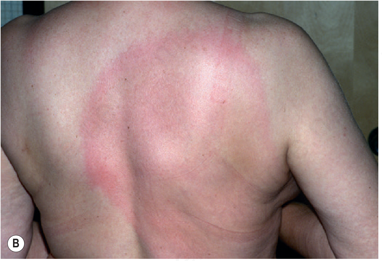
Erythema migrans on the upper back - Andrews' Diseases of the Skin
Stage 2 - Early Disseminated (Weeks to months)
- Musculoskeletal: Migratory arthralgia, myalgia
- Neurologic (Lyme neuroborreliosis): Stiff neck, headache, meningitis, cranial nerve palsies (Bell palsy is classic), radiculopathy, peripheral neuropathy, cognitive deficits, vestibular neuronitis
- Cardiac: Fluctuating degrees of AV block or complete heart block (most often in young men), lasting 3 days to 6 weeks; dilated cardiomyopathy in European cases
- Skin: Multiple secondary EM lesions
In Europe: Bannworth syndrome - focal radicular pains + lymphocytic meningitis + cranial nerve paralysis.
Stage 3 - Late Disseminated (Months to years)
- Chronic arthritis - especially of the knees; ~10% of untreated US patients; leads to severe disability in half
- Acrodermatitis chronica atrophicans (ACA) - almost exclusively in Europe (B. afzelii); diffuse reddish-blue, paper-thin atrophic skin on extremities; progressive and never fully involutes
- Neurologic sequelae
Diagnosis
- Clinical: Erythema migrans is the most sensitive evidence of early infection - diagnose clinically without waiting for serology
- Serology: Two-tier testing:
- Screening: ELISA (89% sensitive, 72% specific)
- Confirmation: Western blot (IgM: 2/3 bands positive; IgG: 5/10 bands positive)
- Serology limitations: Only 27% positive if symptoms <7 days, 41% at 7-14 days, 88% if >2 weeks
- PCR: Specific but insensitive; not widely available
- False positives occur in: syphilis, yaws, pinta, leptospirosis, relapsing fever, infectious mononucleosis, autoantibody-related diseases (note: VDRL is negative in B. burgdorferi infection)
Treatment
| Situation | Treatment |
|---|---|
| Early localized/disseminated (adults) | Doxycycline 100 mg twice daily x 21 days (first choice; also covers Ehrlichia) |
| Adults (alternative) | Amoxicillin 500 mg TID x 21 days OR Cefuroxime axetil 500 mg BID x 21 days |
| Children <9 years | Amoxicillin 20 mg/kg/day in divided doses |
| Pregnant (localized) | Amoxicillin |
| Pregnant (disseminated) / Immunodeficient | IV Penicillin G or IV Ceftriaxone |
| Carditis / Neurologic / Arthritic involvement | More aggressive regimens; parenteral antibiotics often indicated |
Prevention:
- Avoid tick-infested areas; inspect for ticks after outdoor activity
- Nymphs are tiny ("the freckle that moves") - harder to spot
- Post-exposure prophylaxis: Single dose of doxycycline 200 mg within 72 hours of a bite from a partially engorged I. scapularis in a high-incidence area is 87% effective
Key Differences at a Glance
| Feature | DKA | Lyme Disease |
|---|---|---|
| Cause | Insulin deficiency + counter-regulatory excess | Borrelia burgdorferi spirochete infection |
| Transmission | Metabolic / endocrine | Tick bite (Ixodes spp.) |
| Key sign | Kussmaul breathing, fruity breath, altered mental status | Erythema migrans (bull's-eye rash) |
| Key lab | pH <7.3, elevated ketones, anion gap acidosis | ELISA + Western blot serology |
| Acute treatment | IV fluids, insulin, electrolyte replacement | Doxycycline (oral) |
| Mortality | ~4% (DKA), up to 20% (HHS) | Low if treated early; complications if untreated |
Sources: Goldman-Cecil Medicine (DKA); Andrews' Diseases of the Skin and Medical Microbiology 9e (Lyme disease)
This is a shared conversation. Sign in to Orris to start your own chat.