Search some dermatology images

Asking for Preferences
Searching Images

eczema atopic dermatitis skin rash

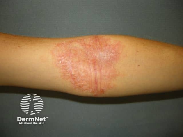
Clinical photography of an inflammatory dermatosis involving the forearm. Imaging modality: standard digital dermatology photograph. The primary subject is human skin with erythematous, edematous plaques demonstrating fine surface scaling and lichenification consistent with atopic dermatitis (eczema). Lesions are localized to the ventral forearm with a slightly linear distribution and central clearing absent. The skin appears mildly dry with fissuring in fissures near the flexural creases. Color variation includes bright red patches interspersed with pale desquamation. The pattern is typical of inflammatory dermatitis rather than infectious etiologies, though secondary infection cannot be excluded without clinical history. The image captures typical features: pruritic rash, chronic scratching marks, layering of erythema, and xerosis. In a clinical setting, this presentation supports a diagnosis of atopic dermatitis or eczema flare, particularly in patients with personal or family history of atopy, asthma, or allergic rhinitis. Differential diagnoses include contact dermatitis, seborrheic dermatitis, psoriasis, tinea corporis, or nummular eczema. Diagnostically, the image is useful for educational purposes, for documenting disease severity, and for correlating with treatment response to emollients and topical corticosteroids. This photographic record aids trainee clinicians in recognizing pattern recognition of eczema and in differentiating from other inflammatory dermatoses. Images support diagnosis, education, patient guidance.
Clinical photography of an inflammatory dermatosis involving the forearm. Imaging modality: standard digital dermatology photograph. The primary subject is human skin with erythematous, edematous plaques demonstrating fine surface scaling and lichenification consistent with atopic dermatitis (eczema). Lesions are localized to the ventral forearm with a slightly linear distribution and central clearing absent. The skin appears mildly dry with fissuring in fissures near the flexural creases. Color variation includes bright red patches interspersed with pale desquamation. The pattern is typical of inflammatory dermatitis rather than infectious etiologies, though secondary infection cannot be excluded without clinical history. The image captures typical features: pruritic rash, chronic scratching marks, layering of erythema, and xerosis. In a clinical setting, this presentation supports a diagnosis of atopic dermatitis or eczema flare, particularly in patients with personal or family history of atopy, asthma, or allergic rhinitis. Differential diagnoses include contact dermatitis, seborrheic dermatitis, psoriasis, tinea corporis, or nummular eczema. Diagnostically, the image is useful for educational purposes, for documenting disease severity, and for correlating with treatment response to emollients and topical corticosteroids. This photographic record aids trainee clinicians in recognizing pattern recognition of eczema and in differentiating from other inflammatory dermatoses. Images support diagnosis, education, patient guidance.

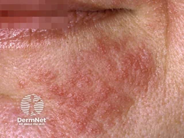
Clinical photograph of facial skin showing erythematous, scaly plaques predominantly on the left cheek with mild surface desquamation and fissuring along the perioral region. The epidermis appears dry with accentuated roughness and fine white scale at the margins of the patches. The involved area shows diffuse erythema and indistinct borders, lacking frank vesiculation or purpura. No obvious edema or secondary infection is evident in this field of view. The presentation is consistent with inflammatory dermatitis of the face; possibilities include atopic dermatitis (eczema), seborrheic dermatitis, or irritant/contact dermatitis. The pattern is unilateral here but may be bilateral in other settings. The lesion lacks the classic ring-shaped border of tinea faciei and does not show pustules or crusting. A dermal inflammation with epidermal hyperkeratosis and xerosis is suggested by the dry, scaly skin. This image is useful for illustrating recognition of facial dermatitis patterns, differentiation from infectious etiologies, and for educative comparison across chronic inflammatory dermatoses. Clinically relevant keywords: dermatitis, eczema, atopic dermatitis, seborrheic dermatitis, contact dermatitis, facial rash, scaling, erythema, xerosis, inflammatory skin disease, differential diagnoses, patch testing, topical corticosteroids, emollients, skin barrier disruption, diagnosis and management planning. Clinical utility includes guiding topical therapy, emollient use, and monitoring response progress.
Clinical photograph of facial skin showing erythematous, scaly plaques predominantly on the left cheek with mild surface desquamation and fissuring along the perioral region. The epidermis appears dry with accentuated roughness and fine white scale at the margins of the patches. The involved area shows diffuse erythema and indistinct borders, lacking frank vesiculation or purpura. No obvious edema or secondary infection is evident in this field of view. The presentation is consistent with inflammatory dermatitis of the face; possibilities include atopic dermatitis (eczema), seborrheic dermatitis, or irritant/contact dermatitis. The pattern is unilateral here but may be bilateral in other settings. The lesion lacks the classic ring-shaped border of tinea faciei and does not show pustules or crusting. A dermal inflammation with epidermal hyperkeratosis and xerosis is suggested by the dry, scaly skin. This image is useful for illustrating recognition of facial dermatitis patterns, differentiation from infectious etiologies, and for educative comparison across chronic inflammatory dermatoses. Clinically relevant keywords: dermatitis, eczema, atopic dermatitis, seborrheic dermatitis, contact dermatitis, facial rash, scaling, erythema, xerosis, inflammatory skin disease, differential diagnoses, patch testing, topical corticosteroids, emollients, skin barrier disruption, diagnosis and management planning. Clinical utility includes guiding topical therapy, emollient use, and monitoring response progress.

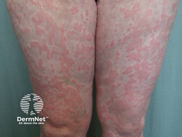
This is a high-resolution clinical photograph of the anterior aspects of both thighs and legs showing widespread erythematous, coalescing patches with fine scaling distributed bilaterally on extensor surfaces and proximal lower limbs. The skin appears dry with diffuse erythema; borders are variably defined and some plaques show mild lichenification. No vesicles or crusts are clearly visible. The pattern is symmetric and patchy, more pronounced on the thighs and distal legs near the knees, with faint residual hyperpigmentation in older lesions. The image suggests a chronic inflammatory dermatosis such as atopic dermatitis (eczema) or contact dermatitis, though psoriasis or fungal infection (tinea) could mimic the appearance. Absence of clear vesiculation or pustulation argues against acute dermatitis. Etiology may relate to irritant or allergen exposure, scratching, or a systemic inflammatory condition. In clinical practice, this pattern would warrant correlation with symptoms (pruritus, swelling), exposure history, and potential skin biopsy if diagnostic uncertainty persists. This image is valuable for education on diffuse eczematous eruptions, differential diagnosis of extensor-surface dermatitis, and assessment of therapeutic response to topical corticosteroids, emollients, or phototherapy. Relevant search terms: dermatitis, eczema, atopic dermatitis, contact dermatitis, psoriasis, tinea, dermatology, lower extremity rash, bilateral leg lesions, chronic skin disease. for reference.

This is a high-resolution clinical photograph of the anterior aspects of both thighs and legs showing widespread erythematous, coalescing patches with fine scaling distributed bilaterally on extensor surfaces and proximal lower limbs. The skin appears dry with diffuse erythema; borders are variably defined and some plaques show mild lichenification. No vesicles or crusts are clearly visible. The pattern is symmetric and patchy, more pronounced on the thighs and distal legs near the knees, with faint residual hyperpigmentation in older lesions. The image suggests a chronic inflammatory dermatosis such as atopic dermatitis (eczema) or contact dermatitis, though psoriasis or fungal infection (tinea) could mimic the appearance. Absence of clear vesiculation or pustulation argues against acute dermatitis. Etiology may relate to irritant or allergen exposure, scratching, or a systemic inflammatory condition. In clinical practice, this pattern would warrant correlation with symptoms (pruritus, swelling), exposure history, and potential skin biopsy if diagnostic uncertainty persists. This image is valuable for education on diffuse eczematous eruptions, differential diagnosis of extensor-surface dermatitis, and assessment of therapeutic response to topical corticosteroids, emollients, or phototherapy. Relevant search terms: dermatitis, eczema, atopic dermatitis, contact dermatitis, psoriasis, tinea, dermatology, lower extremity rash, bilateral leg lesions, chronic skin disease. for reference.

Searching Images

psoriasis plaque skin lesion

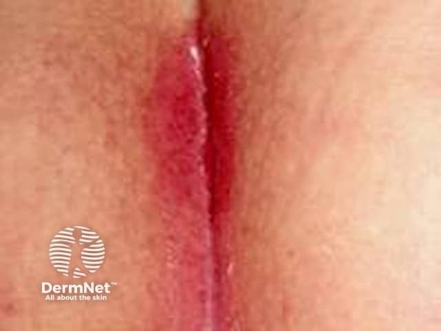
Clinical photograph of a flexural skin fold lesion consistent with plaque psoriasis manifested in an intertriginous region. This high-resolution image depicts a single, erythematous plaque with well-defined edges, located within a skin crease. The plaque exhibits fissuring, mild crusting, and surrounding rim of erythema, with minimal scaling attributable to occlusion in folds. The presentation is characteristic of flexural psoriasis, which commonly affects the antecubital and inguinal regions, producing smooth, shiny, red plaques that may merge with adjacent folds. The image emphasizes the anatomic context (intertriginous skin) and the morphological features (fissuring, plaque formation, erythema) that help distinguish psoriasis from eczematous dermatitis, candidal intertrigo, and irritant contact dermatitis. Clinically, flexural psoriasis indicates T-cell mediated hyperproliferation of keratinocytes with parakeratosis and Munro microabscesses on histology, though histology is not shown here. The diagnostic significance lies in recognizing the pattern specific to intertriginous areas, guiding treatment with potent topical corticosteroids, vitamin D analogs, or calcineurin inhibitors, and considering systemic therapy in extensive disease. This image is relevant for dermatology education, differential diagnosis practice, and clinical decision support in intertriginous skin disorders. Keywords: plaque psoriasis, intertriginous psoriasis, flexural psoriasis, psoriasis in folds, inverse psoriasis, dermatitis mimicry, psoriasis differential, topical therapy, phototherapy, systemic therapy considerations.
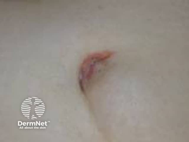
Clinical photograph of a flexural skin fold lesion consistent with plaque psoriasis manifested in an intertriginous region. This high-resolution image depicts a single, erythematous plaque with well-defined edges, located within a skin crease. The plaque exhibits fissuring, mild crusting, and surrounding rim of erythema, with minimal scaling attributable to occlusion in folds. The presentation is characteristic of flexural psoriasis, which commonly affects the antecubital and inguinal regions, producing smooth, shiny, red plaques that may merge with adjacent folds. The image emphasizes the anatomic context (intertriginous skin) and the morphological features (fissuring, plaque formation, erythema) that help distinguish psoriasis from eczematous dermatitis, candidal intertrigo, and irritant contact dermatitis. Clinically, flexural psoriasis indicates T-cell mediated hyperproliferation of keratinocytes with parakeratosis and Munro microabscesses on histology, though histology is not shown here. The diagnostic significance lies in recognizing the pattern specific to intertriginous areas, guiding treatment with potent topical corticosteroids, vitamin D analogs, or calcineurin inhibitors, and considering systemic therapy in extensive disease. This image is relevant for dermatology education, differential diagnosis practice, and clinical decision support in intertriginous skin disorders. Keywords: plaque psoriasis, intertriginous psoriasis, flexural psoriasis, psoriasis in folds, inverse psoriasis, dermatitis mimicry, psoriasis differential, topical therapy, phototherapy, systemic therapy considerations.

Clinical photograph of the umbilical region demonstrating a localized skin plaque. Modality: clinical photography, digital high-resolution anterior view; close-up of umbilicus. The lesion sits within the periumbilical skin, along the midline abdomen. The plaque is erythematous with well-demarcated yet irregular margins. Overlying desquamation and light scaling are evident; a slight fissure or crust may be present at the margin. No surrounding edema; mild perifollicular involvement; skin around appears normal. This appearance is characteristic of psoriasis vulgaris when presenting as localized plaque psoriasis; however, differential includes chronic dermatitis, eczema, or fungal infection (tinea) in intertriginous areas. The image alone cannot confirm histology or systemic involvement. Clinically, psoriasis at the umbilicus is uncommon but plausible; the lesion may reflect Koebnerization or friction-induced psoriasis in skin folds. Diagnostic significance lies in recognizing psoriatic plaque in atypical sites, guiding therapy (topical corticosteroids, vitamin D analogs, calcipotriol, coal tar preparations) and prompting evaluation for psoriatic arthritis if joint symptoms exist. Potential clinical use cases include teledermatology triage, educational illustration of plaque psoriasis morphology, and differential diagnosis exercises for dermatology trainees. If biopsy or dermoscopy is available, histopathology would show epidermal hyperplasia with parakeratosis and neutrophilic Munro microabscesses; lack of fungal elements would favor psoriasis.
Clinical photograph of the umbilical region demonstrating a localized skin plaque. Modality: clinical photography, digital high-resolution anterior view; close-up of umbilicus. The lesion sits within the periumbilical skin, along the midline abdomen. The plaque is erythematous with well-demarcated yet irregular margins. Overlying desquamation and light scaling are evident; a slight fissure or crust may be present at the margin. No surrounding edema; mild perifollicular involvement; skin around appears normal. This appearance is characteristic of psoriasis vulgaris when presenting as localized plaque psoriasis; however, differential includes chronic dermatitis, eczema, or fungal infection (tinea) in intertriginous areas. The image alone cannot confirm histology or systemic involvement. Clinically, psoriasis at the umbilicus is uncommon but plausible; the lesion may reflect Koebnerization or friction-induced psoriasis in skin folds. Diagnostic significance lies in recognizing psoriatic plaque in atypical sites, guiding therapy (topical corticosteroids, vitamin D analogs, calcipotriol, coal tar preparations) and prompting evaluation for psoriatic arthritis if joint symptoms exist. Potential clinical use cases include teledermatology triage, educational illustration of plaque psoriasis morphology, and differential diagnosis exercises for dermatology trainees. If biopsy or dermoscopy is available, histopathology would show epidermal hyperplasia with parakeratosis and neutrophilic Munro microabscesses; lack of fungal elements would favor psoriasis.

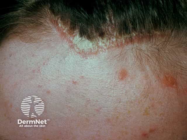
Clinical photography of the scalp and frontal hairline illustrating a clear plaque psoriasis lesion. Modality: clinical color photograph; frontal/anterior view focused on the forehead and perihairline skin. Primary subject: an erythematous, well-circumscribed plaque with coarse, adherent scale arising at the anterior hairline and extending forward onto the forehead. Color is orange-red with silvery-white scale at the margins, demonstrating classic plaque morphology. The lesion is well demarcated from adjacent nonlesional skin, with mild surface desquamation and subtle surrounding erythema. Hair-bearing scalp adjacent skin shows dry, fine scale; no secondary lesions are evident in this field. The image captures typical psoriasis features: sharply demarcated edge, plaque elevation, and hyperkeratosis. This supports a clinical diagnosis of plaque psoriasis involving the scalp and frontal scalp/forehead region; differential diagnoses include seborrheic dermatitis, atopic dermatitis, tinea capitis, candidiasis, and irritant contact dermatitis. The diagnostic significance lies in recognizing chronic plaque psoriasis pattern, guiding topical therapy with corticosteroids, vitamin D analogs, or coal tar preparations, and prompting evaluation for additional site involvement. Clinically correlates with pruritus, flaking, and potential Koebnerization; useful for education, documentation, and treatment monitoring in dermatology research and medical education. This image serves as an educational reference for residents and students studying plaque psoriasis morphology on the hairy scalp.

Clinical photography of the scalp and frontal hairline illustrating a clear plaque psoriasis lesion. Modality: clinical color photograph; frontal/anterior view focused on the forehead and perihairline skin. Primary subject: an erythematous, well-circumscribed plaque with coarse, adherent scale arising at the anterior hairline and extending forward onto the forehead. Color is orange-red with silvery-white scale at the margins, demonstrating classic plaque morphology. The lesion is well demarcated from adjacent nonlesional skin, with mild surface desquamation and subtle surrounding erythema. Hair-bearing scalp adjacent skin shows dry, fine scale; no secondary lesions are evident in this field. The image captures typical psoriasis features: sharply demarcated edge, plaque elevation, and hyperkeratosis. This supports a clinical diagnosis of plaque psoriasis involving the scalp and frontal scalp/forehead region; differential diagnoses include seborrheic dermatitis, atopic dermatitis, tinea capitis, candidiasis, and irritant contact dermatitis. The diagnostic significance lies in recognizing chronic plaque psoriasis pattern, guiding topical therapy with corticosteroids, vitamin D analogs, or coal tar preparations, and prompting evaluation for additional site involvement. Clinically correlates with pruritus, flaking, and potential Koebnerization; useful for education, documentation, and treatment monitoring in dermatology research and medical education. This image serves as an educational reference for residents and students studying plaque psoriasis morphology on the hairy scalp.

Searching Images

urticaria hives allergic skin reaction
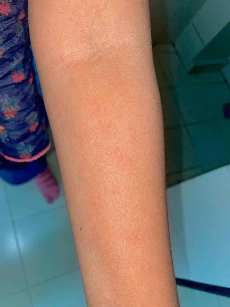
This clinical photograph displays a close-up view of a human upper extremity, specifically the forearm and elbow region, exhibiting a diffuse dermatological reaction. The skin presents with a generalized erythematous (reddish) hue and is covered by an extensive, confluent rash. Visible features include numerous small, raised, erythematous papules and classic wheals characteristic of urticaria (hives). The distribution is widespread across the limb, with a notable concentration of skin inflammation and swelling around the antecubital fossa (inner elbow). This presentation is clinically significant as it illustrates a hypersensitivity reaction or angioedema-like presentation, which in this medical context is associated with an adverse drug reaction following olanzapine administration. The image serves as an educational tool for identifying medication-induced cutaneous manifestations and acute allergic skin responses in clinical practice.
This clinical photograph displays a close-up view of a human upper extremity, specifically the forearm and elbow region, exhibiting a diffuse dermatological reaction. The skin presents with a generalized erythematous (reddish) hue and is covered by an extensive, confluent rash. Visible features include numerous small, raised, erythematous papules and classic wheals characteristic of urticaria (hives). The distribution is widespread across the limb, with a notable concentration of skin inflammation and swelling around the antecubital fossa (inner elbow). This presentation is clinically significant as it illustrates a hypersensitivity reaction or angioedema-like presentation, which in this medical context is associated with an adverse drug reaction following olanzapine administration. The image serves as an educational tool for identifying medication-induced cutaneous manifestations and acute allergic skin responses in clinical practice.

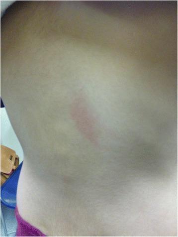
This clinical photograph displays a focal skin lesion on the mid-back of a pediatric patient. The primary finding is a single, vertically-oriented, erythematous plaque or wheal. The lesion is characterized by distinct borders, central redness, and a slightly raised morphology relative to the surrounding healthy skin, which appears otherwise smooth and normochromic. This clinical presentation is characteristic of urticaria (hives), specifically an immediate hypersensitivity reaction. The image demonstrates a localized cutaneous manifestation of a Type I allergic reaction, which in this clinical context occurred 20 minutes following an oral challenge with amoxicillin. This visual serves as an educational example of drug-induced immediate-type allergy in pediatric dermatology and immunology, highlighting the characteristic appearance of a positive oral provocation test result.

This clinical photograph displays a focal skin lesion on the mid-back of a pediatric patient. The primary finding is a single, vertically-oriented, erythematous plaque or wheal. The lesion is characterized by distinct borders, central redness, and a slightly raised morphology relative to the surrounding healthy skin, which appears otherwise smooth and normochromic. This clinical presentation is characteristic of urticaria (hives), specifically an immediate hypersensitivity reaction. The image demonstrates a localized cutaneous manifestation of a Type I allergic reaction, which in this clinical context occurred 20 minutes following an oral challenge with amoxicillin. This visual serves as an educational example of drug-induced immediate-type allergy in pediatric dermatology and immunology, highlighting the characteristic appearance of a positive oral provocation test result.

Here is a collection of dermatology images across three common inflammatory skin conditions:

Atopic Dermatitis (Eczema)

1. Forearm eczema - classic presentation
Atopic dermatitis on forearm
Erythematous, scaly plaques on the ventral forearm with lichenification and fissuring near flexural creases. Hallmark features: pruritic rash, xerosis, and chronic scratching marks.

2. Facial eczema / seborrheic dermatitis
Facial dermatitis
Erythematous, scaly plaques on the cheek with perioral fissuring. Differential includes atopic dermatitis, seborrheic dermatitis, and contact dermatitis.

3. Bilateral leg eczema
Bilateral eczema on legs
Widespread coalescing patches on both thighs and lower legs, with mild lichenification and residual hyperpigmentation from older lesions.

Psoriasis

4. Flexural (inverse) psoriasis
Inverse psoriasis in skin fold
Smooth, shiny erythematous plaque within a skin crease. Minimal scaling (due to occlusion), with well-defined edges and fissuring - typical of inverse/flexural psoriasis.

5. Psoriasis at the umbilicus
Periumbilical psoriasis
Localized plaque at an atypical site. Erythematous with well-demarcated margins and light scaling. May represent Koebnerization from friction.

6. Scalp / frontal hairline psoriasis
Scalp psoriasis at hairline
Classic plaque psoriasis at the anterior hairline - orange-red with coarse, adherent silvery-white scale and a sharply demarcated edge extending onto the forehead.

Urticaria (Hives)

7. Drug-induced urticaria - forearm
Urticaria on forearm
Diffuse erythematous wheals and papules on the forearm following olanzapine administration. Wheals are confluent with concentration around the antecubital fossa.

8. Pediatric urticaria - amoxicillin reaction
Pediatric urticaria on back
Single vertically-oriented erythematous wheal on the mid-back of a child, occurring 20 minutes after oral amoxicillin challenge - a textbook Type I hypersensitivity reaction.

Key differentiators at a glance:
FeatureEczemaPsoriasisUrticaria
BordersIndistinctWell-demarcatedDistinct (transient)
ScaleFine, dryCoarse, silveryNone
DistributionFlexural, faceExtensor, scalpAnywhere
DurationChronicChronicMinutes to hours
ItchIntenseModerateIntense
This is a shared conversation. Sign in to Orris to start your own chat.