
Cvc spleen Cvc lung Cvc liver
- Spleen: Becomes enlarged (congestive splenomegaly), with sinusoidal dilation and eventual fibrosis (Gamna-Gandy bodies in chronic cases).
- Lung: "Brown induration" - capillary congestion, alveolar hemorrhage, and hemosiderin-laden macrophages ("heart failure cells").
- Liver: "Nutmeg liver" - centrilobular congestion and necrosis with periportal sparing, giving a mottled appearance.
nutmeg liver chronic venous congestion centrilobular
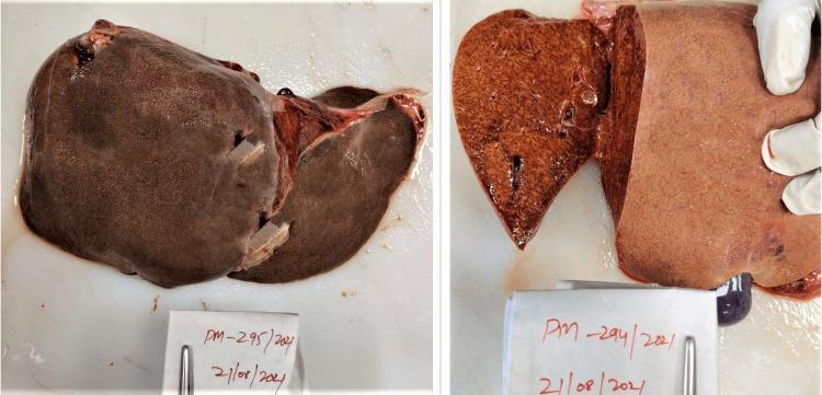
This clinical photograph displays a gross pathology specimen of a human liver obtained during a postmortem examination. The image is divided into two panels: the left shows the intact liver surface, while the right displays a transverse cut section of the hepatic parenchyma. The organ demonstrates prominent yellowish discoloration and a distinctive mottled appearance. This pattern, frequently referred to as a 'nutmeg liver,' is characterized by alternating dark reddish-brown areas, representing chronic venous congestion in the centrilobular regions, and paler yellowish areas, representing fatty change or normal parenchyma in the periportal regions. The specimen illustrates the macroscopic effects of chronic passive congestion, often secondary to congestive heart failure or toxic insults. The educational focus is on the gross recognition of hepatic congestion and jaundice, which are critical diagnostic indicators in forensic and clinical pathology.
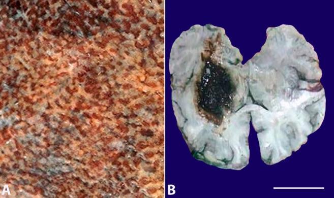
This composite clinical photograph displays two gross pathological specimens (labeled A and B) from a medical autopsy. Image A shows a close-up detail of a cut liver surface presenting a classic 'nutmeg liver' appearance. This is characterized by a variegated, mottled pattern with alternating red-brown areas (representing centrilobular congestion and necrosis) and lighter tan or yellowish-brown areas (representing relatively normal or fatty periportal parenchyma). Image B is a transverse cut section of a brain specimen showing a large, irregularly shaped, dark brown-to-black hematoma localized within the left basal ganglia. The lesion shows clear contrast against the paler surrounding cerebral tissue and demonstrates extension toward the ventricular system. A white scale bar representing 7 cm is provided in the bottom right corner of image B. These images illustrate gross manifestations of chronic passive venous congestion in the liver and a massive intracranial hemorrhage, which may be associated with systemic complications such as hemophagocytic lymphohistiocytosis (HLH) or severe infections like H1N1 influenza.
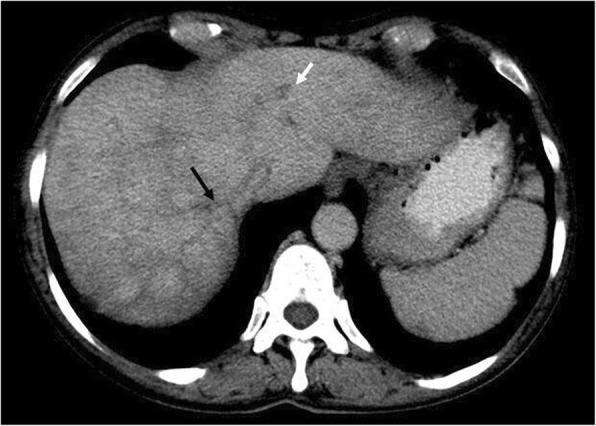
This diagnostic image is an axial computed tomography (CT) scan of the upper abdomen obtained during the portovenous phase. The primary finding is Budd-Chiari syndrome in a patient with underlying myelofibrosis. The image reveals occlusion and lack of contrast enhancement in the middle hepatic vein (indicated by a black arrow) and the left hepatic vein (indicated by a white arrow). A characteristic 'nutmeg liver' appearance—a mottled, reticular pattern of enhancement—is clearly visible within the liver parenchyma, particularly in segment 7. This heterogeneous enhancement pattern is a hallmark of hepatic venous outflow obstruction, resulting from chronic passive congestion and centrilobular necrosis. The clinical significance of these findings demonstrates splanchnic vein thrombosis, a common and serious complication of myeloproliferative neoplasms. This visual is an essential educational tool for identifying the radiographic signs of hepatic venous congestion and vascular occlusion in the context of systemic hematologic disorders.
heart failure cells hemosiderin lung brown induration
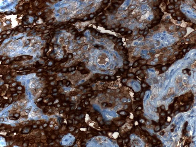
Immunohistochemistry image of lung tissue demonstrating epithelial membrane antigen (EMA) immunoreactivity in a sclerosing pneumocytoma. The tissue section is stained with EMA (brown DAB chromogen) and counterstained with hematoxylin (blue), visualized under bright-field light microscopy. The specimen is a pulmonary lesion composed of two characteristic cell populations: surface epithelial–like cuboidal cells forming papillary structures, and underlying round stromal cells embedded in fibrous or sclerotic stroma. In this image EMA expression is evident on the membranes of both surface and stromal cells, producing a continuous brown membranous outline along cell borders. The combination of EMA positivity in both compartments supports the neoplastic nature of the lesion and aligns with the dual-cell phenotype seen in sclerosing pneumocytoma, formerly called sclerosing hemangioma. The notable histologic context includes papillary folds, sclerosis, and occasional hemosiderin-laden macrophages, but without overt cytologic atypia or high mitotic activity. Clinically, EMA positivity helps distinguish this entity from adenocarcinoma and other primary lung neoplasms, particularly in the setting of ambiguous morphology. EMA positivity highlights diagnostic utility of IHC for differential diagnosis, emphasizing surface and stromal immunophenotype, and supports the benign behavior and favorable prognosis after surgical excision. Pathorama.ch provided caption and licensing.
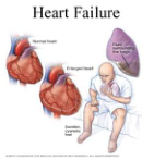
Summary : This medical illustration explains heart failure by comparing a normal heart to an enlarged heart, showing associated symptoms and complications. photo: Scene Overview : • The main subjects are two anatomical heart diagrams (normal and enlarged), a diagram of a lung with fluid, and a person sitting on a bed appearing short of breath. • The illustration uses a mix of realistic and schematic styles, with red and brown tones for the hearts, purple for the lung, and a pale blue/white background. • The person is depicted holding their chest and breathing with difficulty, suggesting distress. Technical Details : • Labels identify “Normal heart,” “Enlarged heart,” “Swollen, cyanotic feet,” and “Fluid surrounding the lungs.” • The heart diagrams show the normal heart as smaller and the enlarged heart as larger and more rounded. • The lung diagram highlights fluid accumulation around the lungs. • The person’s feet are shown as swollen and cyanotic (bluish), indicating poor circulation. Spatial Relationships : • The normal and enlarged hearts are placed side by side for direct comparison. • The lung diagram is positioned above and to the right, with a label pointing to the fluid. • The person is seated in the foreground, with visual emphasis on their feet and facial expression. Analysis : • The figure visually communicates that heart failure leads to an enlarged heart, fluid buildup around the lungs, and swelling/cyanosis in the feet, resulting in difficulty breathing and physical distress.
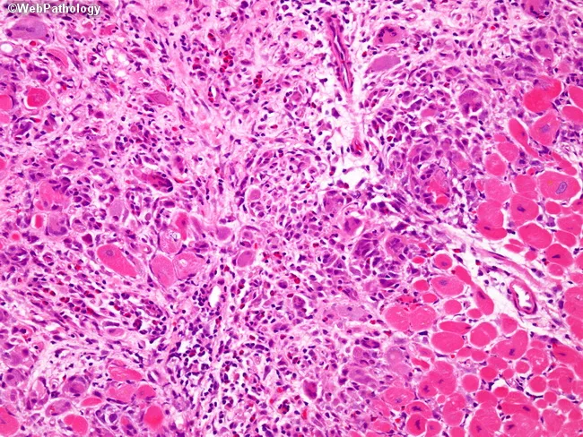
This histopathology image derives from cardiac muscle tissue subjected to light microscopy after hematoxylin and eosin staining. The section interrogates myocardial wall with preserved vascular framework showing dense inflammatory infiltration. The dominant cellular response comprises multinucleated giant cells intermixed with dense bands of lymphocytes, eosinophils, and histiocytes occupying the interstitium and peri-vascular spaces. Within the myocardium there is focal loss and fragmentation of myofibers, replaced by inflammatory cells and necrotic debris, a pattern compatible with myocarditis. The giant cells are derived from activated macrophages and are arranged in clusters adjacent to singleton foci of myocyte necrosis. Eosinophilic breakdown products and hemosiderin may be present in scattered macrophages. The tissue architecture shows patchy to confluent involvement of the ventricular myocardium, with occasional adipose tissue representing pericardial/epicardial infiltration at the margins. The image demonstrates a classic giant cell myocarditis phenotype—a rare, fulminant inflammatory cardiomyopathy associated with rapid heart failure and arrhythmias. The diagnostic significance lies in distinguishing giant cell myocarditis from lymphocytic myocarditis or granulomatous disease; findings guide immunosuppression and transplant considerations. This image is educational for cardiovascular pathology and graduate-level medical education, illustrating cellular diversity within myocarditis and the importance of multinucleated giant cells as a key diagnostic clue.
congestive splenomegaly chronic venous congestion spleen histology
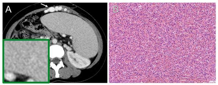
A composite image consisting of a diagnostic radiological scan and a histopathological photomicrograph illustrating congestive splenomegaly. Image (A) is an axial contrast-enhanced CT scan of the abdomen showing significant, diffuse, homogeneous enlargement of the spleen (splenomegaly). The splenic parenchyma maintains a uniform enhancement pattern. Prominent portosystemic varices are visible along the anterior abdominal wall (indicated by a white arrow), suggesting portal hypertension. A green-bordered inset provides a magnified view of the uniform splenic texture. Image (B) is a light microscopy photomicrograph of splenic tissue stained with Hematoxylin and Eosin (H-E). The histology demonstrates features of chronic passive congestion, characterized by marked engorgement of the splenic sinuses and parenchyma with red blood cells (erythrocytes). The cellular architecture remains largely intact but displays a dense, congested appearance. This educational material correlates gross radiological findings of organomegaly and vascular shunting with microscopic evidence of vascular congestion, typically associated with conditions like portal hypertension or congestive heart failure.
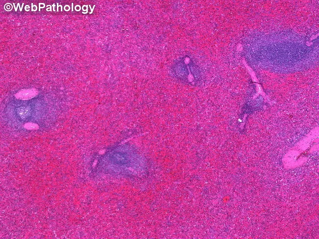
Histopathology of splenic tissue from a patient with autoimmune hemolytic anemia. Imaging modality: light microscopy of an H&E-stained section of spleen; micrograph shows gross splenomegaly with a deep red cut surface. The microscopic fields reveal marked congestion of the red pulp with dilated sinusoids and widened splenic cords (Billroth cords) within the red pulp. White pulp is not markedly expanded. The architecture appears largely preserved apart from venous congestion; erythrocyte pooling produces a hyperemic, reddish appearance. There is no evident infarction or necrosis in the depicted fields. The prominent vascular congestion reflects hypersplenism and increased sequestration/destruction of red cells in the setting of autoimmune hemolytic anemia. Pathologic findings are dominated by red pulp hyperemia and congested sinusoids, consistent with congestive splenomegaly. Clinically, this morphology correlates with ongoing hemolysis, anemia, and potential hypersplenism; splenic pooling can contribute to decreased circulating red cells and altered retic count. Differential considerations include portal hypertension-related splenomegaly, infectious mononucleosis, or other causes of splenic congestion, but the clinical context (AIHA) supports splenic vascular congestion as the primary lesion. This image is relevant for educational discussions of splenic pathology, hypersplenism, AIHA-associated splenomegaly, and examples of congestive splenic changes. This description supports radiologists and pathologists in correlating clinical data.
heart failure cells hemosiderin laden macrophages alveoli lung histology
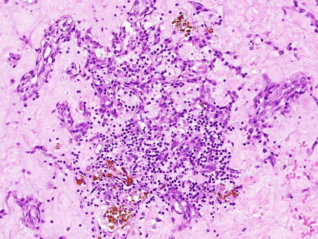
This case depicts a hematoxylin-eosin stained histologic section of a cardiac myxoma, displaying a prominent myxoid stroma rich in mucopolysaccharide matrix with scattered tumor cells. The cellular component comprises small, oval to stellate myxoma cells arranged in intricate, cribriform or arborizing patterns around vascular channels, a morphology that can mimic metastatic carcinoma when arranged in pseudo-glandular architectures. Background shows chronic inflammatory cells and hemosiderin-laden macrophages within the loose myxoid matrix, consistent with prior microhemorrhage or prior inflammation. The tissue architecture ranges from loose, hyaline-like regions to more cellular whorled areas, underscoring the heterogeneity of myxoid tumors. Diagnostic significance lies in recognizing classic myxoma features—mucinous stroma, sparsely distributed cells, and vessel-adjacent patterns—to distinguish from carcinomatous metastases and other myxoid neoplasms. Differential diagnoses include metastatic adenocarcinoma, myxoid liposarcoma, angiosarcoma, and fibromyxoid sarcomas. Clinically, this histology should be integrated with cardiac imaging findings (echocardiography, cardiac MRI) to confirm mass origin in the heart; surgical excision is typically curative, and pathologic assessment must assess margins and exclude metastasis. The cribriform and arborizing patterns around vascular channels are focal; overall architecture remains myxoid. Immunohistochemistry, when performed, shows positivity for vimentin and negativity for cytokeratins, aiding distinction from metastatic carcinomas. This image serves as an instructive reference for pathology education and multidisciplinary tumor-board correlation.
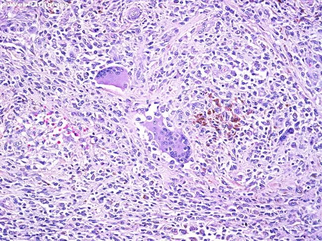
This is a light microscopy image of a tissue biopsy stained with hematoxylin and eosin (H&E). The slide shows granulomatous inflammation with multinucleated giant cells and scattered hemosiderin-laden macrophages embedded in a dense inflammatory background. Epithelioid histiocytes predominate, forming nodular aggregates with elongated nuclei, abundant cytoplasm, and indistinct cell borders. Multinucleated giant cells are evident within the granulomas, sometimes with a peripheric arrangement suggestive of Langhans-type morphology. Intermixed small lymphocytes and plasma cells contribute to the chronic inflammatory milieu. Brown to golden-brown hemosiderin pigment within macrophages is conspicuous and suggests prior hemorrhage or vascular congestion; occasional clusters of iron-laden macrophages are dispersed in the interstitium. Mild edema and vascular congestion may be present; necrosis is not a prominent feature in this field. The tissue architecture reflects organized granulomatous reaction rather than acute suppurative inflammation. No overt microbial organisms are visible at this magnification; however, special stains or molecular tests are required to identify potential infectious etiologies. Clinically, these findings raise differential diagnoses including mycobacterial and fungal infections, sarcoidosis, foreign body granulomatous reaction, or hemorrhagic granulomatous response. This image is valuable for diagnostic pathology review, education on granulomatous histology, and research into macrophage activation and iron handling in chronic inflammation process.
Chronic Venous Congestion (CVC) - Spleen, Lung & Liver
1. CVC - LUNG
Cause
Gross
- Lungs become firm, heavy, and brown ("brown induration")
- Brown color = hemosiderin deposits from old hemorrhages
- Firm/indurated texture = fibrosis of alveolar walls
Microscopy
| Phase | Features |
|---|---|
| Acute | Engorged alveolar capillaries + alveolar septal edema + focal intraalveolar hemorrhage |
| Chronic | Septal thickening + fibrosis + numerous hemosiderin-laden macrophages in alveolar spaces |
- "Heart failure cells" = alveolar macrophages laden with hemosiderin (golden-brown pigment), derived from phagocytosed red cells that leaked from congested capillaries. Positive on Prussian Blue (Perls') stain for iron.
- In severe/long-standing congestion (e.g., mitral stenosis): abundant heart failure cells + fibrosis of alveolar walls = soggy, firm lungs
Clinical
- Dyspnea, orthopnea, PND
- Pink frothy sputum (in acute pulmonary edema)
- Fine crepitations at lung bases
2. CVC - LIVER
Cause
Gross
- Congestive hepatomegaly (liver enlarged and heavy)
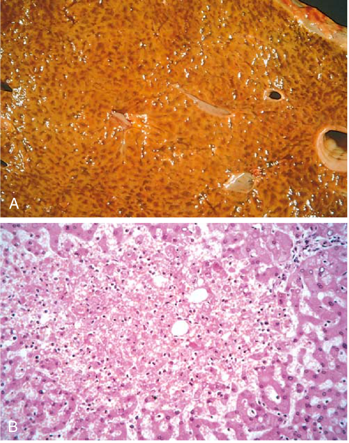
- Cut section shows "nutmeg liver": congested red-brown centrilobular areas alternating with paler, non-congested periportal parenchyma - resembling the cut surface of a nutmeg
Why this pattern?
- The centrilobular zone (zone 3) is at the distal end of hepatic blood supply - most vulnerable to ischemia
- The periportal zone (zone 1) is better oxygenated by hepatic arterioles - relatively spared, may show fatty change
Microscopy
| Phase | Features |
|---|---|
| Acute | Central vein + sinusoids distended with blood; centrilobular hepatocyte necrosis (ischemic); periportal fatty change |
| Chronic | Centrilobular congestion + hemorrhage + hemosiderin-laden macrophages + hepatocyte dropout |
| Long-standing | Centrilobular fibrosis → "cardiac cirrhosis" (central-to-central bridging fibrosis) |
Additional features (with concurrent LVF)
- Severe central hypoxia → centrilobular necrosis (in addition to sinusoidal congestion)
Clinical
- Tender hepatomegaly
- Raised JVP, pulsatile liver (in tricuspid regurgitation)
- Eventually: ascites, jaundice, abnormal LFTs
3. CVC - SPLEEN
Cause
- Right-sided heart failure → portal hypertension → elevated splenic vein pressure
- Or direct portal hypertension (cirrhosis, portal vein thrombosis)
Gross
- Congestive splenomegaly: tense, enlarged spleen (can reach 500-1000g in chronic cases vs. normal ~150g)
- Cut surface appears congested, dark red
Microscopy
| Phase | Features |
|---|---|
| Acute | Sinusoidal dilation, congestion of red pulp |
| Chronic | Fibrosis of sinusoidal walls (fibrosiderotic nodules) + hemosiderin deposits |
- Gamna-Gandy bodies (in long-standing cases): foci of organized hemorrhage containing hemosiderin, calcium, and fibrous tissue - appear as small brownish nodules ("tobacco-fleck spleen")
- White pulp may be compressed/atrophic
Consequences
- Thrombocytopenia (platelet sequestration - hypersplenism)
- Anemia, leukopenia (pancytopenia of hypersplenism)
- Edema of bowel wall (when portal hypertension severe enough) → may impair nutrient absorption
Summary Comparison Table
| Feature | Lung (CVC) | Liver (CVC) | Spleen (CVC) |
|---|---|---|---|
| Primary cause | Left heart failure | Right heart failure | Portal hypertension / RHF |
| Gross name | Brown induration | Nutmeg liver | Congestive splenomegaly |
| Key micro finding | Heart failure cells (hemosiderin macrophages) | Centrilobular necrosis + nutmeg pattern | Sinusoidal congestion + Gamna-Gandy bodies |
| Acute change | Engorged capillaries, intraalveolar hemorrhage | Central vein/sinusoid distension, centrilobular necrosis | Sinusoidal dilation |
| Chronic change | Fibrosis, thickened septa | Cardiac cirrhosis | Fibrosiderosis, white pulp atrophy |
| Color change | Brown (hemosiderin) | Red-brown center / pale periphery | Dark red congested |
| End-stage | Fibrotic (firm) lungs | Cardiac cirrhosis | Fibrosiderotic spleen |