Here is the full, exam-ready answer for MD Anaesthesia - 50 marks.
SHOCK: Definition, Classification, Types, Features, and Management
[MD Anaesthesia - Long Essay, 50 Marks]
I. DEFINITION
Shock is defined as inadequate tissue perfusion resulting in insufficient oxygen delivery (DO₂) relative to oxygen demand (VO₂), causing cellular hypoxia and organ dysfunction.
The classic Gross definition (1872): "A manifestation of the rude unhinging of the machinery of life."
From the anaesthesia perspective (Miller's Anesthesia, 10e):
Shock is the inability to provide adequate oxygen to the tissues. The condition depends on the balance between oxygen supply (DO₂) and oxygen demand (VO₂).
Key equations:
- CaO₂ (mL/dL) = (1.34 × Hb × SaO₂) + (PaO₂ × 0.003) → normal ≈ 20 mL/dL
- DO₂ (mL/min) = CaO₂ × CO × 0.01
- VO₂ = DO₂ × O₂ Extraction (CaO₂ - CvO₂)/CaO₂
When DO₂ falls below the critical threshold, VO₂ becomes delivery-dependent → anaerobic metabolism → lactic acidosis.
II. CLASSIFICATION OF SHOCK
Four major classes (Harrison's 22e, Miller's 10e, Sabiston Surgery):
| Type | Primary Mechanism | CVP | PCWP | CO | SVR |
|---|
| Hypovolemic | Reduced preload | ↓ | ↓ | ↓ | ↑ |
| Cardiogenic | Pump failure | ↑ | ↑ | ↓ | ↑ |
| Distributive | Vasodilation / maldistribution | ↓ | ↓ | ↑ | ↓↓ |
| Obstructive | Extracardiac outflow block | ↑ | ↓/↑ | ↓ | ↑ |
Source: Harrison's Principles of Internal Medicine 22e, Table 314-2
Schwartz Surgery further subdivides to 6 types: hypovolemic, septic (vasodilatory), neurogenic, cardiogenic, obstructive, and traumatic shock.
III. STAGES OF SHOCK
All types progress through three phases:
- Compensated (early) shock: Neurohumoral responses maintain BP via sympathoadrenal activation, tachycardia, increased SVR, ADH/RAAS activation. BP maintained; subtle signs only.
- Decompensated (progressive) shock: Compensatory mechanisms fail; progressive hypotension, anaerobic metabolism, lactic acidosis, organ hypoperfusion.
- Irreversible (refractory) shock: Irreversible cell death, mitochondrial failure, multiple organ dysfunction syndrome (MODS), death inevitable despite resuscitation.
IV. TYPES OF SHOCK - DETAILED
1. HYPOVOLEMIC SHOCK
Definition: Shock due to absolute reduction in circulating blood/fluid volume causing decreased preload and CO.
Causes:
- Hemorrhagic: Trauma (most common), GI bleeding (upper > lower), surgical blood loss, ruptured ectopic pregnancy/AAA
- Non-hemorrhagic: Vomiting, diarrhea, osmotic diuresis (DKA, DI), burns (fluid redistribution), severe pancreatitis, nephrotic syndrome
ATLS Classification of Hemorrhagic Shock:
| Class | Blood Loss | HR | BP | RR | Urine Output | CNS |
|---|
| I | <15% (<750 mL) | <100 | Normal | 14-20 | >30 mL/h | Normal |
| II | 15-30% (750-1500 mL) | 100-120 | Normal | 20-30 | 20-30 mL/h | Anxious |
| III | 30-40% (1500-2000 mL) | 120-140 | ↓ | 30-40 | 5-15 mL/h | Confused |
| IV | >40% (>2000 mL) | >140 | ↓↓ | >35 | <5 mL/h | Lethargic/Unconscious |
Signs & Symptoms:
- Hypotension, tachycardia, narrow pulse pressure
- Cold, clammy, pale skin (vasoconstriction)
- Decreased urine output (oliguria <0.5 mL/kg/h)
- Altered consciousness: anxiety → confusion → coma
- Increased respiratory rate (compensatory)
- Collapsed JVP/neck veins
- Dry mucous membranes (non-hemorrhagic)
- Metabolic acidosis (elevated lactate)
Pathophysiology:
- Volume loss → ↓ venous return → ↓ preload → ↓ SV → ↓ CO
- Baroreceptor activation → sympathoadrenal surge → tachycardia, vasoconstriction
- RAAS activation → aldosterone → Na⁺ and water retention
- ADH release → water conservation
- If untreated: microcirculatory failure → cellular hypoxia → lactic acidosis → MODS
Management:
- Airway & Breathing: Secure airway; O₂ supplementation; intubate if needed
- Hemorrhage control: Direct pressure, tourniquet, surgical intervention; damage control surgery principle
- IV access: Two large-bore peripheral IVs (14-16G); intraosseous if needed
- Fluid resuscitation:
- Class I/II: Crystalloid (0.9% NaCl or Ringer's Lactate) 1-2 L bolus
- Class III/IV: Massive transfusion protocol - 1:1:1 ratio (PRBC: FFP: Platelets)
- Permissive hypotension (MAP 50-65 mmHg) in penetrating trauma until surgical hemostasis
- Vasopressors: Norepinephrine if fluid-unresponsive (secondary measure)
- Tranexamic acid: 1 g IV within 3 hours of traumatic hemorrhage (CRASH-2 trial evidence)
- Warming: Prevent/treat hypothermia (lethal triad: hypothermia + coagulopathy + acidosis)
- Monitor: Urine output, BP, HR, lactate clearance, ABG
2. CARDIOGENIC SHOCK
Definition: Shock due to primary cardiac pump failure resulting in reduced CO despite adequate preload. CO decreased, SVR increased, PCWP elevated.
Causes:
- Myocardial: AMI (most common - especially large anterior STEMI), myocarditis, dilated cardiomyopathy, post-cardiac surgery low output syndrome, Takotsubo (stress) cardiomyopathy
- Mechanical: Acute mitral regurgitation (papillary muscle rupture), acute VSD (post-MI), aortic stenosis, acute aortic insufficiency
- Arrhythmia: Sustained VT, complete heart block, severe bradyarrhythmia
- Obstructive (intrinsic): Critical AS, severe MS
Signs & Symptoms:
- Hypotension (SBP <90 mmHg), narrow pulse pressure
- Tachycardia (compensatory; may be absent if arrhythmia is causative)
- Cold, clammy extremities (↑ SVR, vasoconstriction) - "cold and wet" pattern
- Elevated JVP/distended neck veins (elevated PCWP/CVP)
- Pulmonary crackles/rales (pulmonary edema from elevated LVEDP)
- S3 gallop, new murmur (mechanical cause)
- Oliguria (<0.5 mL/kg/h)
- Altered mental status (cerebral hypoperfusion)
- Elevated NT-proBNP, troponin
- CXR: pulmonary congestion, cardiomegaly
- ECG: ST changes, arrhythmia
Vicious Cycle of Cardiogenic Shock:
Myocardial dysfunction → ↓ CO → ↓ coronary perfusion pressure → further ischemia → more myocardial dysfunction (the "death spiral")
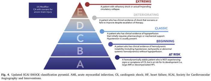
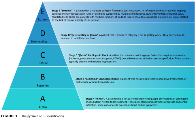
SCAI Classification (2022):
- Stage A: At risk
- Stage B: Beginning (hemodynamic instability without hypoperfusion)
- Stage C: Classic cardiogenic shock
- Stage D: Deteriorating/Doom
- Stage E: Extremis/cardiac arrest
Management:
- Revascularization: Urgent PCI for AMI-related cardiogenic shock (door-to-balloon <90 min)
- Vasopressors/Inotropes:
- Norepinephrine - first-line vasopressor (MAP target ≥65 mmHg)
- Dobutamine - inotrope for low CO; add to norepinephrine when EF reduced
- Dopamine - alternative (higher arrhythmia risk)
- Milrinone (PDE-III inhibitor): inodilator; useful post-cardiac surgery; dose 25-75 mcg/kg load then 0.25-0.75 mcg/kg/min
- Levosimendan - calcium sensitizer; improves contractility without increasing myocardial O₂ consumption; 6-12 mcg/kg load then 0.1-0.2 mcg/kg/min
- Mechanical Circulatory Support (MCS):
- IABP (Intra-aortic Balloon Pump): Counterpulsation; deflates systole (↓ afterload), inflates diastole (↑ coronary perfusion)
- Impella (pLVAD): Microaxial pump; provides up to 5.5 L/min support
- VA-ECMO: Maximum support for refractory cardiogenic shock/cardiac arrest
- TandemHeart: Transseptal LA-to-aorta support
- Treat cause: Percutaneous/surgical repair of mechanical defects, cardioversion for arrhythmia
- Avoid excessive fluids (worsens pulmonary edema)
- Pulmonary artery catheter: Guides hemodynamic optimization in refractory cases
- Avoid/caution: BBs, CCBs acutely (worsen contractility)
3. DISTRIBUTIVE SHOCK
Definition: Shock due to pathological vasodilation with maldistribution of blood flow, causing relative hypovolemia despite normal or elevated CO. SVR markedly reduced.
Three main subtypes:
3A. SEPTIC SHOCK (most common distributive shock)
Definition (Sepsis-3, 2016): Sepsis with persistent hypotension requiring vasopressor support to maintain MAP ≥65 mmHg AND serum lactate >2 mmol/L despite adequate fluid resuscitation.
Pathophysiology:
- Pathogen recognition (LPS, gram+/- bacteria, fungi, viruses) → pattern recognition receptors (TLRs) → massive cytokine release (TNF-α, IL-1, IL-6)
- Endothelial activation → ↑ vascular permeability → "third spacing"
- iNOS upregulation → excessive NO → profound vasodilation
- Myocardial depression (sepsis-induced cardiomyopathy in ~40%)
- Microcirculatory dysfunction → oxygen extraction failure
- Coagulation activation → DIC possible
Hemodynamics: "Warm/hyperdynamic" initially - ↑ CO, ↓ SVR, ↓ CVP, bounding pulses; later "cold/hypodynamic" if myocardial depression develops.
Signs & Symptoms:
- Fever (>38°C) or hypothermia (<36°C) - sepsis
- Tachycardia (HR >90 bpm)
- Tachypnea (RR >20 or PaCO₂ <32)
- Leukocytosis (>12,000) or leukopenia (<4,000)
- Warm, flushed skin (vasodilation; "warm shock")
- Bounding pulses; wide pulse pressure
- Hypotension despite fluid resuscitation
- Oliguria, rising creatinine
- Altered mentation, encephalopathy
- Petechiae/purpura (meningococcemia, DIC)
- Elevated lactate (>2 mmol/L = organ hypoperfusion)
- SOFA score ≥2 (organ dysfunction)
Surviving Sepsis Campaign 2021 - Key Management (Hour-1 Bundle):
- Measure lactate (re-measure if >2 mmol/L)
- Blood cultures ×2 BEFORE antibiotics
- Antibiotics: Broad-spectrum within 1 hour of recognition (MANDATORY - each 1 h delay → 7-8% ↑ mortality)
- Fluid resuscitation: 30 mL/kg IV crystalloid within 3 hours for hypoperfusion (liberal vs. restrictive trial CLOVERS showed equipoise; current SSC favors 30 mL/kg then reassess)
- Vasopressors:
- Norepinephrine (1st line) - target MAP ≥65 mmHg
- Vasopressin 0.03 units/min (2nd line, add to NE; may improve kidney outcomes)
- Epinephrine (3rd line)
- Corticosteroids: Hydrocortisone 200 mg/day IV if ongoing vasopressor requirement (refractory septic shock)
- Source control: Drain abscess, remove infected catheter/device; surgical debridement as needed
- Add dobutamine to norepinephrine OR switch to epinephrine if septic shock + reduced LVEF
- Antibiotic stewardship: Reassess and narrow at 48-72 h based on cultures
- Supportive: LPV for ARDS (6 mL/kg IBW), glucose control (target <180 mg/dL), VTE prophylaxis, early enteral nutrition
Sepsis Biomarkers: PCT (procalcitonin), CRP, lactate, IL-6
3B. ANAPHYLACTIC SHOCK
Definition: Severe, life-threatening systemic IgE-mediated hypersensitivity reaction causing massive histamine/mediator release.
Causes: Medications (penicillin, NSAIDs, contrast media, neuromuscular blockers - most common in OR), latex, insect stings, food (nuts, shellfish)
Anaphylaxis is HIGHLY RELEVANT for anaesthesiologists - NMBAs account for ~60% of perioperative anaphylaxis.
Pathophysiology:
- IgE cross-linking on mast cells/basophils → degranulation → histamine, tryptase, leukotrienes, prostaglandins
- Profound vasodilation + increased vascular permeability (up to 35% of plasma volume can extravasate within 10 min)
- Bronchospasm (histamine, leukotrienes on airways)
- Urticaria, angioedema
Signs & Symptoms (Triad: Cardiovascular + Respiratory + Cutaneous):
- Cardiovascular: Hypotension, tachycardia, distributive shock, cardiovascular collapse
- Respiratory: Bronchospasm (wheezing), laryngeal edema, stridor, respiratory distress, hypoxia
- Cutaneous: Urticaria, angioedema, flushing, pruritus (may be absent in ~20%)
- Nausea, vomiting, abdominal cramps
- Under anaesthesia: sudden bronchospasm + hypotension + ↑ airway pressures may be only clues
NAP6 Grading (Ring & Messmer):
- Grade I: Cutaneous only
- Grade II: Mild systemic (hypotension + tachycardia + bronchospasm)
- Grade III: Severe life-threatening (cardiovascular/respiratory collapse)
- Grade IV: Cardiac arrest
Management:
- REMOVE TRIGGER (stop offending drug, remove latex)
- Epinephrine IM 0.5 mg (1:1000) - absolute first-line; IM into lateral thigh; repeat every 5 min if needed
- IV epinephrine 0.1-1 mcg/kg/min infusion for severe/refractory cases
- Position: Supine with legs elevated (Trendelenburg); if airway compromise - semi-recumbent
- Airway: High-flow O₂; early intubation if airway edema (before it closes)
- IV fluids: Rapid bolus 1-2 L crystalloid (massive third-spacing)
- Antihistamines: Chlorphenamine 10 mg IV (H1 blocker) + ranitidine/famotidine (H2 blocker)
- Corticosteroids: Hydrocortisone 200 mg IV (prevents biphasic reaction; NOT acute emergency treatment)
- Bronchospasm: Salbutamol nebulization/IV; MgSO₄ if severe
- Vasopressors: Norepinephrine or vasopressin for refractory hypotension
- Glucagon: 1-5 mg IV if on beta-blockers (epinephrine-resistant anaphylaxis)
- Post-event: Serum tryptase at 1h, 4h, 24h; allergy/immunology referral
3C. NEUROGENIC SHOCK
Definition: Shock from loss of sympathetic tone below the level of a spinal cord injury (typically T6 and above), causing vasodilation and bradycardia without compensatory tachycardia.
Causes: High spinal cord injury, spinal anaesthesia (high spinal block), autonomic neuropathy
Differentiate from Spinal Shock: Spinal shock = loss of all spinal cord function (motor, sensory, reflexes) below injury - not the same as neurogenic shock.
Hemodynamics: ↓ SVR, ↓ HR (bradycardia - loss of cardiac accelerators T1-T4), ↓ CO → hypotension
Signs & Symptoms:
- Hypotension + Bradycardia (classic triad differentiates from hypovolemic shock where tachycardia predominates)
- Warm, dry skin (no vasoconstriction despite hypotension - paradoxical; no sweating below injury)
- Neurological deficits (paraplegia/quadriplegia, sensory loss)
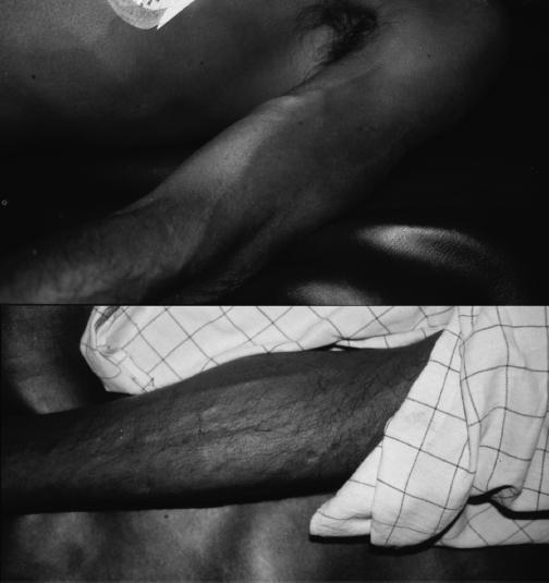
- Priapism (males - loss of sympathetic inhibition)
- Loss of temperature regulation (poikilothermia)
- Absent reflexes below injury (flaccid paralysis acutely)
- Diaphragmatic breathing only (if above C4)
Management:
- Airway/Immobilization: Cervical spine immobilization; intubation with inline stabilization if needed
- IV fluids: Cautious crystalloid (avoid over-hydration - pulmonary edema risk)
- Vasopressors:
- Phenylephrine (pure alpha agonist) for hypotension without bradycardia
- Norepinephrine (alpha + beta) if bradycardia present
- Atropine 0.5-1 mg IV for symptomatic bradycardia
- Dopamine as alternative
- MAP target: ≥85-90 mmHg for 7 days post-SCI (to optimize spinal cord perfusion - ASIA/ACS guidelines)
- Temperature regulation: Active warming (no shivering)
- Methylprednisolone: Controversial; no longer routinely recommended for acute SCI
- DVT prophylaxis (paralyzed limbs → high risk)
- Bladder/bowel management: Urinary catheter, bowel care
4. OBSTRUCTIVE SHOCK
Definition: Shock due to a mechanical obstruction to blood flow extrinsic to the heart, impairing ventricular filling or outflow, resulting in reduced CO.
Causes:
- Impaired venous return (inflow obstruction):
- Tension pneumothorax
- Cardiac tamponade
- Constrictive pericarditis
- Severe positive-pressure ventilation (↑ intrathoracic pressure)
- Outflow obstruction:
- Massive pulmonary embolism
- Severe pulmonary hypertension
- Aortic dissection
- Air embolism, fat embolism
Signs & Symptoms:
Cardiac Tamponade (Beck's Triad):
- Muffled heart sounds
- Elevated JVP (↑ CVP)
- Hypotension
- Pulsus paradoxus (>10 mmHg drop in SBP during inspiration)
- Tachycardia
- ECG: Electrical alternans, low voltage
- Echo: Pericardial effusion, diastolic RV collapse
Tension Pneumothorax:
- Respiratory distress, tachypnea
- Absent breath sounds ipsilateral
- Tracheal deviation (contralateral - late sign)
- Hypotension, tachycardia, elevated JVP
- Haemodynamic collapse (obstructive shock)
Massive Pulmonary Embolism:
- Acute dyspnea, pleuritic chest pain, hemoptysis
- Hypotension, tachycardia, tachypnea
- Elevated JVP, signs of RV failure (right heart strain)
- ECG: S1Q3T3 pattern, right heart strain, sinus tachycardia
- Echo: dilated RV, D-sign (septal flattening)
- CTPA: confirmatory
Management:
| Cause | Immediate Treatment |
|---|
| Tension PTX | Needle decompression (2nd ICS MCL) → chest drain |
| Cardiac Tamponade | Pericardiocentesis (or surgical pericardial window) |
| Massive PE | Systemic thrombolysis (rtPA 100 mg over 2h), ± surgical embolectomy, catheter-directed therapy |
| Constrictive Pericarditis | Pericardiectomy (surgical) |
| Air Embolism | Left lateral Trendelenburg (Durant's maneuver), aspirate via CVP |
- All obstructive shock: IV fluids cautiously (increase preload to maintain CO), vasopressors (norepinephrine), O₂/intubation
- Do NOT use vasodilators (worsen hypotension)
- Inotropes for RV failure in massive PE (dobutamine)
V. MIXED SHOCK
Mixed shock occurs when a patient has more than one type simultaneously. Example:
- Septic shock can involve distributive (vasodilation) + cardiogenic (sepsis-induced cardiomyopathy) + hypovolemic (third spacing) components
- Harrison's 22e: "The types of shock are not mutually exclusive - not uncommonly a patient will present with more than one type"
VI. GENERAL PRINCIPLES OF SHOCK MANAGEMENT
Immediate Assessment (ABC)
- Airway: Secure patent airway; intubate if GCS <8 or respiratory failure
- Breathing: O₂ supplementation; mechanical ventilation if needed; treat tension PTX
- Circulation: IV access × 2 large bore; fluid bolus; assess hemodynamics
- Disability: GCS, pupils, glucose
- Exposure: Look for source, injuries, rashes, temperature
Monitoring in Shock
| Parameter | Target |
|---|
| MAP | ≥65 mmHg (≥85-90 in neurogenic SCI) |
| Heart Rate | Trend toward normal (<100/min) |
| Urine Output | ≥0.5 mL/kg/h |
| Lactate | <2 mmol/L; clearance >10%/2h |
| CVP | 8-12 mmHg (not reliable sole guide) |
| PCWP | 12-15 mmHg (cardiogenic: 15-18) |
| ScvO₂ | >70% |
| Arterial pH | ≥7.35 |
Diagnostic Evaluation
- Lactate - marker of tissue hypoperfusion; serial measurement guides therapy
- ABG - metabolic acidosis (pH, base deficit, lactate)
- ECG - AMI, arrhythmia, PE (S1Q3T3), tamponade (electrical alternans)
- CXR - pulmonary edema (cardiogenic), PTX, effusion, infiltrates (sepsis)
- POCUS/Echo - most rapid bedside tool:
- RUSH protocol (Rapid Ultrasound for Shock and Hypotension)
- ACES protocol (Abdominal and Cardiothoracic Evaluation)
- Assess LV/RV function, pericardial effusion, IVC collapsibility, PTX
- CBC, BMP, coags, cultures, troponin, BNP, procalcitonin
Vasopressors and Inotropes Summary
| Drug | Receptors | Use | Key Points |
|---|
| Norepinephrine | α1>β1 | 1st line most shock | Vasoconstriction, mild inotropy |
| Vasopressin | V1R | 2nd line septic shock | Add to NE; 0.03 units/min; steroid-sparing |
| Epinephrine | α+β1+β2 | Anaphylaxis (1st line), 3rd vasopressor | Bronchodilation, tachycardia, ↑ lactate |
| Dopamine | DA, β1, α | Alternative vasopressor | Higher arrhythmia risk vs NE |
| Dobutamine | β1>β2 | Cardiogenic shock low CO | Inotrope/chronotrope, vasodilation |
| Phenylephrine | α1 | Neurogenic, high-spinal | Pure vasopressor; no inotropy; bradycardia |
| Milrinone | PDE-III | Post-cardiac surgery | Inodilator; avoid if hypotensive |
| Levosimendan | Troponin C | Cardiogenic, post-cardiac surgery | No ↑ myocardial O₂ consumption |
VII. SHOCK IN ANAESTHESIA - SPECIAL CONSIDERATIONS
- Perioperative Anaphylaxis: Most common triggers: NMBAs (rocuronium > suxamethonium), latex, antibiotics (penicillin), colloids; NAP6 (UK audit) - 1:10,000 anaesthetics; management as above
- High Spinal/Epidural: Neurogenic shock - treat with ephedrine/phenylephrine; atropine for bradycardia; position supine + legs elevated
- Anaesthetic agents in shock: Ketamine preferred (sympathomimetic), etomidate (hemodynamically stable); reduce doses of propofol/thiopentone (vasodilatation worsens); volatile agents reduce SVR and CO - use with extreme caution
- Goal-Directed Therapy (GDT): Intraoperative optimization of DO₂ using fluid responsiveness (PLR, SVV, PPV) and targeted vasopressor/inotrope therapy - reduces postoperative morbidity
- Intraoperative monitoring: Invasive arterial line (gold standard for BP in shock), CVP, pulmonary artery catheter (selected cases), cardiac output monitoring (LiDCO, PiCCO, PAC)
- TEG/ROTEM: Guide coagulation management in hemorrhagic/traumatic shock
- Lethal Triad in Trauma: Hypothermia + Acidosis + Coagulopathy - break this cycle; DCR + DCS
VIII. ENDPOINTS OF RESUSCITATION
- Lactate clearance (≥10% per 2 hours) is more reliable than static hemodynamic parameters
- Normalization of base deficit (target BD > -6)
- ScvO₂ >70% (or SvO₂ >65%)
- Urine output ≥0.5 mL/kg/h
- Normothermia, INR, and platelet count normalization in hemorrhagic shock
IX. COMPLICATIONS OF SHOCK
- Acute Kidney Injury (AKI) - most common
- ARDS - acute respiratory distress syndrome
- MODS (Multiple Organ Dysfunction Syndrome)
- DIC - especially septic and traumatic shock
- Intestinal ischemia - bacterial translocation → further sepsis
- Acute Liver Failure - "shock liver" (centrilobular necrosis)
- Myocardial Ischemia - demand-supply mismatch
- Adrenal Insufficiency - relative, in critical illness
SUMMARY TABLE
| Feature | Hypovolemic | Cardiogenic | Septic | Anaphylactic | Neurogenic | Obstructive |
|---|
| HR | ↑↑ | ↑ | ↑↑ | ↑ | ↓ or N | ↑ |
| BP | ↓ | ↓ | ↓ | ↓ | ↓ | ↓ |
| SVR | ↑↑ | ↑↑ | ↓↓ | ↓↓ | ↓↓ | ↑ |
| CO | ↓↓ | ↓↓ | ↑ | ↑ | ↓ | ↓↓ |
| CVP/JVP | ↓ | ↑ | ↓ | ↓ | ↓ | ↑ |
| Skin | Cold/clammy | Cold/clammy | Warm/flushed | Flushed/urticaria | Warm/dry | Cold |
| 1st Rx | Fluids + control bleed | Inotropes + revascularize | Abx + fluids + NE | Epinephrine IM | NE/phenylephr. | Treat cause |
SCAI staging of cardiogenic shock (A-E)
References (Authoritative Textbooks):
- Harrison's Principles of Internal Medicine 22e (2025), Chapter 314-315 (Shock and Sepsis)
- Miller's Anesthesia 10e, Chapter 75 (Shock)
- Sabiston Textbook of Surgery - Chapter on Shock
- Surviving Sepsis Campaign Guidelines 2021 (Evans L et al, Crit Care Med 2021)
- Fuster & Hurst's The Heart 15e - Cardiogenic Shock Chapter