
Relations of uterus
Reading File
The uterus is a midline pelvic organ with complex anatomical relations on all sides - anteriorly, posteriorly, superiorly, laterally, and inferiorly. Let me search the medical library for a detailed, textbook-level answer.
Searching sources...
Finding Sources
Finding Sources
Finding Sources
Reading File
Reading File
Finding Sources
Reading File
Finding Sources
Reading File
Finding Sources
Reading File
Loading Image
Loading Image
Loading Image
Loading Image
Here is a complete, textbook-level account of the Relations of the Uterus, compiled from the Color Atlas of Human Anatomy Vol. 2 and Berek & Novak's Gynecology.
Relations of the Uterus
The uterus is a thick-walled, anteroposteriorly flattened, pear-shaped muscular organ situated near the center of the lesser (true) pelvis, between the urinary bladder anteriorly and the rectum posteriorly. In the nulliparous adult female it is 7-8 cm long and weighs 50-70 g.
Anterior Relations
- The anterior surface of the body of the uterus faces the urinary bladder.
- The peritoneum passes from the anterior abdominal wall over the superior surface of the bladder and reflects onto the uterus at the junction of the cervix and body (uterovesical/vesicouterine junction), forming the vesicouterine pouch (uterovesical pouch).
- When the bladder is empty, this pouch is shallow and may virtually obliterate; when the bladder fills, it pushes the uterus posteriorly.
- The anterior surface (vesical surface) of the uterine body is flat and covered by peritoneum (perimetrium) above the vesicouterine reflection.
- The anterior surface of the cervix (supravaginal part) is separated from the bladder by loose connective tissue - the paravesical space - with no intervening peritoneum at this level.
Posterior Relations
- The posterior (intestinal) surface of the uterus is convex and covered entirely by peritoneum.
- The peritoneum descends from the posterior wall of the uterus all the way down to the posterior vaginal fornix, then reflects superiorly onto the rectum, forming the rectouterine pouch (Pouch of Douglas / cul-de-sac).
- The pouch of Douglas is the deepest point of the female peritoneal cavity and is bounded laterally by the rectouterine folds (which contain the uterosacral/sacrouterine ligaments and autonomic nerve fibres of the inferior hypogastric plexus).
- The posterior surface is therefore related to loops of small intestine and sigmoid colon (which rest in the pouch of Douglas), and below to the rectum.
Clinical note: Pathological fluid (blood, pus, ascites) gravitates into the pouch of Douglas; it can be drained by posterior colpotomy (puncture through the posterior vaginal fornix) or confirmed by ultrasound.
Lateral Relations
The lateral borders of the uterus are related to several key structures:
-
Broad ligament - A double fold of peritoneum extending from each lateral border of the uterus to the lateral pelvic wall. It divides the female pelvic peritoneal cavity into anterior (vesicouterine pouch) and posterior (rectouterine pouch) compartments. Within the broad ligament run vessels, nerves, and the uterine tube (mesosalpinx), ovary (mesovarium), and round ligament (mesometrium).
-
Uterine artery - Runs in the base of the broad ligament (parametrium), crosses over the ureter before reaching the lateral wall of the uterus.
-
Ureter - The ureter descends on the lateral pelvic wall, enters the base of the broad ligament (cardinal ligament), and then passes under the uterine artery ("water under the bridge") approximately 2 cm lateral to the cervix, then turns medially to reach the bladder. This is the site most vulnerable to ureteral injury during hysterectomy.
-
Parametrium / Paracervix - Dense connective tissue alongside the cervix and lower uterus that carries the uterine vessels and contains the ureter.
-
Ovaries and uterine tubes - The adnexa lie lateral and superior to the uterus within the broad ligament.
-
Internal iliac vessels and obturator nerve/vessels - On the lateral pelvic sidewall beyond the broad ligament.
Superior Relations
- The fundus of the uterus projects above the uterine horns where the uterine tubes join.
- Superiorly, the fundus is related to loops of small intestine and occasionally sigmoid colon (especially when the uterus is in its normal anteverted/anteflexed position).
- The round ligaments arise near the uterine horns and pass laterally toward the internal inguinal rings.
Inferior Relations
- The cervix projects inferiorly and posteriorly.
- The vaginal part (portio) of the cervix protrudes into the vagina, surrounded by the vaginal fornix (anterior, posterior, and lateral fornices).
- The supravaginal part of the cervix is above the vagina and is related anteriorly to the bladder, posteriorly to the rectum, and laterally to the ureters and parametrium.
- The pelvic floor muscles (levator ani, especially the pubococcygeus/puborectalis) provide the main mechanical support of the uterus inferiorly.
Peritoneal Relations - Summary Table
| Surface | Peritoneal covering | Pouch formed |
|---|---|---|
| Anterior (body) | Yes - above vesicouterine reflection | Vesicouterine pouch (anteriorly) |
| Anterior (cervix) | No - below the reflection | - |
| Posterior | Yes - entire surface + posterior fornix | Rectouterine pouch (Pouch of Douglas) |
| Fundus | Yes (perimetrium continuous) | - |
| Lateral | Forms the broad ligament | - |
Supports of the Uterus
The uterus is held in position primarily by the pelvic floor and secondarily by peritoneal folds and ligaments:
| Ligament | Attachment | Function |
|---|---|---|
| Round ligament | Uterine horns → inguinal canal → labia majora | Maintains anteversion |
| Broad ligament | Lateral uterine borders → lateral pelvic wall | Holds uterus and adnexa in place |
| Cardinal ligament (Mackenrodt) | Cervix → lateral pelvic wall | Main support of cervix and upper vagina |
| Uterosacral (rectouterine) ligament | Cervix/supravaginal cervix → sacrum | Posterior support of cervix |
| Pelvic floor (levator ani) | - | Primary mechanical support |
Key Clinical Points
- Vesicouterine pouch is shallow and is less clinically significant than the pouch of Douglas.
- Pouch of Douglas is the most dependent part of the peritoneal cavity - site of fluid collection, endometriosis deposits, and ovarian cyst rupture.
- The ureter's proximity to the uterine artery (2-3 cm lateral to the cervix) makes it vulnerable during hysterectomy - always identify it before clamping the cardinal ligament.
- Anteversion + anteflexion is the normal uterine position. Retroversion may cause the uterus to lie closer to the rectum, altering posterior relations.
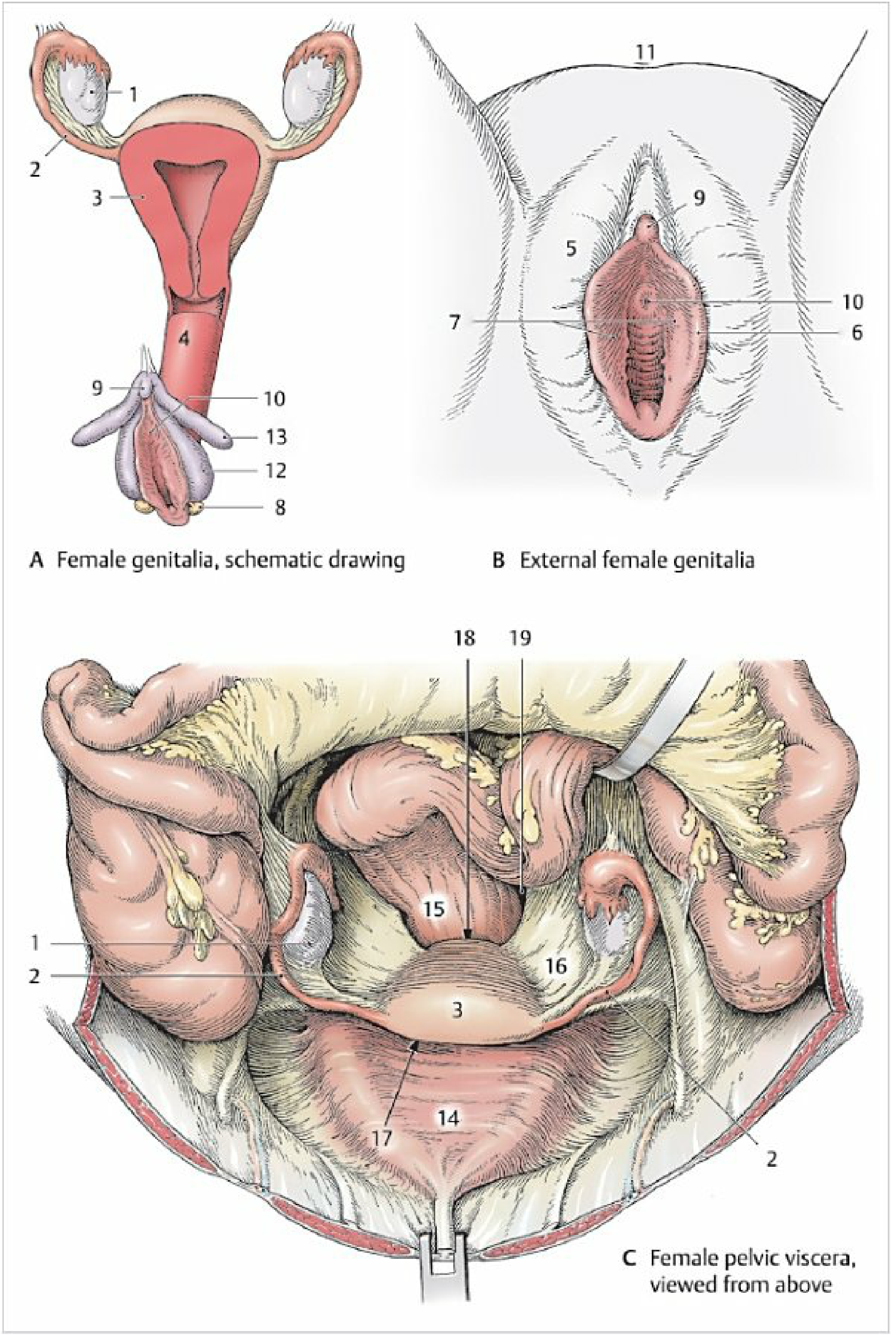
Female pelvic viscera viewed from above - Color Atlas of Human Anatomy Vol. 2, Internal Organs
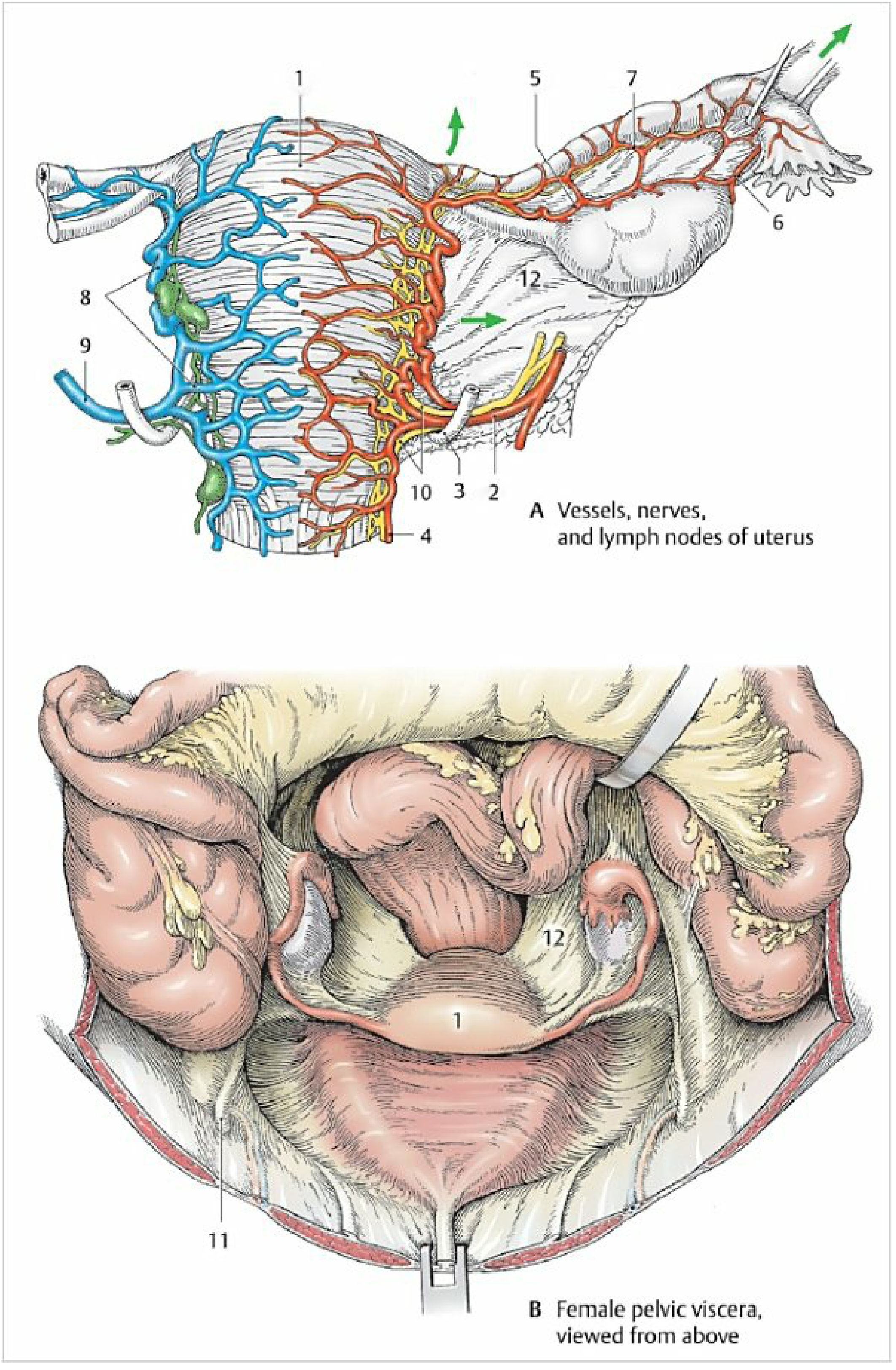
Vessels, nerves, and uterine suspensory ligaments - Color Atlas of Human Anatomy Vol. 2, Internal Organs
Sources:
- Color Atlas of Human Anatomy Vol. 2, Internal Organs - Peritoneal Relations of the Female Pelvis, pp. 268/280; Support of the Uterus, p. 461
- Berek & Novak's Gynecology - Peritoneal Cavity, p. 207; Ureter relations, pp. 207-208
This is a shared conversation. Sign in to Orris to start your own chat.