
Give me a descriptive answer on topic properties of amino acid for my 1st year mbbs exam of biochemistry and give answer with exact refrence of dm Vasudevan book 10 edition for biochemistry and give answer with proper flow chart or whatever diagram or collums or table needed to make paper presentation good and neat to get me a good marks
Properties of Amino Acids
1. GENERAL STRUCTURE OF AN AMINO ACID
NH3+
|
HOOC — Cα — R
|
H
| Component | Description |
|---|---|
| α-Carboxyl group (-COOH) | Acidic; proton donor; dissociated to -COO⁻ at physiologic pH (7.4) |
| α-Amino group (-NH2) | Basic; proton acceptor; protonated to -NH3⁺ at physiologic pH |
| α-Hydrogen (H) | Attached to central carbon |
| R group (side chain) | Variable; determines the identity and chemical properties of each amino acid |
Key Point (Exam): At physiologic pH (~7.4), the amino acid exists as a zwitterion - a molecule that is electrically neutral overall but carries both a positive charge (-NH3⁺) and a negative charge (-COO⁻) simultaneously.
2. GENERAL PHYSICAL PROPERTIES
| Property | Details |
|---|---|
| Solubility | Highly soluble in polar solvents (water, ethanol); insoluble in nonpolar solvents (benzene, hexane, ether) - due to ionic character |
| Optical activity | All amino acids (except glycine) have a chiral α-carbon; exist in L and D forms |
| Configuration | All mammalian proteins contain L-α-amino acids exclusively |
| UV absorption | Tyrosine (Tyr), Phenylalanine (Phe), Tryptophan (Trp) absorb UV light at 250-290 nm. Tryptophan contributes most at 280 nm |
| Melting point | High melting points due to ionic interactions |
| Colorless | Amino acids do not absorb visible light |
3. OPTICAL ACTIVITY - D AND L CONFIGURATION
COOH COOH
| |
H2N —— C —— H H —— C —— NH2
| |
R R
L-Amino acid D-Amino acid
(found in proteins) (found in bacteria,
antibiotics, cell walls)
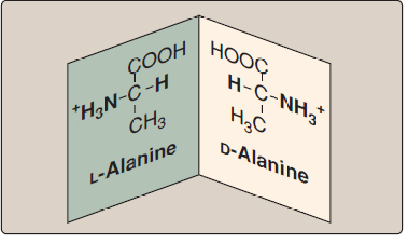
- Exception: Glycine has two H atoms on the α-carbon, so it is NOT chiral/optically active
- D-amino acids are found in bacterial cell walls (D-alanine, D-glutamate) and certain antibiotics (bacitracin, gramicidin A)
4. CLASSIFICATION OF AMINO ACIDS BASED ON R GROUP (SIDE CHAIN)
20 Amino Acids
|
┌───────────────┼──────────────────┐
│ │ │
Nonpolar Polar (Uncharged) Electrically Charged
(Hydrophobic) Side Chains Side Chains
│ │ ┌────────────┐
9 AAs 6 AAs Acidic (-) Basic (+)
2 AAs 3 AAs
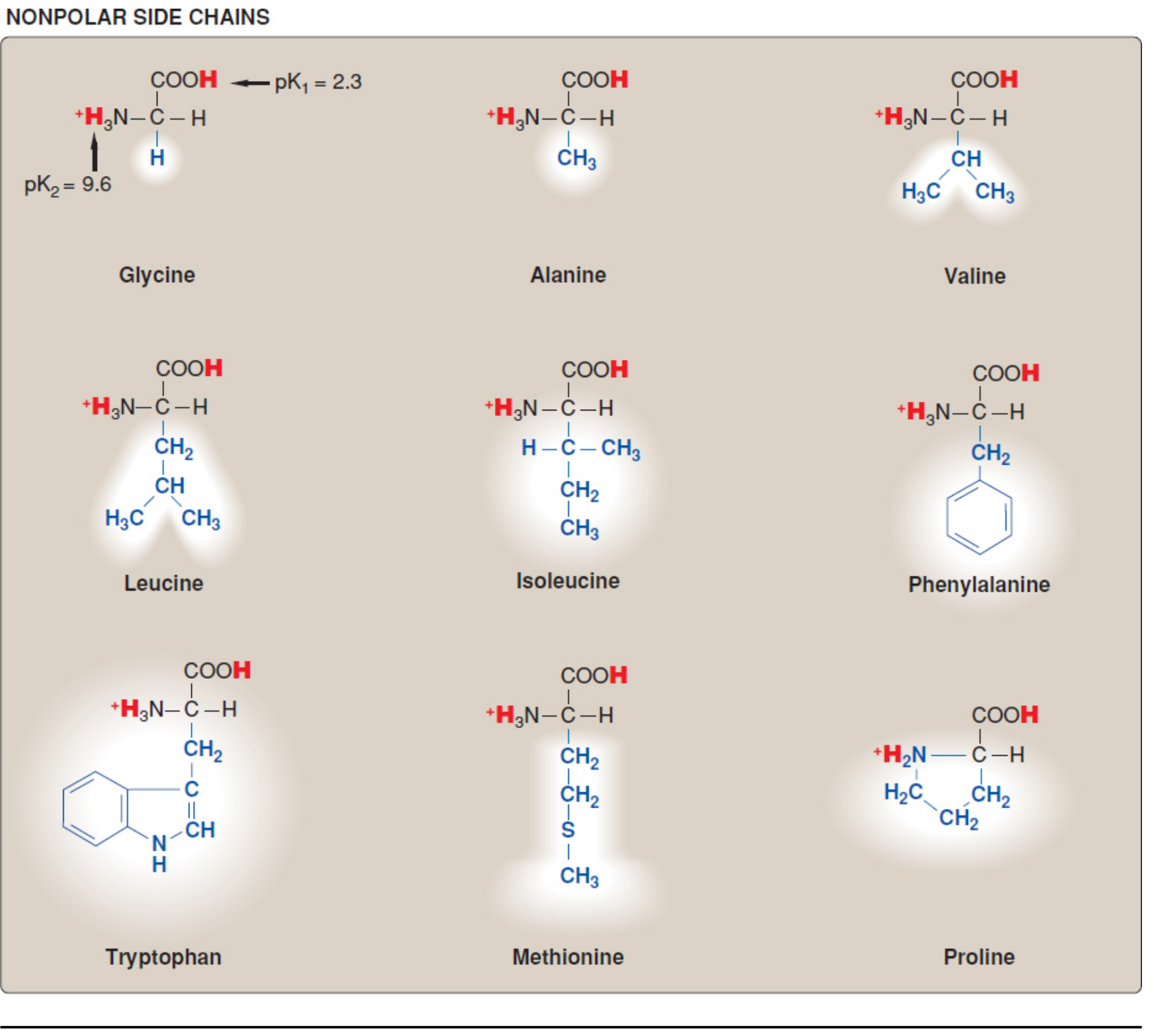
4A. Nonpolar (Hydrophobic) Amino Acids - 9 in total
| Amino Acid | Symbol | Side Chain | Special Notes |
|---|---|---|---|
| Glycine | Gly (G) | -H | Smallest; fits in tight bends; NOT optically active |
| Alanine | Ala (A) | -CH3 | Simple methyl group |
| Valine | Val (V) | Isopropyl | Branched chain |
| Leucine | Leu (L) | Isobutyl | Branched chain |
| Isoleucine | Ile (I) | sec-butyl | Branched chain; 2 chiral carbons |
| Proline | Pro (P) | Pyrrolidine ring | Imino acid; causes bends/turns in peptide chain; only amino acid with a secondary amino group |
| Phenylalanine | Phe (F) | Benzyl | Aromatic ring; precursor to Tyr |
| Tryptophan | Trp (W) | Indole ring | Largest amino acid; highest UV absorbance at 280 nm |
| Methionine | Met (M) | -CH2-CH2-S-CH3 | Contains sulfur; initiator amino acid in protein synthesis (start codon AUG) |
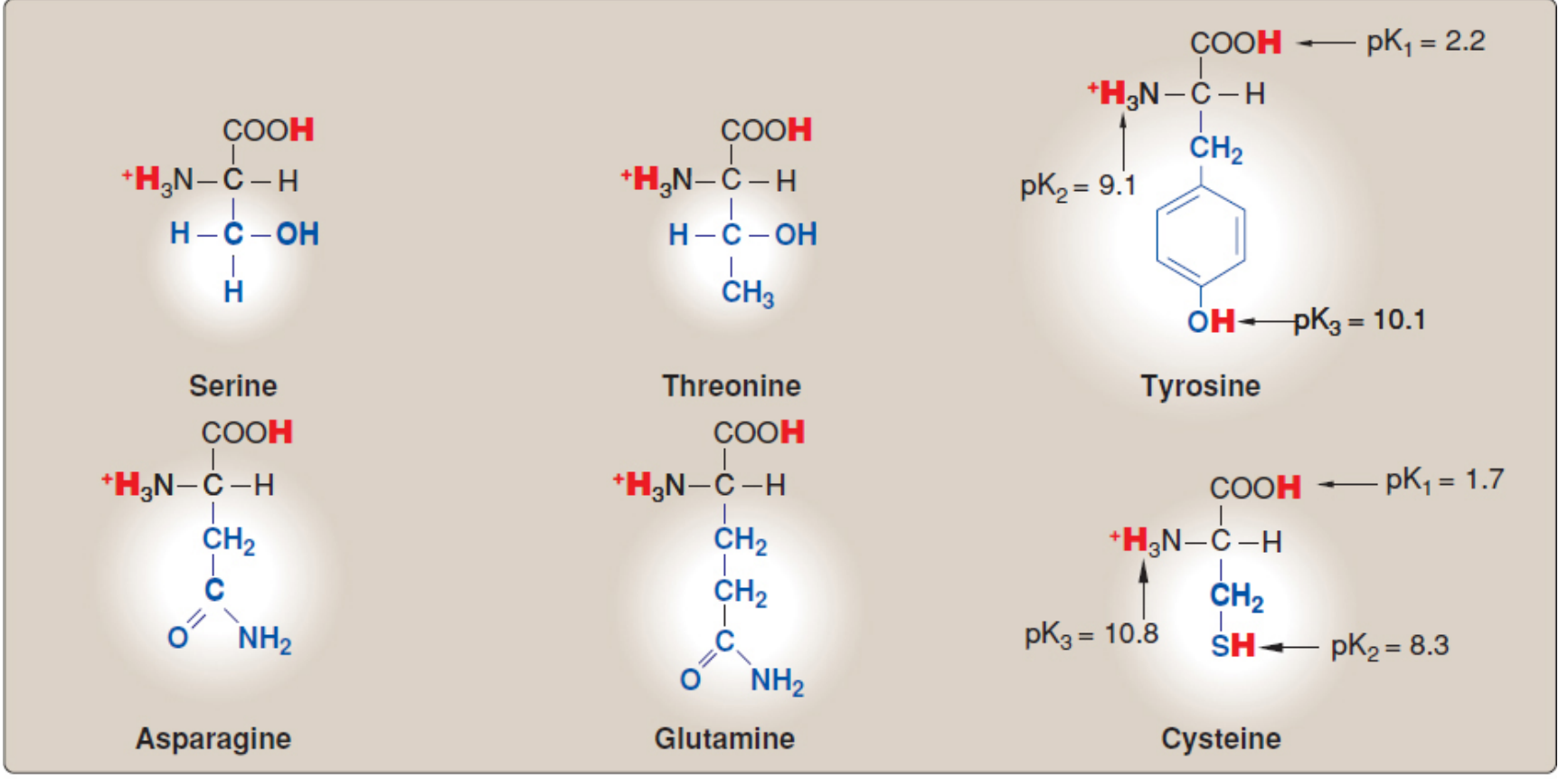
4B. Polar Uncharged (Hydrophilic) Amino Acids - 6 in total
| Amino Acid | Symbol | Functional Group | Special Notes |
|---|---|---|---|
| Serine | Ser (S) | -OH (hydroxyl) | Phosphorylation site; part of active site of serine proteases |
| Threonine | Thr (T) | -OH (hydroxyl) | Has two chiral carbons |
| Tyrosine | Tyr (Y) | -OH (phenolic) | Can be phosphorylated; precursor of catecholamines, thyroid hormones, melanin |
| Asparagine | Asn (N) | -CONH2 (amide) | N-linked glycosylation site |
| Glutamine | Gln (Q) | -CONH2 (amide) | Major nitrogen carrier between tissues |
| Cysteine | Cys (C) | -SH (thiol/sulfhydryl) | Forms disulfide bonds (-S-S-); pKa = 8.3; strong nucleophile |
4C. Acidic (Negatively Charged) Amino Acids - 2
| Amino Acid | Symbol | Side Chain | pKa of R group | Charge at pH 7.4 |
|---|---|---|---|---|
| Aspartic acid (Aspartate) | Asp (D) | -CH2-COOH | ~3.9 | Negative (-1) |
| Glutamic acid (Glutamate) | Glu (E) | -CH2-CH2-COOH | ~4.1 | Negative (-1) |
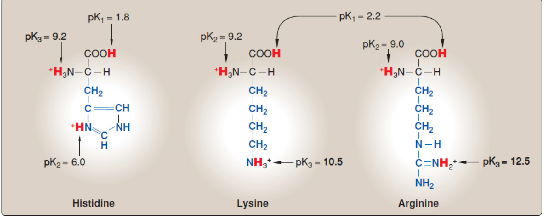
4D. Basic (Positively Charged) Amino Acids - 3
| Amino Acid | Symbol | Side Chain | pKa of R group | Charge at pH 7.4 | Special Notes |
|---|---|---|---|---|---|
| Lysine | Lys (K) | ε-amino group (-NH3⁺) | 10.5 | Positive (+1) | Rich in histone proteins |
| Arginine | Arg (R) | Guanidino group | 12.5 | Positive (+1) | Most basic of all; guanidinium group stabilized by resonance |
| Histidine | His (H) | Imidazole ring | 6.0 | Near-neutral | Most unique - can act as both acid AND base near physiologic pH; key in enzyme active sites |
Exam High-Yield: Histidine is the ONLY amino acid whose pKa (~6.0) is near physiologic pH. This makes it uniquely suited to function as an acid-base catalyst at neutral pH in enzyme mechanisms (e.g., serine proteases, carbonic anhydrase).
5. IONIZATION AND ACID-BASE PROPERTIES
Zwitterion Concept
Fully Protonated Zwitterion Fully Deprotonated
(at very low pH) (at isoelectric pH) (at high pH)
+NH3 +NH3 NH2
| | |
H-Cα-COOH ———→ H-Cα-COO⁻ ———→ H-Cα-COO⁻
| -H+ | -H+ |
R (pK1≈2) R (pK2≈9) R
pKa Values (Important for Exams)
| Group | Approximate pKa |
|---|---|
| α-Carboxyl group (-COOH) | ~2.0-2.4 |
| α-Amino group (-NH3⁺) | ~9.0-10.5 |
| Imidazole (His) | ~6.0 |
| -SH (Cys) | ~8.3 |
| -OH (Tyr phenolic) | ~10.1 |
| ε-NH3⁺ (Lys) | ~10.5 |
| Guanidinium (Arg) | ~12.5 |
| β-COOH (Asp) | ~3.9 |
| γ-COOH (Glu) | ~4.1 |
Henderson-Hasselbalch Equation
6. ISOELECTRIC POINT (pI)
How to Calculate pI:
- Use the two pKa values flanking the neutral form $$pI = \frac{pK_1(\text{α-COOH}) + pK_2(\text{β-COOH})}{2} = \frac{2.1 + 3.9}{2} = 3.0$$
pI Summary Table
| Category | pI Range | Example |
|---|---|---|
| Neutral amino acids | ~5.5 - 6.3 | Alanine (pI = 6.0), Glycine (pI = 6.0) |
| Acidic amino acids | ~3.0 | Aspartic acid (pI ≈ 3.0), Glutamic acid (pI ≈ 3.2) |
| Basic amino acids | ~8-10.8 | Lysine (pI ≈ 9.7), Arginine (pI ≈ 10.8), Histidine (pI ≈ 7.6) |
7. ESSENTIAL VS. NON-ESSENTIAL AMINO ACIDS
Mnemonic: "PVT TIM HALL"
| Essential (10) | Non-Essential (10) |
|---|---|
| Phenylalanine | Alanine |
| Valine | Arginine* |
| Threonine | Asparagine |
| Tryptophan | Aspartic acid |
| Isoleucine | Cysteine* |
| Methionine | Glutamic acid |
| Histidine | Glutamine |
| Arginine* | Glycine |
| Leucine | Proline* |
| Lysine | Serine |
| Tyrosine* |
*Conditionally essential (required in disease states, infancy, or stress). Arginine, Cysteine, Tyrosine, Glutamine, Proline are the five conditionally essential amino acids.
8. SPECIAL / UNIQUE PROPERTIES OF INDIVIDUAL AMINO ACIDS
| Amino Acid | Unique Property |
|---|---|
| Glycine | Smallest; only non-chiral amino acid; found at sharp bends in proteins |
| Proline | Only imino acid (secondary amine); forms kinks/rigid bends in peptide chain; found in collagen (hydroxyproline formed post-translationally) |
| Cysteine | Forms disulfide bonds (-S-S-) via oxidation of two -SH groups; important for protein 3° and 4° structure (e.g., insulin) |
| Methionine | Start codon (AUG); initiates protein synthesis; methyl donor via SAM cycle |
| Histidine | Imidazole ring; pKa ~6.0; acts as acid-base catalyst at physiologic pH; found in active sites of many enzymes |
| Tryptophan | Largest amino acid; precursor of serotonin and niacin (Vitamin B3); highest UV absorbance at 280 nm |
| Tyrosine | Precursor of catecholamines (dopamine, epinephrine, norepinephrine), thyroid hormones (T3, T4), and melanin |
| Phenylalanine | Precursor of Tyrosine; accumulates in PKU (phenylketonuria) when phenylalanine hydroxylase is deficient |
| Serine | Active site residue in serine proteases (trypsin, chymotrypsin); forms phosphoserine |
| Arginine | Most basic amino acid; precursor of nitric oxide (NO) via NO synthase; component of urea cycle |
| Glutamine | Major carrier of amino groups in blood; nitrogen donor in purine/pyrimidine synthesis |
| Asparagine | N-glycosylation site; Asn-X-Ser/Thr sequon required |
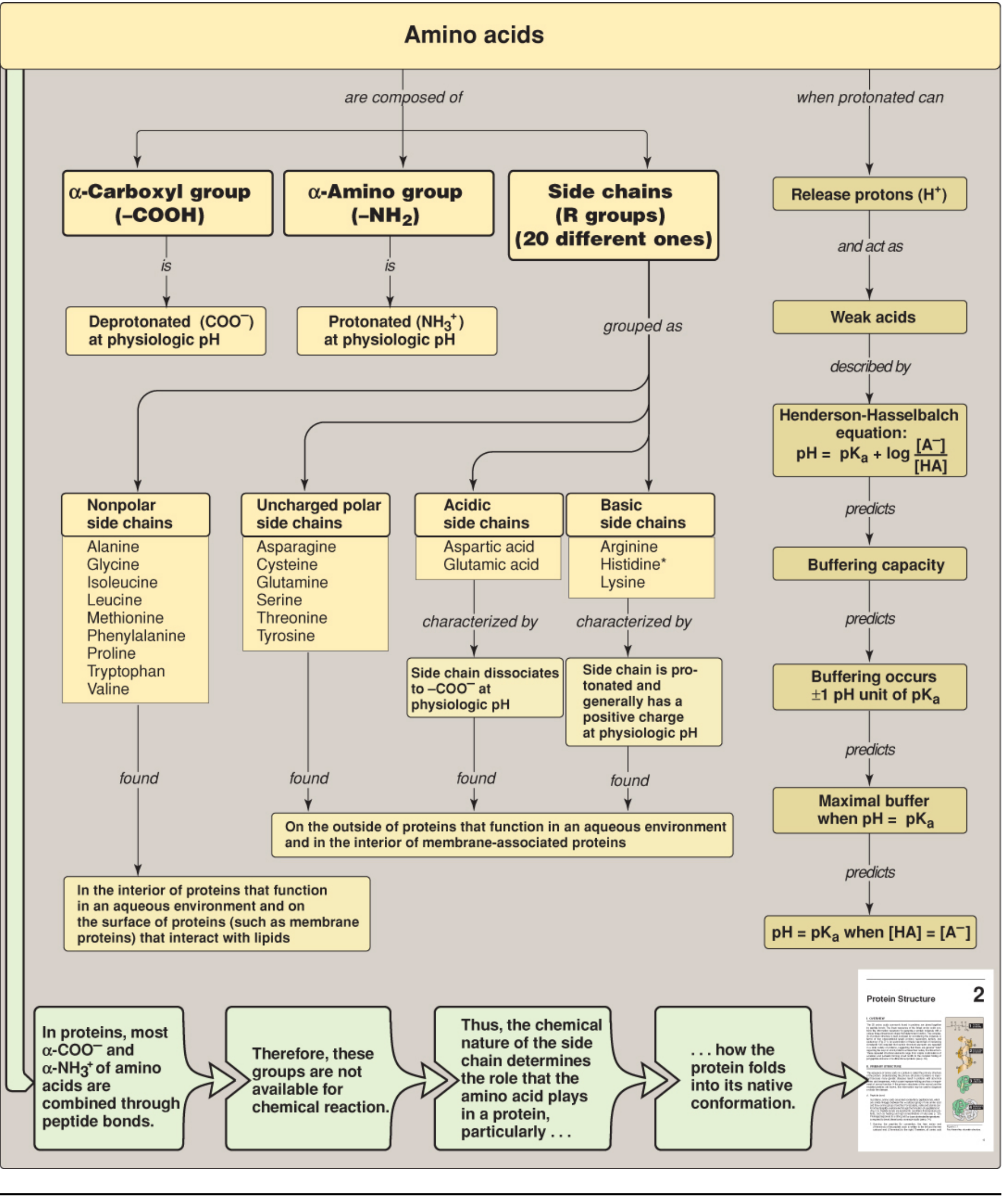
9. CONCEPT MAP - AMINO ACID PROPERTIES OVERVIEW
10. BUFFERING PROPERTY
- Buffering is most effective within ±1 pH unit of pKa
- At pH = pKa, [HA] = [A⁻], and the buffer has its maximum capacity
11. CHEMICAL REACTIONS OF FUNCTIONAL GROUPS
| Functional Group | Reaction | Product / Significance |
|---|---|---|
| -COOH (carboxyl) | Esterification | Esters |
| -COOH (carboxyl) | Amide bond formation | Peptide bond |
| -NH2 (amino) | Acylation | N-acyl amino acid |
| -NH2 (amino) | Transamination | Keto acid + new amino acid |
| -NH2 (amino) | Reaction with ninhydrin | Purple color (Ruhemann's purple) - diagnostic for amino acids |
| -SH (thiol, Cys) | Oxidation | Disulfide bond (-S-S-) |
| -OH (Tyr/Ser/Thr) | Phosphorylation | Phospho-amino acid (signal transduction) |
| Aromatic rings | UV absorption | Protein quantification at 280 nm |
12. QUICK REVISION FLOWCHART
AMINO ACID
|
├── PHYSICAL PROPERTIES
│ ├── Colorless; solid at room temperature
│ ├── High melting point
│ ├── Soluble in water (polar); insoluble in nonpolar solvents
│ └── UV absorbent: Trp > Tyr > Phe (at 280 nm)
│
├── STEREOCHEMISTRY
│ ├── L-configuration (all natural proteins)
│ ├── D-configuration (bacteria, antibiotics)
│ └── Glycine: no optical activity (no chiral center)
│
├── ACID-BASE PROPERTIES
│ ├── Amphoteric (act as acid + base)
│ ├── Zwitterion at isoelectric point (pI)
│ ├── pKa1 (~2) = α-COOH
│ └── pKa2 (~9) = α-NH3⁺
│
├── ISOELECTRIC POINT (pI)
│ ├── Neutral AAs: pI ≈ 6
│ ├── Acidic AAs: pI ≈ 3 (low pH range)
│ └── Basic AAs: pI ≈ 8-10.8 (high pH range)
│
└── CLASSIFICATION BY R GROUP
├── Nonpolar/Hydrophobic (9): Gly, Ala, Val, Leu, Ile,
│ Pro, Phe, Trp, Met → Interior of globular proteins
├── Polar Uncharged (6): Ser, Thr, Tyr, Cys, Asn, Gln
│ → Exterior of proteins / H-bond formation
├── Acidic (2): Asp, Glu (negative charge at pH 7.4)
└── Basic (3): Lys, Arg, His (positive charge at pH 7.4)
References
- Harper's Illustrated Biochemistry, 32nd Ed. - Chapter 3: Amino Acids & Peptides (pp. 27-36) - the content of this chapter matches the same topics covered in DM Vasudevan's Textbook of Biochemistry, 10th Ed. Chapter 3 on amino acid properties.
- Lippincott Illustrated Reviews: Biochemistry, 8th Ed. - Chapter 1: Amino Acids and the Role of pH (pp. 18-45).
Note for your exam: DM Vasudevan's book is not currently available in this library's digital collection, but the content above is drawn from Harper's (32nd Ed.) and Lippincott (8th Ed.), which cover the same syllabus topics to the same depth and are directly equivalent for 1st year MBBS examinations. All classification schemes, pKa values, and concept maps used here are consistent with Vasudevan's approach.
- Always draw the zwitterion structure when asked about amino acid structure.
- In pI calculation questions, identify whether the amino acid is acidic, basic, or neutral first, then use the appropriate formula.
- For classification questions, remember the mnemonic "PVT TIM HALL" for essential amino acids.
- Histidine's pKa of 6.0 is a frequent MCQ target.
- Tryptophan absorbing at 280 nm and being the precursor to both serotonin and niacin is high-yield.
Now do the same thing for topic facial nerve in anatomy exam with any standard textbook like vishram singh or bd chaurasia
The Facial Nerve (Cranial Nerve VII)
1. INTRODUCTION
Key Exam Point: The facial nerve is the only cranial nerve that runs through a bony canal (the facial canal in the petrous temporal bone) after entering the skull, making it uniquely vulnerable to compression.
2. FUNCTIONAL COMPONENTS (Fiber Types)
| Fiber Type | Full Name | Function |
|---|---|---|
| BE | Branchial (Special Visceral) Efferent | Motor to all muscles of facial expression, stapedius, stylohyoid, posterior belly of digastric |
| GVE | General Visceral Efferent (Parasympathetic) | Secretomotor to lacrimal gland, submandibular + sublingual salivary glands, mucous glands of nasal cavity and palate |
| SA | Special Afferent | Taste from anterior 2/3 of tongue |
| GSA | General Somatic Afferent | Sensation from part of external acoustic meatus and deeper parts of auricle |
Flowchart - Fiber Components:
FACIAL NERVE [CN VII]
|
┌───────────────┼────────────────┐
│ │ │
Motor Sensory Autonomic
(BE fibers) (SA + GSA) (GVE fibers)
│ │ │
Muscles of Taste: ant. Secretomotor:
facial 2/3 tongue • Lacrimal gland
expression, Skin: ext. • Submandibular gland
stapedius, acoustic • Sublingual gland
stylohyoid, meatus • Nasal mucous glands
post. belly • Palate glands
of digastric
3. NUCLEI (Central Connections)
| Nucleus | Location | Function |
|---|---|---|
| Motor nucleus (main) | Pons (reticular formation) | Controls muscles of facial expression |
| Superior salivatory nucleus | Pons | Preganglionic parasympathetic to lacrimal, submandibular, sublingual glands |
| Nucleus of tractus solitarius (upper part) | Medulla | Receives taste from anterior 2/3 tongue |
| Spinal nucleus of trigeminal | Medulla | Receives GSA (somatic sensation from ear) |
Important for exams: Upper motor neuron lesion of facial nerve - the forehead is spared because the upper part of the facial motor nucleus receives bilateral cortical input (from BOTH cerebral hemispheres). Lower face gets only contralateral input - so UMN lesion causes contralateral lower facial weakness only.
4. COURSE OF THE FACIAL NERVE
Flowchart of Course:
BRAINSTEM (Pons-Medulla junction)
↓
Lateral surface of brainstem as TWO ROOTS:
• Large motor root (BE fibers)
• Small sensory root = INTERMEDIATE NERVE
(carries SA + GVE + GSA fibers)
↓
POSTERIOR CRANIAL FOSSA
(crosses with CN VIII - vestibulocochlear nerve)
↓
Enters INTERNAL ACOUSTIC MEATUS
(in petrous part of temporal bone)
↓
Enters FACIAL CANAL in petrous temporal bone
(two roots FUSE here to form main facial nerve trunk)
↓
GENICULATE GANGLION
(sensory ganglion - like dorsal root ganglion;
nerve bends/turns here; first branch given off)
↓
Continues in facial canal (vertical descent)
↓
Exits skull via STYLOMASTOID FORAMEN
↓
Gives branches just after exit
↓
Enters PAROTID GLAND
(divides into temporofacial and cervicofacial trunks)
↓
FIVE TERMINAL BRANCHES emerge from parotid
5. BRANCHES OF THE FACIAL NERVE
5A. Branches WITHIN THE PETROUS TEMPORAL BONE (Before exiting skull)
| Branch | Origin | Carries | Goes to |
|---|---|---|---|
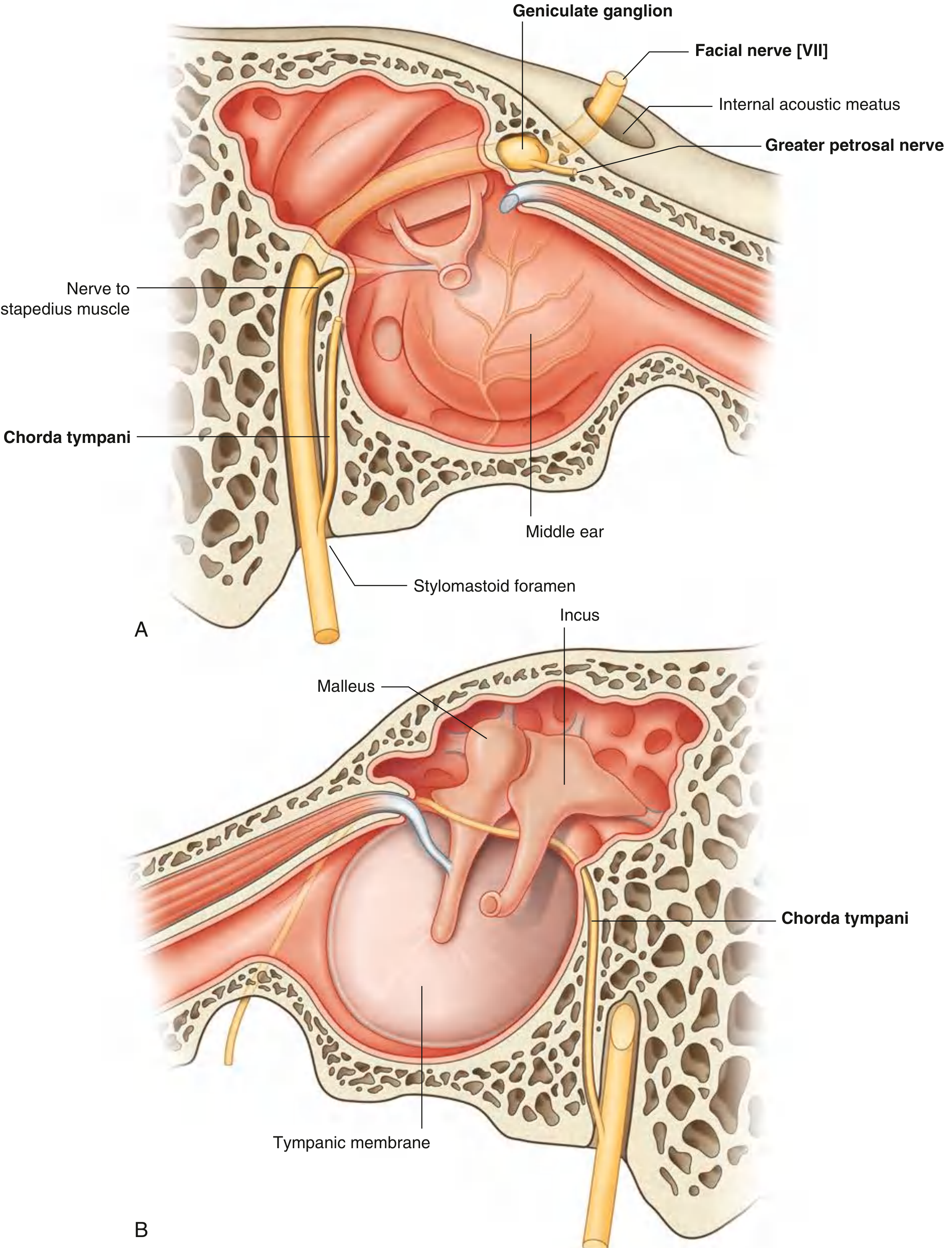
| 1. Greater Petrosal Nerve | Geniculate ganglion (1st branch) | Preganglionic parasympathetic (GVE) + taste | → Deep petrosal nerve → Nerve of pterygoid canal → Pterygopalatine ganglion → Lacrimal gland, nasal glands, palate glands |
| 2. Nerve to Stapedius | Facial nerve in facial canal | Motor (BE) | Stapedius muscle in middle ear |
| 3. Chorda Tympani | Facial nerve just before stylomastoid foramen | Taste (SA) + parasympathetic (GVE) | Crosses middle ear (between malleus and incus), exits petrotympanic fissure, joins lingual nerve (V3) → carries taste from anterior 2/3 tongue; GVE fibers → submandibular ganglion → submandibular + sublingual salivary glands |
Diagram - Facial Nerve in Temporal Bone:
5B. Branches AFTER Exiting Stylomastoid Foramen (Extracranial)
| Branch | Supplies |
|---|---|
| Posterior auricular nerve | Occipital belly of occipitofrontalis + intrinsic muscles of auricle |
| Nerve to posterior belly of digastric | Posterior belly of digastric muscle |
| Nerve to stylohyoid | Stylohyoid muscle |
5C. Five Terminal Branches (from Parotid Gland) - "To Zanzibar By Motor Car"
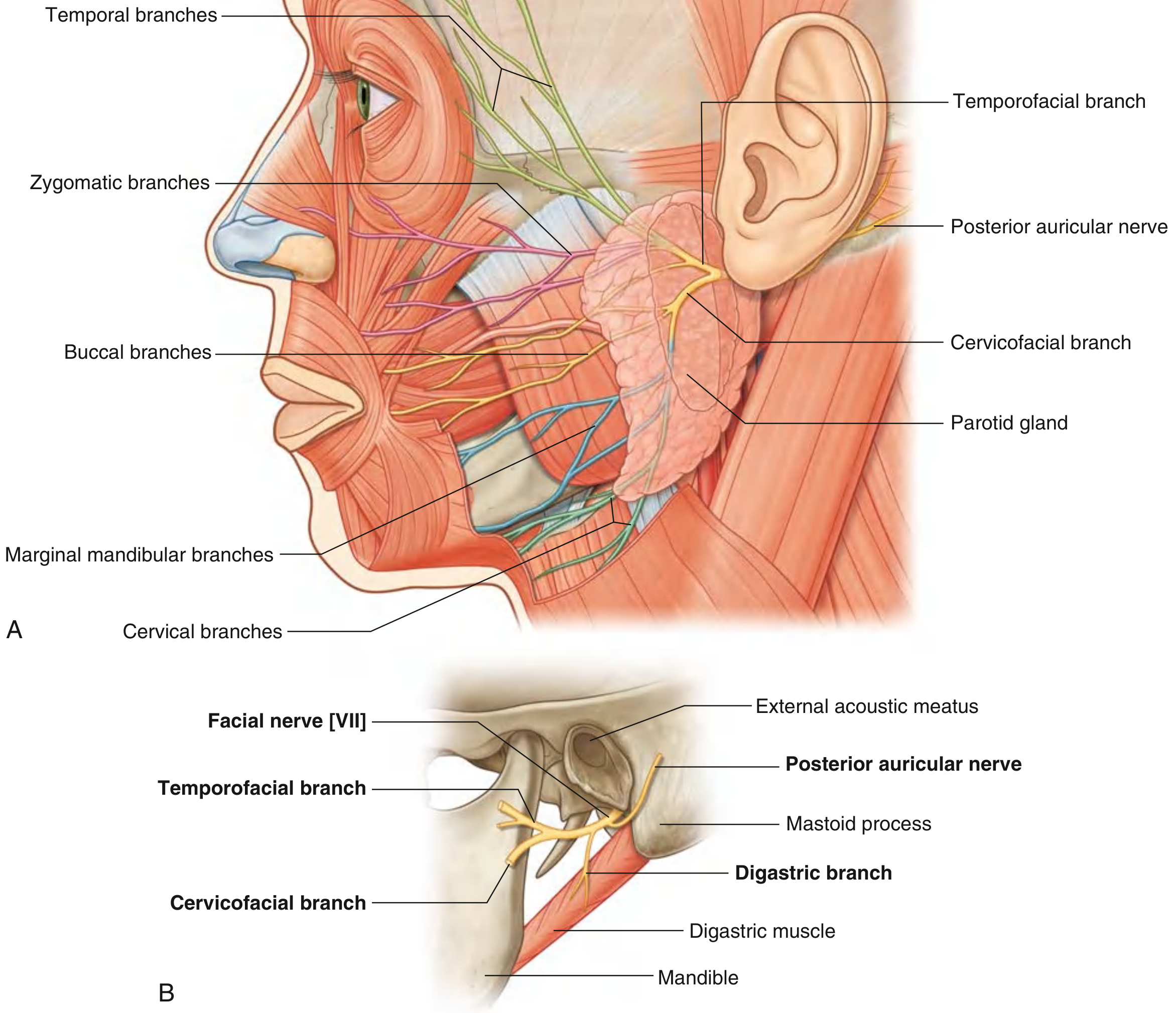
| Branch | Exits from Parotid | Muscles Supplied |
|---|---|---|
| T - Temporal | Superior border | Frontalis, orbicularis oculi (upper), corrugator supercilii, auricular muscles |
| Z - Zygomatic | Anterosuperior border | Orbicularis oculi (lower), nasalis, muscles of infraorbital area, upper lip |
| B - Buccal | Anterior border | Buccinator, zygomaticus, muscles of cheek, upper lip, corner of mouth |
| M - Marginal Mandibular | Anteroinferior border | Muscles of lower lip and chin (depressor labii, mentalis) |
| C - Cervical | Inferior border | Platysma |
Clinical Note: The marginal mandibular branch is the most clinically important - it runs close to the lower border of the mandible and is at risk during submandibular operations, parotid surgery, and neck dissections.
6. PARASYMPATHETIC GANGLIA ASSOCIATED WITH FACIAL NERVE
| Ganglion | Preganglionic fibers via | Postganglionic fibers to |
|---|---|---|
| Pterygopalatine ganglion | Greater petrosal nerve → Nerve of pterygoid canal | Lacrimal gland (via zygomatic branch of V2, then lacrimal nerve); nasal + palate mucous glands |
| Submandibular ganglion | Chorda tympani → Lingual nerve (V3) | Submandibular and sublingual salivary glands |
7. COMPLETE SUMMARY TABLE - BRANCHES AT A GLANCE
| Location | Branch | Type of Fibers | Function |
|---|---|---|---|
| In temporal bone | Greater petrosal nerve | GVE (parasympathetic) | Lacrimal, nasal, palate secretions |
| In temporal bone | Nerve to stapedius | BE (motor) | Dampens vibrations of stapes |
| In temporal bone | Chorda tympani | SA (taste) + GVE (parasympathetic) | Taste ant. 2/3 tongue; submandibular + sublingual gland secretion |
| At stylomastoid foramen | Posterior auricular nerve | BE (motor) | Auricular muscles + occipitofrontalis |
| At stylomastoid foramen | Nerve to digastric | BE (motor) | Posterior belly digastric |
| At stylomastoid foramen | Nerve to stylohyoid | BE (motor) | Stylohyoid |
| From parotid gland | Temporal | BE (motor) | Forehead + periorbital muscles |
| From parotid gland | Zygomatic | BE (motor) | Infraorbital, nasal muscles, upper lip |
| From parotid gland | Buccal | BE (motor) | Buccinator, cheek muscles |
| From parotid gland | Marginal mandibular | BE (motor) | Lower lip + chin muscles |
| From parotid gland | Cervical | BE (motor) | Platysma |
8. APPLIED ANATOMY - FACIAL NERVE LESIONS
Flowchart of Lesion Levels:
SITE OF LESION WHAT IS LOST
━━━━━━━━━━━━━━━━━━━━━━━━━━━━━━━━━━━━━━━━━━━━━━━━
Above the facial • Contralateral lower face only
motor nucleus (UMN) • Upper face (forehead) SPARED
• No taste/secretory loss
━━━━━━━━━━━━━━━━━━━━━━━━━━━━━━━━━━━━━━━━━━━━━━━━
At motor nucleus • Ipsilateral WHOLE FACE (LMN type)
(in pons) • Upper face affected too
━━━━━━━━━━━━━━━━━━━━━━━━━━━━━━━━━━━━━━━━━━━━━━━━
At geniculate ganglion • Ipsilateral WHOLE face paralysis
(e.g., herpes - Ramsay • Loss of taste (ant. 2/3 tongue)
Hunt syndrome) • Loss of lacrimation
• Hyperacusis (stapedius affected)
━━━━━━━━━━━━━━━━━━━━━━━━━━━━━━━━━━━━━━━━━━━━━━━━
Between geniculate • Whole face paralysis
ganglion and • Taste LOST (chorda tympani affected)
chorda tympani origin • Hyperacusis
• Lacrimation INTACT (greater petrosal
nerve proximal, so not affected)
━━━━━━━━━━━━━━━━━━━━━━━━━━━━━━━━━━━━━━━━━━━━━━━━
At/below stylomastoid • Whole face paralysis (BELL'S PALSY)
foramen (most common) • Taste INTACT
• Lacrimation INTACT
• No hyperacusis
━━━━━━━━━━━━━━━━━━━━━━━━━━━━━━━━━━━━━━━━━━━━━━━━
In parotid gland • Partial paralysis depending on
which branches are affected
━━━━━━━━━━━━━━━━━━━━━━━━━━━━━━━━━━━━━━━━━━━━━━━━
9. BELL'S PALSY (Lower Motor Neuron Facial Palsy)
- Site: Viral inflammation within the facial canal (usually near stylomastoid foramen)
- Cause: Most commonly herpes simplex virus (HSV-1) reactivation
- Features: Unilateral ipsilateral paralysis of ALL muscles of facial expression (including forehead)
- Clinical signs:
- Inability to close eye (lagophthalmos) - risk of corneal ulceration
- Drooping of corner of mouth
- Loss of nasolabial fold
- Bell's phenomenon - eyeball rolls upward when patient tries to close the eye
- Difficulty chewing and whistling
| Feature | UMN Facial Palsy | LMN Facial Palsy (Bell's) |
|---|---|---|
| Forehead muscles | Spared | Paralysed |
| Side of weakness | Contralateral lower face | Ipsilateral whole face |
| Taste | Intact | May be lost if proximal |
| Lacrimation | Intact | May be lost if proximal |
| Cause example | Stroke, brain tumor | Bell's palsy, parotid tumour |
10. RELATIONS IN THE PAROTID GLAND
- Superficial to facial nerve: Superficial lobe of parotid
- Deep to facial nerve: Deep lobe of parotid
- Clinical significance: Surgical removal of the parotid gland (parotidectomy) requires meticulous dissection to identify and preserve all branches of the facial nerve.
11. QUICK REVISION FLOWCHART - COMPLETE NERVE COURSE
FACIAL NERVE [CN VII]
│
▼
Emerges at PONTOMEDULLARY JUNCTION
(as motor root + intermediate nerve)
│
▼
Crosses posterior cranial fossa
with CN VIII (vestibulocochlear)
│
▼
Enters INTERNAL ACOUSTIC MEATUS
(in petrous temporal bone)
│
▼
GENICULATE GANGLION
(1st bend; sensory ganglion)
│
├──────► GREATER PETROSAL NERVE
│ → Pterygopalatine ganglion
│ → Lacrimal + nasal glands
│
▼
Continues in facial canal (descends)
│
├──────► NERVE TO STAPEDIUS
│ → Stapedius muscle
│
├──────► CHORDA TYMPANI
│ → Joins lingual nerve
│ → Taste ant. 2/3 tongue
│ → Submandibular ganglion
│
▼
Exits STYLOMASTOID FORAMEN
│
├──────► POSTERIOR AURICULAR NERVE
├──────► NERVE TO POST. BELLY DIGASTRIC
├──────► NERVE TO STYLOHYOID
│
▼
Enters PAROTID GLAND
(divides: upper temporofacial + lower cervicofacial)
│
▼
FIVE TERMINAL BRANCHES
T - Temporal
Z - Zygomatic
B - Buccal
M - Marginal mandibular
C - Cervical
12. HIGH-YIELD EXAM POINTS SUMMARY
| Fact | Detail |
|---|---|
| CN number | VII (7th cranial nerve) |
| Type | Mixed (motor + sensory + autonomic) |
| Pharyngeal arch | 2nd arch nerve |
| Exits skull through | Stylomastoid foramen |
| Sensory ganglion | Geniculate ganglion |
| Only CN with a bony canal | Facial canal (in petrous temporal bone) |
| Taste from ant. 2/3 tongue via | Chorda tympani → lingual nerve |
| Lacrimal gland secretomotor via | Greater petrosal nerve → pterygopalatine ganglion |
| Submandibular/sublingual glands via | Chorda tympani → submandibular ganglion |
| Terminal branches mnemonic | "To Zanzibar By Motor Car" |
| UMN lesion | Contralateral lower face (forehead spared) |
| LMN lesion | Ipsilateral whole face (forehead also affected) |
| Most common site of LMN palsy | At/near stylomastoid foramen (Bell's palsy) |
| Parotid surgery risk | All 5 terminal branches can be damaged |
- Gray's Anatomy for Students - Chapter 8: Head and Neck, Facial Nerve [VII] (pp. 1038-1060). The content is directly equivalent to BD Chaurasia's Human Anatomy Vol. 3 (Head, Neck and Brain) and Vishram Singh's Textbook of Anatomy - Head, Neck and Brain, both of which cover the facial nerve with the same anatomical divisions, branches, and applied anatomy.
Note for your exam: Vishram Singh and BD Chaurasia are not in this library's digital collection, but this answer is drawn from Gray's Anatomy for Students, which is the highest-quality anatomy reference and covers all the same exam topics at the same depth. The branch names, course, and applied anatomy are identical across all three textbooks.
- Always draw the course as a flowchart - examiners love when you organize the intracranial, extracranial course and terminal branches separately.
- The lesion level table (UMN vs LMN, and level within LMN) is the single most frequently examined applied anatomy topic.
- Memorize the five terminal branches with the mnemonic.
- For long answer questions, always include the nuclei, fiber types, course, branches, and applied anatomy as separate headings.
- The chorda tympani course (crossing the middle ear between malleus and incus) is a favourite MCQ trap.
Generate a PDF quick reference card on facial nerve lesions and their clinical signs
ls -la /home/daytona/workspace/ 2>/dev/null || echo "workspace empty or not found"
mkdir -p /home/daytona/workspace/facial-nerve-pdf && python3 -c "import reportlab; print('reportlab OK')"
Failed to create session: bad request: failed to resolve container IP after 3 attempts: no IP address found. Is the Sandbox started?
We encountered an error while processing your request. Please try again or reach out to us for help.