
Cystic fibrosis
"cystic fibrosis"[MeSH Terms] AND treatment
cystic fibrosis lung pathology CFTR mutation
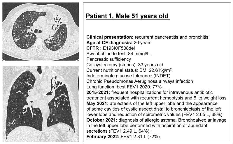
This composite educational graphic features diagnostic imaging and a clinical summary for a 51-year-old male with cystic fibrosis (CFTR genotype E193K/F508del). On the left, axial and coronal CT pulmonary scan slices demonstrate advanced respiratory pathology. Visible features include diffuse, bilateral bronchiectasis characterized by bronchial wall thickening and luminal dilation. Notably, the left upper lobe shows signs of significant atelectasis and volume loss, while the left lower lobe displays cystic cavities distal to the bronchiectatic airways. Heterogeneous lung density suggests areas of chronic inflammation and mucus plugging. The right panel provides a detailed clinical timeline and physiological data, including a sweat chloride test of 84 mmol/L, pancreatic sufficiency status, and chronic Pseudomonas aeruginosa infection. It tracks a longitudinal decline in lung function, noting an FEV1 decrease to 64–72% alongside episodes of recurrent hemoptysis and significant weight loss. This visual material serves as an educational case study on the radiological progression and clinical manifestation of specific CFTR mutations in adult patients.
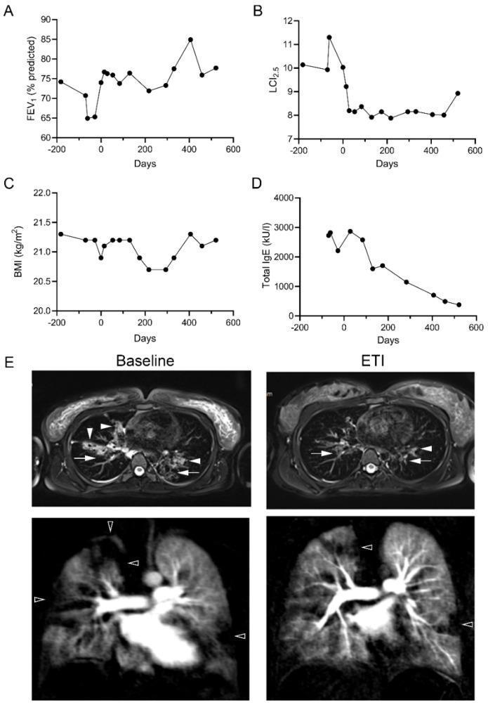
This composite educational graphic illustrates clinical and radiological changes in a Cystic Fibrosis (CF) patient carrying the G85E mutation following elexacaftor/tezacaftor/ivacaftor (ETI) therapy. Panels A-D present longitudinal line graphs showing clinical metrics from 200 days pre-initiation to 600 days post-initiation: (A) FEV1 % predicted increases, (B) Lung Clearance Index (LCI2.5) decreases, (C) Body Mass Index (BMI) remains stable, and (D) Total Serum IgE (kU/l) shows a marked decline. Panel E displays representative axial T2-weighted and perfusion MRI scans comparing 'Baseline' and 'ETI' treatment states. At baseline, white arrows indicate significant bronchial wall thickening and bronchiectasis, while white arrowheads highlight mucus plugging. Black arrowheads in the perfusion MRI denote regions of impaired lung perfusion. Post-ETI MRI images demonstrate notable resolution of mucus plugging and a decrease in perfusion abnormalities, signifying improved airway clearance and pulmonary vascular distribution. This material is designed for medical professionals studying CFTR modulator efficacy and pulmonary imaging manifestations in cystic fibrosis.
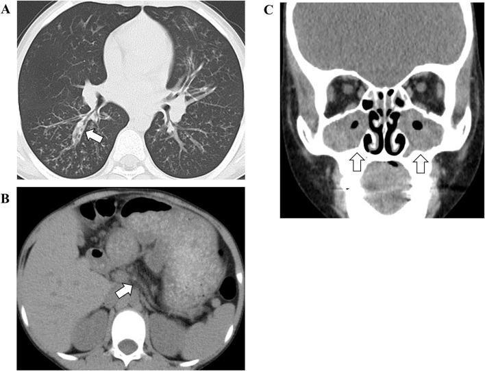
This composite of computerized tomography (CT) images illustrates multi-organ manifestations of cystic fibrosis. Image A (Axial Chest CT) shows significant pulmonary pathology, including bilateral thickened bronchial walls and bronchiectasis (white arrow), with underlying lung hyperinflation indicative of chronic obstructive airway disease. Image B (Axial Abdominal CT) reveals an atrophic pancreas characterized by fatty infiltration (white arrow), a hallmark of exocrine pancreatic insufficiency. Image C (Coronal Facial CT) demonstrates paranasal sinus involvement with bilateral opacification and distinct gas-fluid levels (white arrows), accompanied by internal gas bubbles suggesting acute-on-chronic sinusitis. Together, these diagnostic images provide a comprehensive clinical overview of the respiratory, gastrointestinal, and ENT manifestations associated with CFTR gene mutations.
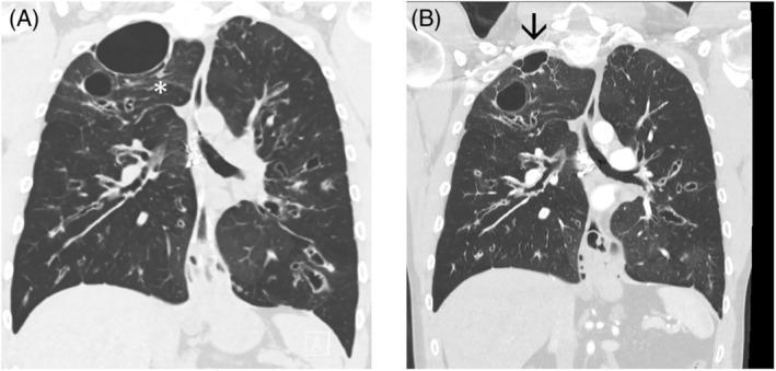
Coronal High-Resolution Computed Tomography (HRCT) of the chest comparing lung morphology before (A) and after (B) cystic fibrosis transmembrane conductance regulator (CFTR) modulator therapy. Image A demonstrates advanced bronchiectasis with prominent bronchial wall thickening, mucous plugging, and multiple cystic structures. A large, dominant air-filled cyst is visible at the right lung apex, exerting a mass effect and compression on the adjacent lung parenchyma (indicated by an asterisk). Image B, obtained 18 months post-therapy, shows a significant reduction in the size of the large apical cyst (arrow) and improvement in the expansion of surrounding lung tissue. There is a visible decrease in bronchial wall thickening and the size of varicose airways throughout both lung fields. These findings illustrate the structural lung remodeling and reduction in hyperinflation and cystic manifestations achievable with targeted pharmacotherapy in cystic fibrosis patients.
Cystic Fibrosis (CF)
Overview
Gene and Protein
- Two membrane-spanning domains (MSD1, MSD2) - 12 transmembrane segments form the chloride channel pore
- Two nucleotide (ATP)-binding domains (NBD1, NBD2) - ATP hydrolysis opens/closes the channel
- One regulatory (R) domain - phosphorylation sites control channel activity
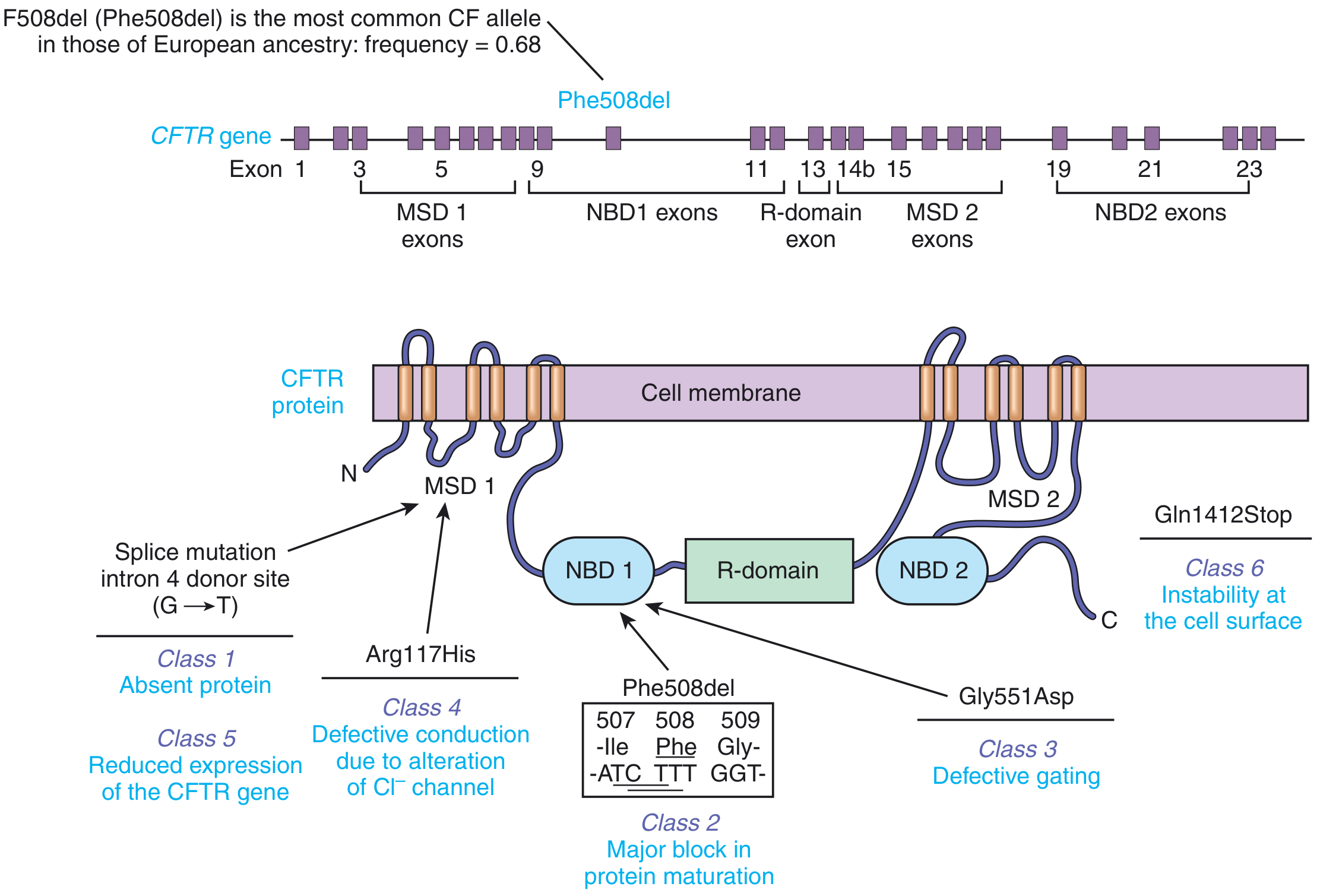
Mutations - Six Functional Classes
| Class | Defect | Example | Consequence |
|---|---|---|---|
| 1 | No protein produced (null alleles, premature stop codons) | Splice mutation intron 4 (G→T) | Absent CFTR |
| 2 | Protein misfolding - cannot exit ER | F508del (most common, ~70%) | Protein degraded before reaching cell surface |
| 3 | Protein reaches surface but channel gating is defective | Gly551Asp | Non-functional channel |
| 4 | Defective chloride conduction (MSD mutations) | Arg117His | Reduced Cl⁻ transport |
| 5 | Reduced CFTR transcript production | Splice site variants | Fewer functional channels |
| 6 | Normal synthesis but unstable at cell surface | Gln1412Stop | Rapid protein degradation |
Pathophysiology
- Sweat glands: Loss of CFTR function prevents chloride reabsorption in the ductal epithelium → elevated Na⁺ and Cl⁻ in sweat (the basis of the sweat chloride test; diagnostic threshold >60 mmol/L)
- Lungs: Hyperabsorption of sodium + reduced chloride secretion → depleted airway surface liquid → mucus becomes thick and adherent → impaired mucociliary clearance → chronic infection (especially Pseudomonas aeruginosa) and inflammation → progressive bronchiectasis
- Pancreas: Obstruction of ductal outflow by viscid secretions → autodigestion → exocrine pancreatic insufficiency (PI) in ~85-90% of patients
Clinical Features
Pulmonary (dominant cause of morbidity/mortality - 67% of deaths)
- Chronic productive cough, recurrent pulmonary infections
- Progressive bronchiectasis, hyperinflation, air trapping
- Chronic infection with Pseudomonas aeruginosa, Staphylococcus aureus, Burkholderia cepacia
- Respiratory failure (end-stage)
Gastrointestinal / Pancreatic
| Organ | Manifestation | Frequency |
|---|---|---|
| Pancreas | Exocrine insufficiency (steatorrhea, fat maldigestion) | 85-90% |
| Pancreas | Abnormal glucose tolerance | 20-30% |
| Pancreas | CF-related diabetes (CFRD) | 4-7% overall; 30% by age 20, 40% by age 30 |
| Intestine | Meconium ileus (neonates) | 10-25% |
| Intestine | Distal intestinal obstruction syndrome (DIOS) | 3% (18% in adults) |
| Liver | Focal biliary cirrhosis / portal hypertension | 2-3% |
| Biliary | Gallstones | 8-25% |
| Esophagus | GERD | ~80% in adults |
Other Features
- Genitourinary: >98% of males are infertile (congenital bilateral absence of vas deferens - CBAVD); females have reduced fertility from thickened cervical mucus
- Skeletal: Osteopenia, hypertrophic pulmonary osteoarthropathy, retarded bone age
- Salt loss: Excessive NaCl loss through skin - risk of heat stroke and hyponatremia
- Sinuses: Chronic sinusitis, nasal polyps
Diagnosis
- Newborn screening - immunoreactive trypsinogen (IRT) assay; over 60% of US CF patients diagnosed this way. Elevated IRT is followed by CFTR mutation analysis or sweat chloride testing.
- Sweat chloride test (gold standard):
- ≥60 mmol/L = diagnostic for CF
- 30-59 mmol/L = borderline (repeat or genetic testing)
- <30 mmol/L = normal
- CFTR genetic testing - used to confirm diagnosis in borderline sweat chloride cases, identify mutations for targeted therapy eligibility
- Nasal potential difference - for atypical presentations with normal sweat chloride
Genotype-Phenotype Correlations
- Pancreatic phenotype: Strongly predicted by genotype. F508del homozygotes and null allele homozygotes are almost always pancreatic insufficient (PI). Alleles with partial function (e.g., Arg117His) tend to be pancreatic sufficient (PS).
- Pulmonary phenotype: Poorly predicted by CFTR genotype. Severity is heavily influenced by modifier genes including TGFβ1 (tissue remodeling), IFRD1, and IL8 (infection tolerance).
- Genocopy: Mutations in SCNN1 (epithelial sodium channel) can produce a CF-like phenotype in individuals without CFTR mutations.
Treatment
CFTR Modulator Therapy (disease-modifying)
| Drug | Class | Target Mutation |
|---|---|---|
| Ivacaftor (Kalydeco) | Potentiator | Class 3 gating mutations (e.g., Gly551Asp) |
| Lumacaftor/ivacaftor (Orkambi) | Corrector + potentiator | F508del homozygous (Class 2) |
| Tezacaftor/ivacaftor (Symdeko) | Corrector + potentiator | F508del homozygous or heterozygous with select mutations |
| Elexacaftor/tezacaftor/ivacaftor (Trikafta/Kaftrio) | Triple combo | F508del (one or two copies) - covers ~90% of CF patients |
Pulmonary Management
- Airway clearance techniques (chest physiotherapy, oscillatory PEP devices, vest therapy)
- Inhaled mucolytics: Dornase alfa (DNase - breaks down extracellular DNA in mucus), hypertonic saline
- Inhaled antibiotics: Tobramycin, aztreonam (for chronic P. aeruginosa suppression)
- Oral azithromycin (anti-inflammatory, reduces exacerbations)
- Systemic antibiotics for acute pulmonary exacerbations
- Lung transplantation (bilateral sequential) for end-stage disease
Pancreatic / GI Management
- Pancreatic enzyme replacement therapy (PERT): Lipase/trypsin/chymotrypsin supplements taken with every meal and snack
- Fat-soluble vitamin supplementation: A, D, E, K (malabsorbed due to EPI)
- High-calorie, high-fat diet (>100% of recommended daily intake)
- CFRD: Insulin therapy (oral hypoglycemics are generally not first-line); multidisciplinary team approach
- Meconium ileus (neonates): Gastrografin enema (non-operative, ~36-39% success); surgery for complicated cases
- DIOS (adults): Polyethylene glycol (PEG) laxatives, Gastrografin enema, N-acetylcysteine
Nutrition
Prognosis
- Median predicted survival for patients born 2012-2016 in the USA: ~42.7 years
- Patients who reach age 40 have median predicted survival approaching 60 years
- Primary cause of death: chronic pulmonary / cardiorespiratory failure (67% of deaths)
- CF-related diabetes worsens both respiratory and nutritional outcomes and increases mortality
- Pancreatic-sufficient patients have better pulmonary status and longer survival than pancreatic-insufficient patients
Screening and Carrier Testing
- All US states include CF in newborn screening programs (associated with improved outcomes)
- All women should be offered CF carrier screening preconceptionally or prenatally regardless of ethnicity (ACOG recommendation), though standard panels have lower sensitivity in non-European populations
Clinical Images

