
Torsades De pointes
Torsades de Pointes ECG polymorphic ventricular tachycardia
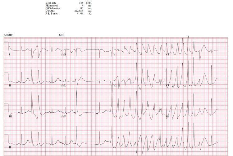
A 12-lead electrocardiogram (ECG/EKG) demonstrating a polymorphic ventricular tachycardia known as Torsades de Pointes. The tracing shows rapid, irregular, wide-complex QRS morphology characterized by a distinctive 'twisting' or undulating pattern of the QRS peaks around the isoelectric baseline. The amplitude of the QRS complexes continuously waxes and wanes, creating a spindle-like appearance. Quantitative data at the top left indicates a ventricular rate of 145 BPM and a significantly prolonged corrected QT interval (QTc) of 655 ms, which is a critical precursor to this specific arrhythmia. The precordial leads (V1-V6) clearly display the bizarre morphology and large, shifting T-waves associated with the tachyarrhythmia. This diagnostic image illustrates a life-threatening cardiac event often triggered by electrolyte imbalances or QT-prolonging medications in susceptible individuals, serving as an educational example of malignant ventricular rhythms.
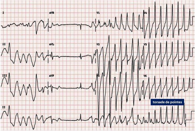
This diagnostic image is a 12-lead electrocardiogram (ECG) strip demonstrating a polymorphic ventricular tachycardia known as Torsades de Pointes (TdP). The tracing shows wide-complex tachycardia characterized by a cyclical variation in the QRS amplitude and axis, giving the classic appearance of the complexes 'twisting' around the isoelectric baseline. The rhythm is highly irregular with no discernible P waves. In the precordial leads (specifically V4, V5, and V6), the morphology exhibits rapid, spindle-shaped oscillations where the peaks of the QRS complexes shift direction. The onset is preceded by a 'short-long-short' RR interval sequence, a common trigger for this arrhythmia. This clinical finding is critical for cardiology and emergency medicine, often associated with a prolonged QT interval and potentially leading to ventricular fibrillation. The ECG is presented on standard grid paper for the evaluation of cardiac rate, rhythm, and interval measurements.
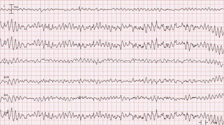
This diagnostic image is an electrocardiogram (ECG) rhythm strip displaying Torsades de Pointes (TdP), a specific form of polymorphic ventricular tachycardia. The tracing shows a continuous, irregular rapid rhythm characterized by the hallmark visual feature of QRS complexes 'twisting' around the isoelectric baseline. This manifest as cyclical variations in QRS amplitude and morphology, where the complexes wax and wane in size and periodically flip their polarity. The ECG lacks discernible P waves, normal QRS complexes, or T waves, indicating an absence of organized atrial or ventricular repolarization. Multiple leads (I, II, III, V, AVR, AVL, and AVF) are shown on a standard grid, with the polymorphic nature and shifting axis most prominent in the limb leads. Clinically, this pattern is often associated with a prolonged QT interval and represents a life-threatening arrhythmia requiring immediate intervention, such as magnesium sulfate or defibrillation.
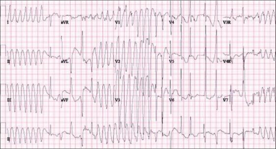
This diagnostic image is a 12-lead surface electrocardiogram (ECG) demonstrating a classic presentation of Torsades de Pointes (TdP), a specific form of polymorphic ventricular tachycardia. The tracing shows rapid, irregular QRS complexes that continuously vary in morphology, axis, and amplitude. A hallmark 'twisting of the points' is visible, where the peaks of the QRS complexes appear to rotate around the isoelectric baseline in a sinusoidal pattern. In leads I, II, III, and V1-V3, the arrhythmia exhibits rhythmic waxing and waning of amplitudes. Conversely, leads V4-V7 show highly disorganized, low-amplitude polymorphic activity. This pattern is clinically significant as it often arises in the setting of prolonged QT intervals (LQTS), such as those caused by congenital channelopathies (LQT1, LQT2, LQT3) or electrolyte imbalances. The image serves as a critical educational tool for identifying life-threatening ventricular arrhythmias that can lead to syncope or sudden cardiac death.
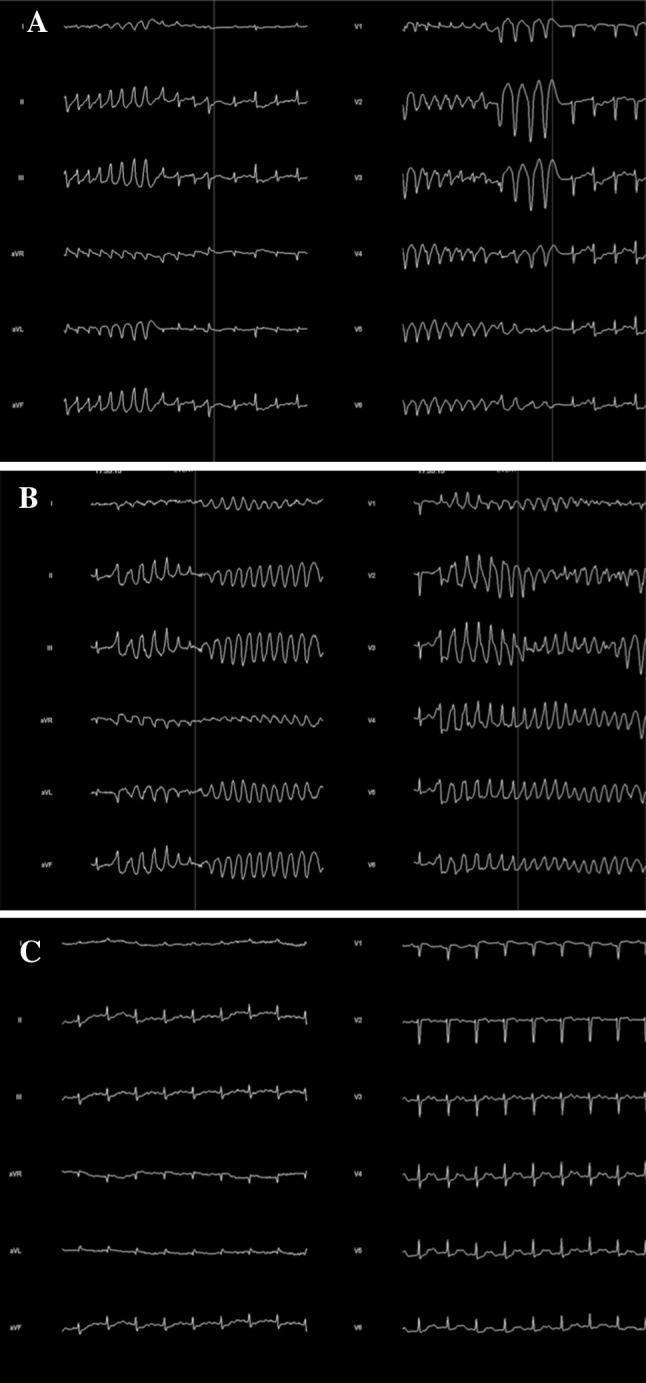
A three-panel 12-lead electrocardiogram (ECG) tracing demonstrating the evolution and features of polymorphic ventricular tachycardia. Panel A shows the initiation phase with a short-long-short R-R interval sequence, a common precursor to malignant arrhythmias. Panel B displays Torsades de Pointes (TdP), characterized by a rapid, irregular rhythm with wide QRS complexes that appear to 'twist' around the isoelectric line, varying in amplitude and morphology across leads II, III, aVF, and the precordial leads V2-V6. Panel C represents a baseline or post-event rhythm showing significant QT interval prolongation (long QT syndrome), which predisposes the patient to these ventricular events. The image illustrates critical electrophysiological findings relevant to cardiovascular medicine and emergency critical care, specifically the relationship between repolarization abnormalities and the onset of life-threatening polymorphic ventricular tachycardia.

This diagnostic image is an electrocardiogram (ECG) rhythm strip illustrating Torsades de Pointes (TdP), a specific form of polymorphic ventricular tachycardia. The tracing demonstrates a characteristic 'twisting' morphology of the QRS complexes around the isoelectric baseline. Key visual features include a rapid, irregular rate with QRS complexes that continuously vary in amplitude, polarity, and morphology. The rhythm exhibits cyclical oscillations where the peaks of the complexes appear to spiral around the baseline, alternating between larger and smaller deflections. This finding is a critical clinical indicator of a medical emergency often associated with prolonged QT intervals. The image serves as a quintessential educational example for medical students and clinicians to recognize malignant ventricular arrhythmias in cardiology and emergency medicine contexts.
"torsades de pointes" AND management
Torsades de Pointes (TdP)
Definition and ECG Features
- Ventricular rate >200 beats/min (range 120-300 bpm)
- Undulating QRS axis - the polarity of complexes appears to twist/shift around the isoelectric baseline
- Paroxysms typically <90 seconds - often self-terminating bursts of up to 30 cycles
- ROSEN's Emergency Medicine, p. 1063
Pathophysiology
Congenital vs. Acquired TdP
| Feature | Congenital (LQTS) | Acquired |
|---|---|---|
| Frequency | Rare (~1 in 2000 births) | Much more common |
| Trigger | Sympathetic excess, tachycardia, exertion | Slow heart rate, pauses (pause-dependent) |
| Setting | Children, young adults | Any age; often multifactorial |
| Mechanism | Ion channel gene mutations | Drugs, electrolytes, disease states |
Congenital Long QT Syndrome (LQTS)
- LQT1 - Mutation in cardiac potassium channel (KCNQ1; IKs current); triggered by exercise/swimming
- LQT2 - Mutation in cardiac potassium channel (HERG/KCNH2; IKr current); triggered by sudden noises/stress
- LQT3 - Mutation in cardiac sodium channel (SCN5A); TdP often occurs at rest/during sleep
- Tintinalli's Emergency Medicine, p. 158
Causes of Acquired QT Prolongation / TdP
Electrolyte Disturbances
- Hypokalemia (most common)
- Hypomagnesemia
- Hypocalcemia
Drugs (the most common cause)
- Class IA antiarrhythmics: Quinidine ("quinidine syncope"), procainamide, disopyramide
- Class IC antiarrhythmics: Flecainide, propafenone
- Class III antiarrhythmics: Sotalol, dofetilide, amiodarone (rarely)
- Antipsychotics: Haloperidol, thioridazine, quetiapine, ziprasidone
- Antidepressants: TCAs, citalopram at high doses
- Antibiotics: Azithromycin, fluoroquinolones, azole antifungals (e.g., fluconazole, ketoconazole)
- Antiemetics: Ondansetron (especially IV), metoclopramide
- Opioids: Methadone (especially at high doses)
Sex hormones modify cardiac ion channels - women have an intrinsically longer QTc and a significantly higher risk of drug-induced TdP than men. Testosterone deficiency in men (e.g., during prostate cancer treatment) also raises risk. - Goodman & Gilman, p. 698
Other Disease States
- Acute coronary syndrome / acute MI
- Severe LV dysfunction / heart failure
- Bradyarrhythmias (high-degree AV block, sick sinus syndrome)
- Hypothyroidism, subarachnoid hemorrhage
- Starvation/liquid protein diets
QT Interval - Normal Values and Risk Stratification
| QTc | Interpretation |
|---|---|
| ≤440 ms (men), ≤460 ms (women) | Normal |
| 480-499 ms | Moderate risk of TdP |
| ≥500 ms | Significantly elevated risk - high concern |
- Tintinalli's Emergency Medicine, p. 158
Clinical Presentation
- Palpitations, dizziness, presyncope, syncope - most common symptoms (often during TdP bursts)
- Cardiac arrest / sudden cardiac death - if TdP degenerates into ventricular fibrillation (VF)
- Episodes are often paroxysmal and self-terminating (bursts <30 cycles)
- Syncope mistaken for seizures - a documented diagnostic pitfall (seen in ~20% misdiagnosis rate for "nonepileptic seizures") - Kaplan & Sadock's Comprehensive Textbook of Psychiatry
ECG Diagnosis
- Wide-complex (>120 ms) tachycardia at 200-300 bpm
- QRS complexes that twist around the isoelectric line, with constantly changing amplitude and axis
- No discernible P waves during the episode
- Preceded by a prolonged QT interval in the baseline rhythm
- Classic short-long-short R-R initiating sequence
Treatment
Acute / Unstable TdP
| Situation | Treatment |
|---|---|
| Hemodynamic collapse / pulseless | Immediate defibrillation (unsynchronized, since synchronization may not be possible) |
| Active TdP with pulse | IV Magnesium sulfate 1-2 g over 10-30 minutes (first-line, even without documented hypomagnesemia) |
| Bradycardia-dependent/pause-dependent TdP | Overdrive pacing at 100-120 bpm (transvenous or transcutaneous); Isoproterenol infusion to increase HR |
Essential Supportive Measures (ALL patients)
- Discontinue all QT-prolonging drugs immediately
- Correct electrolytes: replete potassium (target K⁺ >4.5 mEq/L), magnesium, calcium
- Treat any underlying bradycardia
- Do NOT use Class IA or IC antiarrhythmics - they will worsen QT prolongation
Congenital LQTS (Chronic / Preventive)
- Beta-blockers are first-line (nadolol, atenolol, metoprolol) - especially effective in LQT1 and LQT2
- ~25% of patients continue to have VT on full beta-blocker doses and require an ICD
- ICD is recommended for all patients with aborted cardiac arrest
- Left cardiac sympathetic denervation (LCSD) substantially reduces TdP and syncope in high-risk patients refractory to beta-blockers, and decreases ICD shock frequency
- Goldman-Cecil Medicine, p. 591