
Complete information about iliostomy and colostomy bag its indication side effects every inch detail about it
ileostomy colostomy stoma bag types
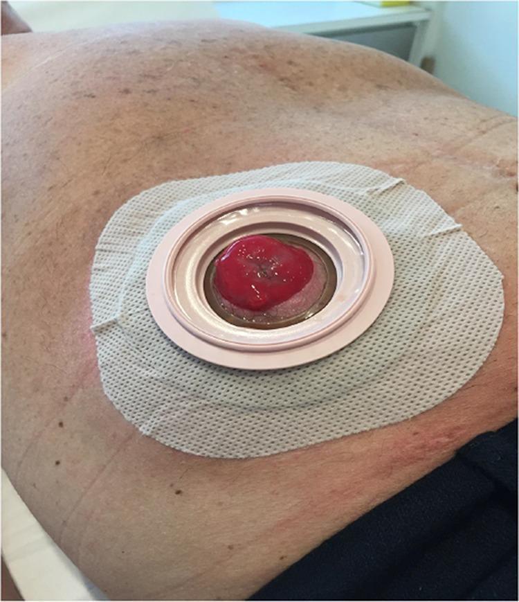
This clinical photograph shows a close-up view of a stoma appliance (wafer or baseplate) securely affixed to a patient's abdomen, 12 months following surgery for stoma prolapse repair. The appliance consists of a square-shaped, white, textured adhesive wafer with rounded corners. At its center is a raised, pale pink circular coupling ring designed to connect to an ostomy bag. The stoma itself is visible through the aperture; it appears healthy, moist, and beefy red in color, indicative of adequate vascularization. The surrounding peristomal skin shows mild erythema and minor irritation near the inferior border of the adhesive, which is common in chronic ostomy management. This image demonstrates the successful long-term maintenance of a stoma after a corrective procedure (such as mesh-reinforced stomal repair) and highlights the typical appearance of a functioning colostomy or ileostomy site with a two-piece pouching system component in place.
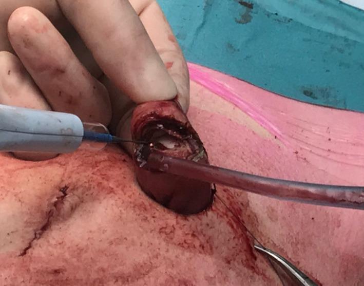
This clinical photograph captures an intraoperative view of a surgical stoma creation (ileostomy or colostomy). The image focuses on the exteriorization and incision of a bowel segment through the abdominal wall. The protruding bowel is reddish-brown and moist, showing a visible staple line from a previous transection. A surgeon's gloved hand stabilizes the tissue while an electrocautery device with a blue cable is used to perform a precision incision at the tip of the exteriorized segment. A clear catheter or suction tube is inserted into the lumen of the bowel to manage contents during the procedure. The surrounding peristomal skin shows signs of surgical preparation, including mild erythema and surgical sutures. A surgical instrument, likely forceps, is visible in the lower right field, assisting in tissue manipulation. This visual illustrates a critical step in stoma maturation, specifically the opening of the bowel wall before full-thickness eversion and fixation to the skin to create a functional spout.
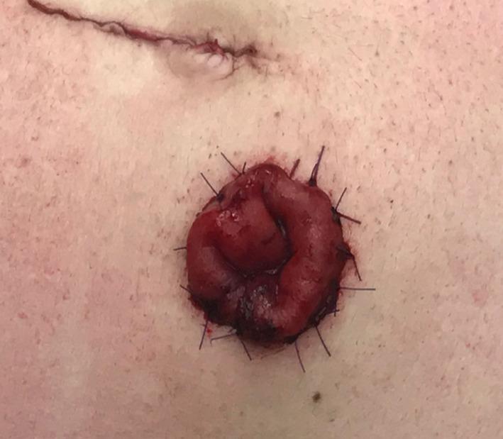
A clinical photograph showing a close-up of a newly created abdominal stoma (likely an ileostomy or colostomy). The stoma is a circular, beefy red, everted spout of bowel tissue protruding from the abdominal wall. It is fixed to the surrounding pale skin at the mucocutaneous junction using multiple interrupted polydioxanone (PDS) sutures, which are visible as dark blue or black threads placed radially. Superior to the stoma, a recent surgical incision scar is visible, appearing as a thin, raised, linear red line. The stoma tissue appears moist and well-vascularized, which is a key clinical indicator of bowel viability post-surgery. This image illustrates the surgical technique of stoma maturation, where the bowel is everted to prevent contact between fecal output and the skin, thereby reducing the risk of parastomal skin irritation and ensuring a proper fit for an ostomy pouching system.
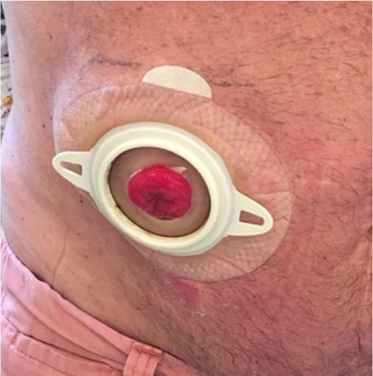
This clinical photograph shows a close-up view of a post-operative abdominal stoma, specifically a colostomy or ileostomy, 28 days following a surgical procedure for stoma prolapse correction. The stoma itself is a healthy, reddish-pink, moist, and rounded protrusion of intestinal mucosa located in the center of the appliance. It is positioned within a white, circular plastic flange of a two-piece ostomy system. The flange is mounted on a beige, textured, skin-protective adhesive wafer (barrier) that adheres to the patient's abdominal skin. The surrounding skin appears mostly intact, with subtle erythema visible near the lower edge of the adhesive patch, possibly indicating minor peristomal skin irritation or post-surgical changes. The image serves as a clinical example of successful stoma management and appliance fitting following the surgical repair of a prolapse using a polypropylene mesh technique.
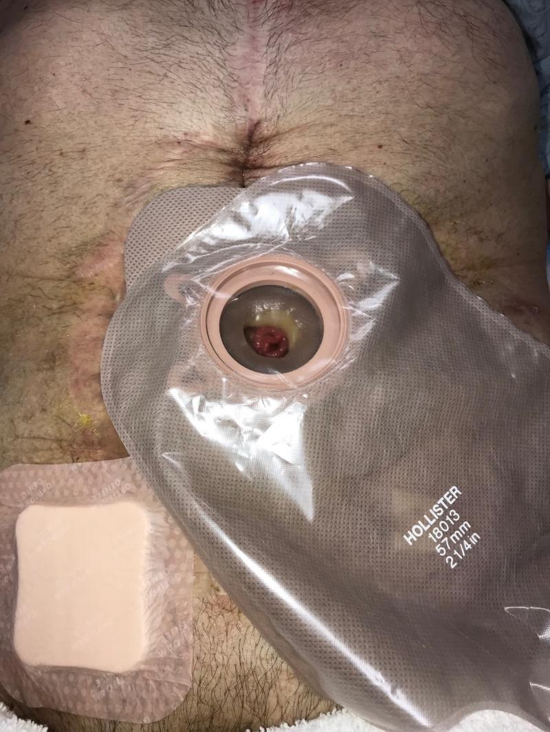
This clinical photograph displays a post-operative adult abdomen showing a healing surgical wound and stoma management. A prominent midline vertical laparotomy scar is visible, extending from the epigastrium toward the umbilical region, indicating a previous major abdominal intervention. Centered on the mid-abdomen is a transparent Hollister (57mm) ostomy appliance containing a visible reddish, fleshy stoma, likely an ileostomy or colostomy, surrounded by some yellowish effluent. To the lower left of the ostomy bag, a square, beige hydrocolloid or foam adhesive dressing is applied over a secondary wound site or skin irritation area. The peristomal skin and surrounding abdominal surface show signs of uneven pigmentation, thinning, and mild erythema, consistent with chronic wound management and previous use of negative pressure wound therapy (NPWT). This image illustrates advanced wound and fistula care in a patient with a history of complex Crohn's disease and abdominal sepsis, demonstrating the transition to post-acute home care management.
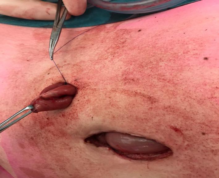
This clinical photograph captures an intraoperative view of a surgical stoma creation, likely an ileostomy or colostomy, following bowel transection. A segment of reddish-pink, viable bowel is being exteriorized through a small circular skin incision in the abdominal wall. Surgical forceps are visible, grasping the tip of the exteriorized bowel segment to facilitate manipulation. A purple monofilament suture, held by a needle driver, is seen at the superior margin of the stoma site, representing the oversewing of a previously stapled line to control the stoma's diameter. Inferior to the primary stoma site, a larger secondary surgical opening reveals a distended loop of bowel beneath the skin surface, illustrating the clinical challenge of creating a stoma from dilated bowel. The surrounding skin exhibits generalized erythema and mottling, consistent with surgical prep and mechanical manipulation. The image demonstrates the 'cone stoma' technique intended to reduce mucocutaneous separation and parastomal complications by limiting the diameter of the exteriorized segment.
Ileostomy and Colostomy: Complete Guide
1. What Are They?
| Feature | Ileostomy | Colostomy |
|---|---|---|
| Bowel used | Terminal ileum (small bowel) | Colon (large bowel) |
| Location | Right iliac fossa (right lower abdomen) | Usually left side of abdomen |
| Stool consistency | Liquid to semi-liquid | Semi-formed to formed |
| Output volume | 500-1500 mL/day | Less, more solid |
| Bag type | Drainable bag (always) | Drainable or non-drainable |
| Electrolyte risk | High - Na, K, bicarbonate loss | Lower risk |
| Spout | Must have a spout 2-3 cm | Flush or slight protrusion |
2. Types
Ileostomy Types
- End (Brooke) ileostomy - Permanent; after proctocolectomy. The terminal ileum is brought through the abdominal wall, everted (spouted 2-3 cm), and sutured to the skin (Brooke technique).
- Loop ileostomy - Temporary; a loop of distal ileum is brought out through the abdominal wall with an enterotomy. Used to protect a distal anastomosis. Can be closed later without a full laparotomy.
- Divided loop ileostomy - A linear stapler divides the distal limb flush with the skin, preventing incomplete diversion that sometimes occurs with a standard loop.
- Continent ileostomy (Kock pouch) - An internal ileal reservoir with a nipple valve; the patient intubates the pouch to drain it. Now rarely done due to high complication rates.
Colostomy Types
- End colostomy - Most common type. End of the colon brought through the abdominal wall. May be paired with a Hartmann's pouch (distal stump left inside) or a mucus fistula.
- Loop colostomy - Less common; a loop of colon is exteriorized. Prone to prolapse, and the bulkiness makes appliance fitting awkward.
- Transverse colostomy - Created in the transverse colon; higher output due to less water absorption.
- Sigmoid/descending colostomy - Preferred when possible; most of the colon remains in circuit, allowing water absorption and producing more formed stool.
- Ascending colostomy - Higher liquid effluent; less commonly used.
3. Indications
Ileostomy Indications
- Proctocolectomy for ulcerative colitis (especially when sphincter preservation is not possible)
- Familial adenomatous polyposis (FAP) with total proctocolectomy
- Colorectal cancer requiring abdominoperineal resection (APR)
- Patient preference after colectomy
- Failed ileo-anal pouch anastomosis (IPAA)
- Low rectal cancer requiring postoperative pelvic radiation
- Protect a low colorectal/ileoanal anastomosis at risk of leakage (in irradiated field, immunocompromised, malnourished patient)
- Emergency operations (e.g., toxic megacolon, perforation)
- Crohn's disease - severe disease requiring bowel rest
- Acute severe ulcerative colitis (ASUC) - total abdominal colectomy with end ileostomy is the operation of choice for urgent cases (toxic megacolon, perforation, medically refractory disease)
Colostomy Indications
- Colorectal cancer with obstruction or perforation
- Diverticular disease - perforated diverticulitis (Hartmann's procedure)
- Trauma to the colon or rectum
- Anorectal injuries and pelvic trauma
- Neonatal anorectal malformations (Hirschsprung's disease)
- Toxic megacolon
- Low rectal cancer requiring APR
- Anal cancer treated with chemoradiation (defunctioning)
- Rectovaginal/enterocutaneous fistulas
- Radiation proctitis with severe bowel dysfunction
- Faecal incontinence unresponsive to other treatment
- Pelvic floor disorders
- Crohn's disease with complex perianal disease
4. The Stoma Bag (Appliance) - Every Detail
Components
- Adhesive sheet that adheres to the peristomal skin
- Has a central hole (aperture) cut to fit the stoma - available in gasket sizes 25-100 mm diameter
- Made of hydrocolloid material (e.g., Stomahesive, Hollister) that protects skin
- Can incorporate convexity to push against a flat/retracted stoma
- Some have karaya gum on the skin side for additional protection
- The bag that collects effluent
- Available in transparent or opaque materials
- Vented or non-vented (vents contain a charcoal filter to reduce odour)
- Immediately post-op: always use a transparent, non-venting bag to inspect stoma viability
One-Piece vs. Two-Piece Systems
| One-Piece | Two-Piece | |
|---|---|---|
| Design | Barrier and bag fused together | Separate flange + bag that snap together |
| Skin contact | Barrier changed with every bag change | Barrier can remain while bag is changed |
| Flexibility | More discreet, flexible | Easier bag changes without disturbing skin |
| Best for | Active patients, low output | High-output stomas, frequent emptying |
Drainable vs. Non-Drainable Bags
- Drainable bag (MANDATORY for ileostomy): Has an open bottom closed with a clip or Velcro mechanism. Left in place for up to 48 hours, emptied 4-6 times/day when 1/3 to 1/2 full.
- Non-drainable bag (suitable for sigmoid colostomy with formed stool): Sealed at the bottom, removed whole to empty, can be reused. Changed 2-3 times per day.
Accessories
| Accessory | Purpose |
|---|---|
| Karaya gum / Stomahesive paste | Fills skin irregularities between stoma and flange; prevents leakage |
| Skin barrier powder | Treats peristomal skin irritation |
| Skin sealant/protector | Applied over powder to ensure good seal |
| Adhesive remover | Safely removes the barrier without skin trauma |
| Deodorizers (powder or spray) | Reduces pouch odour |
| Charcoal filter | Allows gas to escape from the bag without odour |
| Stoma belt / hernia belt | Provides extra appliance security, especially with parastomal hernia |
| Irrigation kit | Used for colostomy irrigation - warm water through a cone catheter regulates output timing |
| Cover bags | Hide the disposable bag under clothing |
| Convex inserts | For flush or retracted stomas |
5. Stoma Siting (Preoperative Marking)
- Marked preoperatively (ideally by a stoma care nurse)
- Within the rectus abdominis muscle (muscle-splitting aperture)
- Away from the umbilicus, bony prominences (anterior superior iliac spine), skin folds, scars, and the waistline
- Ileostomy: Right iliac fossa, just medial to the lateral border of the rectus muscle
- Transverse colostomy: Upper right quadrant
- Visible to the patient (they must be able to see it to manage it)
- Confirmed in sitting and standing positions
6. Physiology
Ileostomy Physiology
- Normal daily output: 500-750 mL (adapted); volumes >1000 mL are abnormal
- Effluent contains proteolytic enzymes that will digest skin if it leaks
- Electrolyte composition: Na 110-120 mmol/L, K 6-12 mmol/L, Cl 40-70 mmol/L, HCO₃ 30-40 mmol/L
- The colon's water-absorbing function is bypassed - patients are chronically at risk of dehydration and electrolyte imbalance
- Adaptation occurs over weeks-months, reducing output somewhat
Colostomy Physiology
- Output depends on location: ascending > transverse > descending/sigmoid
- Left-sided colostomies: most colon in circuit, allowing water absorption, semi-formed to formed stool
- Dehydration risk is much lower than ileostomy
- Skin irritation less common (formed stool is less corrosive than succus entericus)
7. Complications
Early Complications (within days-weeks)
| Complication | Ileostomy | Colostomy | Notes |
|---|---|---|---|
| Necrosis | Yes | Yes | Due to skeletonized bowel or tight fascial defect. Suprafascial: observe. Subfascial: urgent surgery |
| Retraction | Yes (significant) | Yes (less problematic) | Worsened by obesity. May require local revision |
| High output/dehydration | Very common | Rare | Ileostomy output >1500 mL/day; treat with loperamide, Lomotil, octreotide |
| Skin irritation | Common | Less common | Due to leakage of effluent; treat with barrier powder + sealant |
| Bleeding | Yes | Yes | Usually minor mucosal bleeding |
| Mucocutaneous separation | Yes | Yes | Separation of stoma from skin sutures |
Late Complications
| Complication | Details |
|---|---|
| Parastomal hernia | Most common late complication. More common with colostomy. Causes appliance fitting problems, pain, obstruction, strangulation. Simple suture repair = ~100% recurrence. Requires mesh repair, laparoscopic repair, or stoma resiting. Prophylactic mesh at creation reduces hernia risk by ~40% (based on 12 RCTs). |
| Prolapse | Rare; more common with loop colostomy (usually efferent limb). Associated with parastomal hernia |
| Stenosis | Narrowing at skin or fascial level; may need dilatation or revision |
| Fistulation | Tract forms beside the stoma; usually requires surgical correction |
| Food blockage (ileostomy) | Fibrous/poorly chewed food lodges in distal ileum; presents as colicky pain with decreased or watery output. Treat: supine/knee-chest position, abdominal massage, IV hydration, NGT, stoma irrigation |
| Pouchitis | Specific to continent ileostomy/IPAA; bacterial overgrowth or recurrent IBD in ileal reservoir; treat with metronidazole, probiotics, mesalamine |
| Peristomal skin problems | Most common overall complication: eczema, irritant contact dermatitis - 75% of ileostomy patients have some sensitivity postoperatively |
Ileostomy-Specific Complications Summary
- Stoma necrosis, retraction, dehydration with electrolyte abnormalities, skin irritation, obstruction (at stoma or intra-abdominal), parastomal hernia, prolapse
Colostomy-Specific Complications Summary
- Necrosis, retraction, parastomal hernia (most common late complication), prolapse (more so with loop), obstruction (unusual)
8. Peristomal Skin Care
- The appliance is removed by lifting the edge while pushing down on the skin (not pulling away)
- Use warm water + adhesive remover - prevents skin injury and removes residue fully
- Wash skin with warm water and soap; dry thoroughly
- Avoid products with alcohol or oil on the peristomal skin
- Peristomal hair should be shaved or clipped (not waxed)
- Cut the appliance aperture to exactly fit the stoma - even a few mm gap allows effluent to contact skin
- Treat irritated skin with skin barrier powder, then apply skin sealant over the powder to ensure a proper seal
9. Diet and Lifestyle
Immediate Post-op (first 2-6 weeks):
- Low-residue diet
- Chew food well, cut into small pieces
- Avoid high-fibre foods that can cause blockage
Long-term Diet Guide:
| Goal | Foods |
|---|---|
| Thicken output | Pasta, rice, crackers, cheese, yogurt, applesauce, bananas, potatoes, peanut butter, tapioca |
| Increase output (avoid if high output) | Prunes, raisins, leafy vegetables, spicy foods, caffeine, alcohol, chocolate |
| Reduce gas (avoid) | Beans, lentils, cabbage, broccoli, Brussels sprouts, cauliflower, onions, carbonated drinks, chewing gum |
Hydration (Ileostomy):
- Normal ileostomy emptied 4-6 times/day; total output ~1 L/24 hours
- Output >1 L/day = high output; at risk for dehydration
- Use electrolyte-rich drinks (sports drinks), increase thickening foods
- Medications: loperamide, diphenoxylate; rarely codeine or tincture of opium
Colostomy Irrigation:
- Patients with sigmoid/descending colostomies can regulate output timing
- Done with warm tap water through a cone catheter into the stoma opening
- Allows some patients to go without a bag between irrigations, using just a cover
10. Medications and the Ostomy
- Enteric-coated and sustained-release tablets may pass through an ileostomy unabsorbed - prefer liquid or standard-release formulations
- Antidiarrheal agents (loperamide, codeine, tincture of opium) are used therapeutically to slow ileostomy output
- NSAIDs and steroids can worsen mucosal inflammation
- Antibiotics can disrupt gut flora and increase output
- Iron supplements may cause black stools and odour
- Contraceptive pill absorption may be unreliable with high-output ileostomy
11. Psychological and Quality of Life Aspects
- Most patients accept their stoma well; studies consistently show high quality of life post-ileostomy, particularly after surgery for ulcerative colitis or cancer
- Only a small proportion have significant psychological difficulties
- Body image concerns, sexual dysfunction, clothing limitations, and social anxiety about odour are documented issues
- Preoperative counselling and marking by a stoma care nurse significantly improves outcomes
- Support groups and stoma care services (available in most hospitals) provide practical and psychological help
- Long-term quality of life after continent ileostomy (Kock pouch) is similar to Brooke ileostomy or IPAA
12. Closure (Reversal) of Temporary Stomas
Loop Ileostomy Closure:
- Elliptical incision around the stoma; bowel freed from subcutaneous tissue and fascia
- Hand-sewn or stapled anastomosis created and intestine returned to peritoneal cavity
- Usually avoids laparotomy
- Before closure: flexible endoscopy + Gastrografin contrast enema to confirm anastomotic patency and rule out leak
- Optimise nutritional status first
- In cancer patients on adjuvant chemotherapy, usually defer until chemotherapy is completed (timing affects survival)
End Colostomy Closure (Hartmann's reversal):
- Traditionally requires laparotomy
- Stoma dissected free, distal bowel (Hartmann's stump) identified
- End-to-end anastomosis created
- Increasingly done with minimally invasive (laparoscopic) techniques
Clinical Images
Summary Table: Ileostomy vs. Colostomy
| Ileostomy | Colostomy | |
|---|---|---|
| Bowel | Terminal ileum | Colon |
| Position | Right iliac fossa | Usually left side |
| Spout required | Yes (2-3 cm) | 0.5-1 cm or flush |
| Output | Liquid, 500-1500 mL/day | Semi-formed to solid |
| Bag type | Drainable only | Drainable or non-drainable |
| Bag change | Every 48 hours + drain | 2-3 times/day (non-drain) |
| Dehydration risk | High | Low |
| Skin risk | High (proteolytic enzymes) | Lower |
| Main indications | UC, FAP, low rectal ca, protect anastomosis | Colorectal ca, diverticular perf, trauma, anorectal malformation |
| Most common late complication | Parastomal hernia | Parastomal hernia |
| Reversible | Often | Depends on reason |
| Irrigation possible | No | Yes (sigmoid/descending) |