
Pyq about larynx and pharynx in kuhs
Reading File
Searching the Web
KUHS previous year questions larynx pharynx anatomy
Reading Web Page
https://www.scribd.com/document/970511373/KUHS-MBBS-Chapter-…
Finding Sources
Finding Sources
Now let me read the key anatomy sections from Gray's Anatomy for Students (most relevant for KUHS MBBS) for both larynx and pharynx:
Reading File
Reading File
Reading File
Finding Sources
Reading File
Reading File
Reading File
Reading File
Finding Sources
Now I have enough detailed content from Gray's Anatomy for Students to compile a thorough KUHS PYQ-style answer. Let me also fetch one key image.
Loading Image
Loading Image
I now have comprehensive content to give a detailed KUHS PYQ-style answer. Here is the full compiled response:
KUHS PYQ - Larynx & Pharynx (Anatomy)
These are the most frequently asked topics from KUHS (Kerala University of Health Sciences) first MBBS Anatomy papers on larynx and pharynx, with complete exam-ready answers.
PHARYNX
Q1. Describe the pharynx under the headings: extent, subdivisions, muscles, nerve supply, and blood supply. (10 marks)
Definition & Extent
The pharynx is a musculofascial half-cylinder that links the oral and nasal cavities in the head to the larynx and esophagus in the neck. It is attached above to the base of the skull and is continuous below, approximately at the level of vertebra CVI, with the top of the esophagus.
Subdivisions - based on anterior relations, the pharynx is divided into three parts:
| Part | Anterior relation | Communication |
|---|---|---|
| Nasopharynx | Posterior choanae of nasal cavities | Pharyngotympanic (Eustachian) tubes open into its lateral walls |
| Oropharynx | Posterior opening of oral cavity (oropharyngeal isthmus) | Contains palatine and lingual tonsils |
| Laryngopharynx | Superior aperture of larynx (laryngeal inlet) | Continuous below with esophagus at CVI |
The pharyngeal cavity is separated from the posteriorly positioned vertebral column by a thin retropharyngeal space containing loose connective tissue.
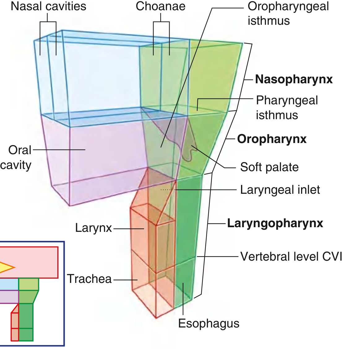
Pharynx showing Nasopharynx, Oropharynx, and Laryngopharynx - Gray's Anatomy for Students
Tonsil Ring (Waldeyer's Ring)
- Lingual tonsil (base of tongue)
- Palatine tonsils (oropharynx lateral walls)
- Pharyngeal tonsil/adenoids (nasopharynx posterior wall)
Soft Palate - "flutter valve"
- Swings up (elevates) to close the pharyngeal isthmus - seals nasopharynx from oropharynx
- Swings down (depresses) to close the oropharyngeal isthmus - seals the oral cavity from the pharynx
Q2. What are the constrictor muscles of the pharynx? Describe their origin, insertion, action, and nerve supply. (5 marks) (Very commonly asked)
The pharyngeal wall consists of three overlapping constrictor muscles:
| Muscle | Origin | Insertion | Action |
|---|---|---|---|
| Superior constrictor | Pterygoid hamulus, pterygomandibular raphe, posterior end of mylohyoid line, side of tongue | Pharyngeal raphe (posterior midline) | Constricts upper pharynx during swallowing |
| Middle constrictor | Greater and lesser horns of hyoid | Pharyngeal raphe | Constricts middle pharynx |
| Inferior constrictor | Oblique line of thyroid cartilage, cricoid cartilage | Pharyngeal raphe | Constricts lower pharynx; lowest fibers (cricopharyngeus) act as upper esophageal sphincter |
Nerve supply: All three constrictors are supplied by the pharyngeal plexus (CN IX + CN X + sympathetics from superior cervical ganglion). Motor supply is via CN X (vagus).
Clinical note: The gap between the superior constrictor and the base of the skull is where the Eustachian tube, levator palati, and ascending palatine artery pass through.
Q3. Piriform fossa - notes (Short note, 3 marks)
- A recess in the laryngopharynx on either side of the laryngeal inlet
- Bounded medially by the aryepiglottic fold and laterally by the thyroid cartilage and thyrohyoid membrane
- The internal branch of the superior laryngeal nerve runs deep to the mucosa of the piriform fossa - this is why local anesthesia injected here can block the laryngeal inlet
- Foreign bodies frequently lodge here
LARYNX
Q4. Describe the larynx under the headings: cartilages, membranes/ligaments, muscles, nerve supply, and blood supply. (10 marks) (Most high-value question)
Introduction
The larynx is a hollow musculoligamentous structure with a cartilaginous framework that caps the lower respiratory tract. It functions as:
- A valve/sphincter to protect the lower respiratory tract
- A sound-producing instrument
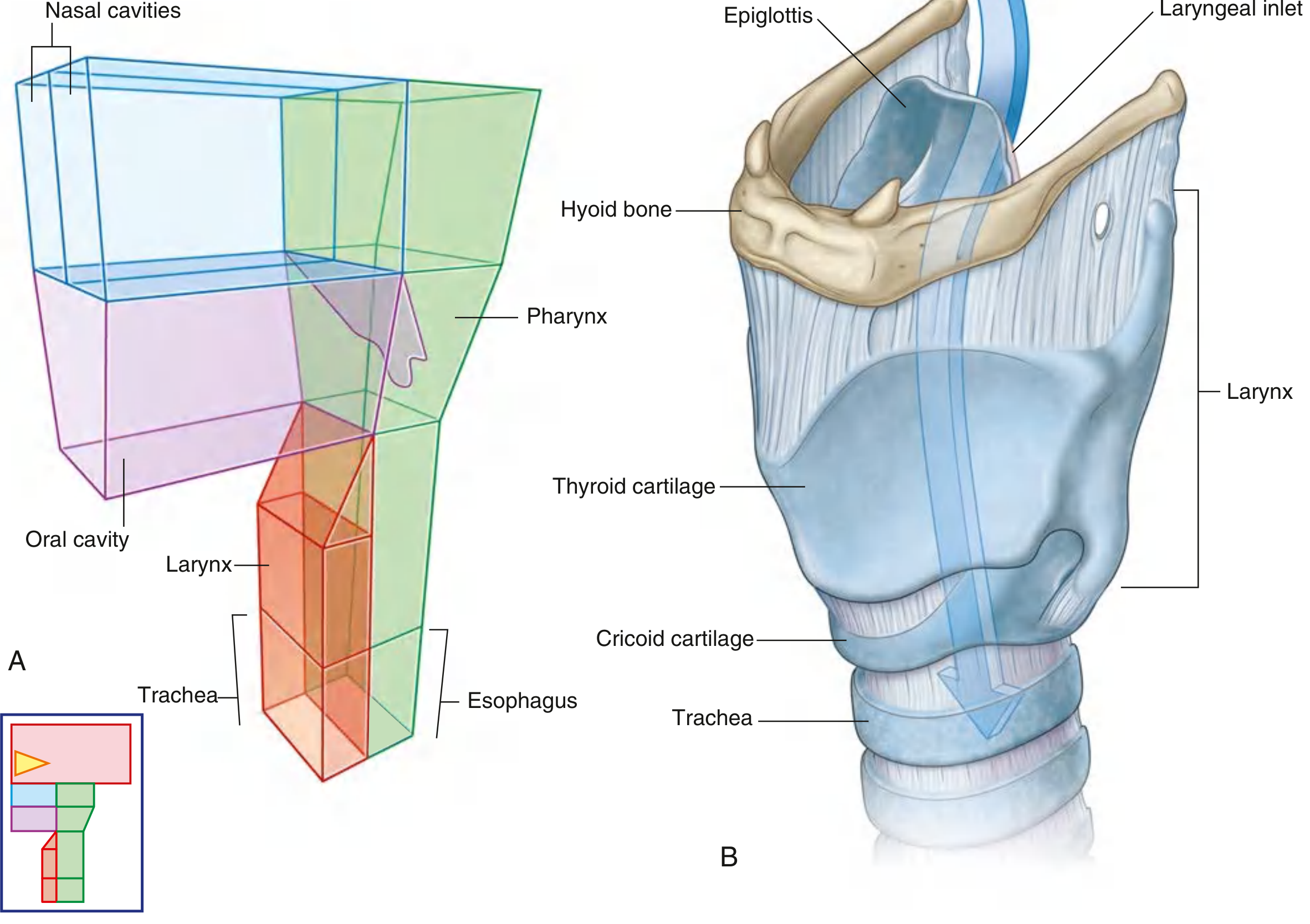
Larynx showing cartilages, hyoid bone, and trachea - Gray's Anatomy for Students
Cartilages of the Larynx
Three unpaired cartilages:
- Thyroid cartilage - largest; two laminae meet anteriorly at the laryngeal prominence (Adam's apple); angle more acute in males (90°) than females (120°); superior and inferior horns; oblique line on outer surface for attachment of sternothyroid, thyrohyoid, and inferior constrictor
- Cricoid cartilage - only complete ring encircling the airway; shaped like a signet ring with a broad lamina posteriorly and narrow arch anteriorly; articulates with arytenoid cartilages (superolateral surface of lamina) and inferior horn of thyroid cartilage
- Epiglottis - leaf-shaped elastic cartilage; attached by petiolus to the angle of the thyroid cartilage; free upper edge faces the pharynx; closes the laryngeal inlet during swallowing
Three paired cartilages:
4. Arytenoid - pyramid-shaped; base articulates with cricoid; vocal process anteriorly (for vocal ligament), muscular process laterally (for arytenoid muscles)
5. Corniculate - sit on apices of arytenoids; form corniculate tubercles
6. Cuneiform - in aryepiglottic folds; form cuneiform tubercles
Membranes and Ligaments
Extrinsic:
- Thyrohyoid membrane - spans thyroid cartilage to hyoid bone; has aperture on each side for superior laryngeal artery, internal branch of superior laryngeal nerve, and lymphatics; thickened median part = median thyrohyoid ligament; thickened posterior borders = lateral thyrohyoid ligaments; may contain triticeal cartilage
- Cricothyroid ligament (membrane) - connects cricoid to thyroid; site of emergency cricothyrotomy
Intrinsic:
- Quadrangular membrane - from arytenoid cartilages to lateral epiglottis; free inferior margin = vestibular ligament (false vocal cord)
- Conus elasticus (cricovocal membrane) - from arch of cricoid to vocal processes; free superior margin = vocal ligament (true vocal cord)
Cavity of the Larynx - Three Regions
Divided by two pairs of folds (vestibular folds above, vocal folds below):
- Vestibule - from laryngeal inlet to vestibular folds (false cords); contains vestibular ligaments
- Middle chamber - between vestibular folds and vocal folds; opens laterally into the laryngeal ventricle (sinus); the laryngeal saccule/appendix extends upward from the anterior part of the ventricle
- Infraglottic space - from vocal folds to inferior opening of larynx (continuous with trachea); completely encircled by cricoid cartilage
Rima glottidis = the gap between the two vocal folds; widest during deep inspiration, narrowed during phonation; largest opening in upper airway
Intrinsic Muscles of the Larynx
All intrinsic muscles are supplied by the recurrent laryngeal nerve (branch of vagus X), EXCEPT the cricothyroid which is supplied by the external branch of the superior laryngeal nerve.
| Muscle | Action |
|---|---|
| Posterior crico-arytenoid (PCA) | Only abductor of vocal folds (opens rima glottidis) |
| Lateral crico-arytenoid | Adducts vocal folds |
| Transverse arytenoid | Adducts arytenoids (closes posterior rima) |
| Oblique arytenoid | Narrows laryngeal inlet; continues as ary-epiglottic muscle |
| Cricothyroid | Tenses/elongates vocal folds (raises pitch) |
| Thyro-arytenoid (including vocalis) | Relaxes/shortens vocal folds; sphincter of vestibule |
| Vocalis | Fine tension adjustment of vocal fold |
| Thyro-epiglottic | Widens laryngeal inlet |
Key KUHS MCQ: Posterior crico-arytenoid is the ONLY abductor - its paralysis (bilateral) causes adduction of cords and respiratory obstruction.
Nerve Supply of the Larynx
Motor and sensory supply is via the vagus nerve [X] through two branches:
-
Superior laryngeal nerve (SLN)
- Internal branch (sensory) - enters through thyrohyoid membrane; sensory to mucosa above the vocal folds; also supplies piriform fossa - used for topical anesthesia
- External branch (motor) - supplies cricothyroid muscle only
-
Recurrent laryngeal nerve (RLN)
- Motor to all intrinsic muscles except cricothyroid
- Sensory to mucosa below vocal folds
- Right RLN hooks around right subclavian artery; Left RLN hooks around arch of aorta (longer course)
- Enters larynx at the cricothyroid joint, in groove between trachea and esophagus
Clinical significance of RLN injury:
- Unilateral injury - hoarseness (cord paralyzed in paramedian position)
- Bilateral injury - respiratory obstruction (both cords adduct)
- Causes: thyroidectomy, carcinoma of thyroid, aortic arch aneurysm (left RLN)
Blood Supply of the Larynx
| Artery | Origin | Entry |
|---|---|---|
| Superior laryngeal artery | Superior thyroid artery (branch of external carotid) | Accompanies internal branch of SLN through thyrohyoid membrane |
| Inferior laryngeal artery | Inferior thyroid artery (from thyrocervical trunk of subclavian) | Accompanies recurrent laryngeal nerve; supplies lower larynx |
Venous drainage mirrors the arteries - to superior and inferior thyroid veins.
Q5. Short notes frequently asked:
Cricothyroid joint - synovial joint between inferior horn of thyroid and lateral surface of cricoid lamina; allows rotation (tilts thyroid forward to increase AP distance between thyroid angle and arytenoid cartilages, thus tensing vocal folds) and gliding movements.
Crico-arytenoid joint - synovial joint between base of arytenoid and superolateral facet of cricoid lamina; allows arytenoids to slide medially/laterally and rotate so vocal processes pivot toward/away from midline - enables abduction and adduction.
Laryngoscopy - procedure to inspect larynx:
- Indirect laryngoscopy: rod-mounted mirror in oropharynx (in conscious patient)
- Direct laryngoscopy: curved laryngoscope blade to displace tongue and epiglottis forward; requires unconscious patient or absent gag reflex
- Fiber-optic nasolaryngoscopy: flexible scope via nasal cavity
Tracheostomy - incision in 2nd and 3rd tracheal rings; performed for laryngeal obstruction (foreign body, anaphylaxis, trauma); strap muscles retracted laterally; thyroid isthmus may need to be divided; long-term tracheostomy patients cannot vocalize (air bypasses vocal folds).
Subglottic space - clinically important in children: the subglottic larynx is the narrowest part of the pediatric airway (unlike in adults where the rima glottidis is the narrowest). Edema here (croup/laryngotracheobronchitis) causes severe stridor.
Q6. Applied/Clinical Questions (asked as short notes or case-based)
Q. A patient presents with hoarseness following thyroidectomy. Explain anatomically.
- During thyroidectomy, the recurrent laryngeal nerve (RLN) is at risk near the cricothyroid joint where it enters the larynx
- Unilateral RLN damage: ipsilateral vocal cord paralyzed in paramedian position - hoarseness
- Left RLN more commonly injured due to its longer course around the aortic arch
Q. How does the larynx protect the airway during swallowing?
- Elevation and anterior movement of larynx under tongue
- Epiglottis tilts down over laryngeal inlet
- Aryepiglottic folds constrict (oblique arytenoid + thyro-epiglottic muscles)
- Thyro-arytenoid muscles act as vestibular sphincter
- True vocal folds adduct (lateral crico-arytenoid + transverse arytenoid)
Q. Where do foreign bodies commonly lodge in the pharynx?
- Piriform fossa (most common in adults) - foreign body lodges on either side of laryngeal inlet
- Valleculae (between tongue base and epiglottis)
- Posterior pharyngeal wall (in children)
Quick Revision Table - Nerve Supply (Frequently tested in KUHS)
| Structure | Nerve |
|---|---|
| All intrinsic laryngeal muscles | Recurrent laryngeal nerve (CN X) |
| Cricothyroid only | External branch of SLN (CN X) |
| Mucosa above vocal folds | Internal branch of SLN (sensory) |
| Mucosa below vocal folds | Recurrent laryngeal nerve (sensory) |
| All pharyngeal muscles (motor) | CN X via pharyngeal plexus |
| Stylopharyngeus | CN IX (glossopharyngeal) - ONLY muscle |
| Sensation to pharynx | CN IX (pharyngeal plexus) |
Source: Gray's Anatomy for Students, 4e - Chapter 8 (Head and Neck)
This is a shared conversation. Sign in to Orris to start your own chat.