
Make notes on “occular syphilis” for md derma theory exam in a easy to remember pattern with preferable flow charts if needed
ocular syphilis uveitis fundus
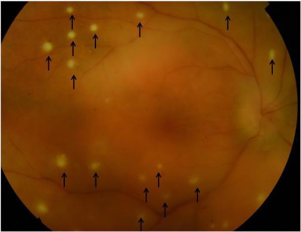
This fundus color photograph illustrates clinical manifestations of posterior syphilitic uveitis. The image shows widespread, small, circular, whitish-yellowish pre-retinal lesions (indicated by black arrows) scattered across the retina. These punctate lesions, sometimes referred to as pre-retinal precipitates, are seen overlying areas of active retinitis and often represent aggregates of inflammatory cells. A generalized haziness is visible across the fundus, characteristic of vitritis (inflammation of the vitreous humor), which obscures the clarity of the underlying retinal vessels and landmarks. The optic disc is visible in the nasal periphery with relatively preserved margins. This presentation is a classic hallmark of ocular syphilis, often termed 'the great mimicker,' where inflammatory cells migrate across the retina into the vitreous. This visual material serves as an educational resource for identifying inflammatory ocular phenotypes and distinguishing pre-retinal precipitates from intra-retinal foci in the context of infectious uveitis.
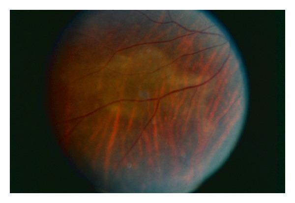
This clinical fundus photograph depicts the right eye of a patient with Acute Posterior Placoid Polymorphous Syphilitic Chorioretinitis (ASPPC), a rare ocular manifestation of secondary syphilis. The image demonstrates a characteristic large, circular, yellowish-white, placoid lesion at the level of the retinal pigment epithelium (RPE) and inner choroid, centrally located within the posterior pole. The surrounding retina displays a reddish-orange hue with visible retinal vasculature. Notably, several retinal vessels traversing the central lesion appear attenuated or partially obscured by the overlying inflammatory process. In the lower half of the fundus, there are linear, reddish streaks and subtle pigmentary changes radiating toward the periphery. The image illustrates the classic 'placoid' variant of syphilitic uveitis, emphasizing the importance of screening for Treponema pallidum in patients presenting with atypical posterior uveitis or placoid chorioretinal lesions.
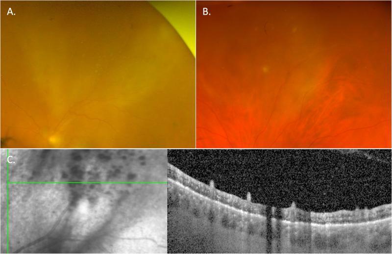
This composite figure illustrates the multimodal clinical presentation of ocular syphilis, specifically punctate inner retinitis.
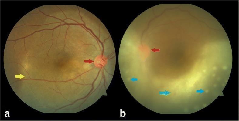
This clinical imaging composite presents two color fundus photographs (a and b) illustrating ocular manifestations of ocular syphilis in a patient with HIV. Image (a) displays the right eye with a hyperemic optic disc (indicated by a red arrow) and a large, creamy-yellow, plaque-like lesion in the posterior pole consistent with posterior placoid retinochoroiditis (yellow arrow). The lesion exhibits a ground-glass appearance. Image (b) shows the left eye with significant media haze caused by dense vitritis (indicated by a green arrow), which obscures clear visualization of retinal details. Despite the haze, a hyperemic optic disc is visible (red arrow), along with multiple, small, punctate yellowish lesions scattered across the posterior pole, identified as superficial retinal precipitates (blue arrows). These clinical findings are characteristic of syphilitic uveitis and retinitis. The imagery demonstrates key diagnostic features including disc edema, active chorioretinitis, and inflammatory vitreous debris, which are critical for the clinical assessment of multi-system infectious diseases.
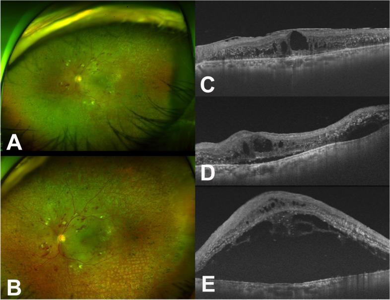
This composite figure displays fundus photography and optical coherence tomography (OCT) of a patient's left eye, illustrating the progression of diabetic retinopathy complicated by syphilitic uveitis. Panels A and B are wide-field fundus photographs showing multiple retinal dot-and-blot hemorrhages, cotton-wool spots, and laser photocoagulation scars. Panel B, taken seven days post-vitrectomy, reveals an increasingly edematous macula compared to the baseline in A. Panels C, D, and E show corresponding OCT cross-sections. Image C (pre-vitrectomy) demonstrates a thickened epiretinal membrane (ERM) and significant intraretinal fluid (IRF) with cystic changes. Image D (post-operative day 1) shows anatomical alterations following ERM peeling. Image E (post-operative day 7) reveals a dramatic progression to massive subretinal fluid (SRF), indicated by a large hyporeflective space beneath the neurosensory retina, which coincided with high VDRL titers. The series illustrates how surgical intervention can trigger or coincide with an acute inflammatory/infectious exacerbation (ocular syphilis) in the setting of existing retinal vascular disease.
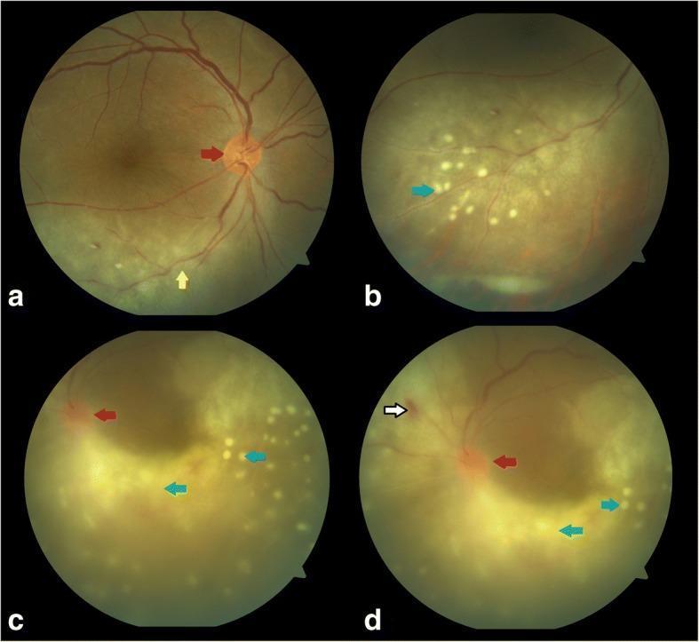
Clinical color fundus photographs of the right (a, b) and left (c, d) eyes demonstrating bilateral ocular syphilis and HIV-related posterior segment inflammation. The right eye (a, b) shows optic disc hyperemia (red arrow) and a widespread area of ground-glass retinochoroiditis (yellow arrow) in the inferior retina. Higher magnification (b) reveals numerous small, discrete, cream-colored superficial retinal precipitates (blue arrows) along the inferotemporal arcade, characteristic of acute syphilitic posterior placoid chorioretinitis. The left eye (c, d) demonstrates more severe inflammation with dense vitritis (green arrow) causing significant haze. Notable findings include a hyperemic optic disc (red arrow), a focal intraretinal hemorrhage superonasal to the disc (white arrow), and scattered superficial retinal precipitates (blue arrows) throughout the posterior pole. The images illustrate key diagnostic features of syphilitic uveitis and retinitis in the context of immunocompromise, highlighting the variation in severity and morphology between eyes, including placoid lesions, vitritis, and multifocal inflammatory precipitates.
"ocular syphilis" diagnosis treatment
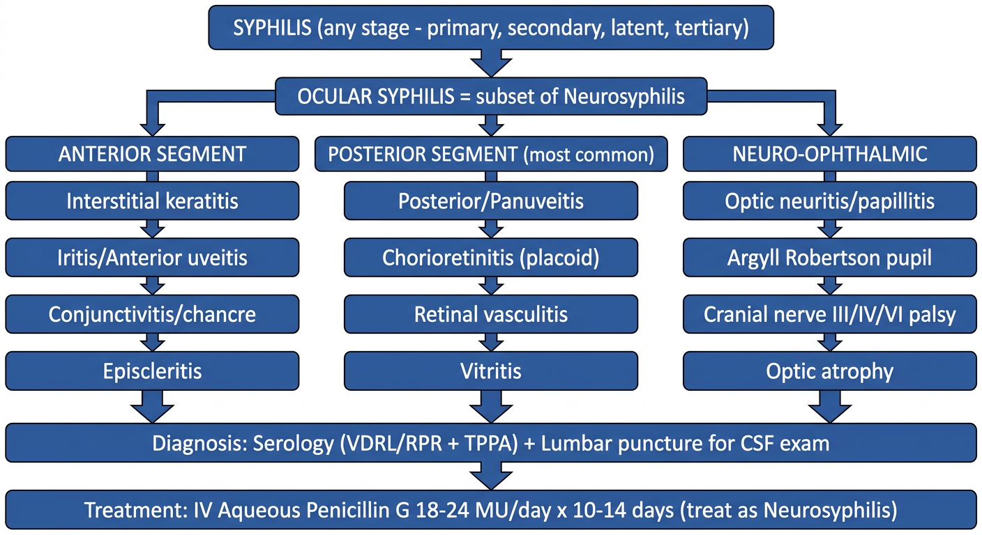
A clean medical education flowchart diagram for "Ocular Syphilis" with the following structure: Top box "SYPHILIS (any stage - primary, secondary, latent, tertiary)" with arrow down to "OCULAR SYPHILIS = subset of Neurosyphilis". From there branch into three columns: (1) "ANTERIOR SEGMENT" with boxes: Interstitial keratitis, Iritis/Anterior uveitis, Conjunctivitis/chancre, Episcleritis; (2) "POSTERIOR SEGMENT (most common)" with boxes: Posterior/Panuveitis, Chorioretinitis (placoid), Retinal vasculitis, Vitritis; (3) "NEURO-OPHTHALMIC" with boxes: Optic neuritis/papillitis, Argyll Robertson pupil, Cranial nerve III/IV/VI palsy, Optic atrophy. Below all three, a merging arrow down to a diagnosis box "Diagnosis: Serology (VDRL/RPR + TPPA) + Lumbar puncture for CSF exam" then arrow down to final box "Treatment: IV Aqueous Penicillin G 18-24 MU/day x 10-14 days (treat as Neurosyphilis)". Use a clean flowchart style with rectangular boxes, arrows, medical blue and white color scheme, clear readable text labels, organized top-to-bottom tree layout suitable for a medical exam study note.
Ocular Syphilis — Exam Notes (MD Derma)
1. One-Line Definition
2. Quick Flowchart (visual summary)
3. Key Facts to Anchor First (highest yield)
| Fact | Detail |
|---|---|
| Can occur | At any stage of syphilis |
| Most common ocular finding | Panuveitis / posterior uveitis |
| Laterality | Bilateral in 50-70% |
| CSF abnormality (neurosyphilis) coexists in | Up to 70% of ocular syphilis cases |
| Category | Treated as a subset of neurosyphilis regardless of CSF findings |
| "The great mimicker" | Because it mimics almost every uveitic pattern - autoimmune, viral, TB |
4. Clinical Features - Organized by Eye Segment (mnemonic: "Front to Back to Nerve")
- Chancre/gumma of conjunctiva
- Conjunctivitis, scleroconjunctivitis
- Interstitial keratitis
- Anterior uveitis/iritis
- Eyelid chancre or condyloma lata (adjacent, not true ocular disease)
- Posterior uveitis / panuveitis (classic)
- Chorioretinitis - including the pathognomonic acute syphilitic posterior placoid chorioretinitis (ASPPC): large yellowish placoid lesion at RPE/inner choroid
- Retinal vasculitis, vitritis ("punctate inner retinitis")
- Retinal detachment, cataract, glaucoma (complications)
- Optic nerve: papillitis, retrobulbar neuritis, optic atrophy, optic nerve gumma
- Argyll Robertson pupil (accommodates but doesn't react to light - classic tabes dorsalis/late neurosyphilis sign)
- III, IV, VI cranial nerve palsies (from syphilitic basilar meningitis)
- Chiasmal syndrome -> bitemporal hemianopia (gumma/meningitis compressing chiasm)
- Stroke syndromes from meningovascular syphilis affecting visual pathways
5. Classic Image to Remember (for spotters)
6. Diagnostic Algorithm (flowchart logic - CDC/Harrison's based)
Patient with syphilis (any stage) + eye symptoms (blurred vision, floaters, pain, red eye)
        |
        v
Serology: nontreponemal (VDRL/RPR) + treponemal (TPPA/FTA-ABS) --> confirm syphilis
        |
        v
Thorough OCULAR exam + full CRANIAL NERVE exam
        |
        v
   Cranial nerve dysfunction present?
   /                              \
 YES                               NO
  |                                 |
  v                                 v
LP for CSF exam MANDATORY      LP not mandatory
  |                                 |
  v                                 v
CSF: cells >5/uL, protein >45,   Still treat as
CSF-VDRL reactive (specific       NEUROSYPHILIS
but insensitive)                  regardless of CSF
  |
  v
Confirms neurosyphilis
7. Treatment (write this as a table - commonly asked directly)
| Regimen | Dose |
|---|---|
| Recommended | Aqueous crystalline Penicillin G 3-4 million units IV q4h (18-24 million units/day) x 10-14 days |
| Alternative | Procaine penicillin 2.4 MU IM daily + Probenecid 500 mg PO QID, both x 10-14 days |
| Penicillin allergy | Ceftriaxone 2 g IM/IV daily x 10-14 days, or desensitize and give penicillin |
8. Special Associations (rapid recall points)
- HIV co-infection: strongly linked; check HIV status in every ocular syphilis case; incidence of ocular/neurosyphilis rising in HIV+ and MSM populations
- RPR titer ≥1:32 or CD4 ≤350/µL in HIV+ patients -> higher risk of neurosyphilis (11-fold in HIV+, 6-fold in HIV-negative) - useful cutoff to remember
- 2014-2015 outbreak reported in San Francisco/Seattle with permanent vision loss in some - historically cited outbreak, sometimes asked in epidemiology questions
- Otosyphilis is the "ear equivalent" - but unlike ocular syphilis, >90% of otosyphilis has NORMAL CSF (key contrast point for exams)
9. One-Line Mnemonic to Remember Everything
- Any Stage = can present at primary/secondary/latent/tertiary syphilis
- Any Segment = anterior, posterior (most common), or neuro-ophthalmic
- Always treat as Neuro = IV penicillin regimen regardless of CSF result