
Csf rhinnorhea
"CSF rhinorrhea" diagnosis management
CSF rhinorrhea skull base defect repair endoscopic
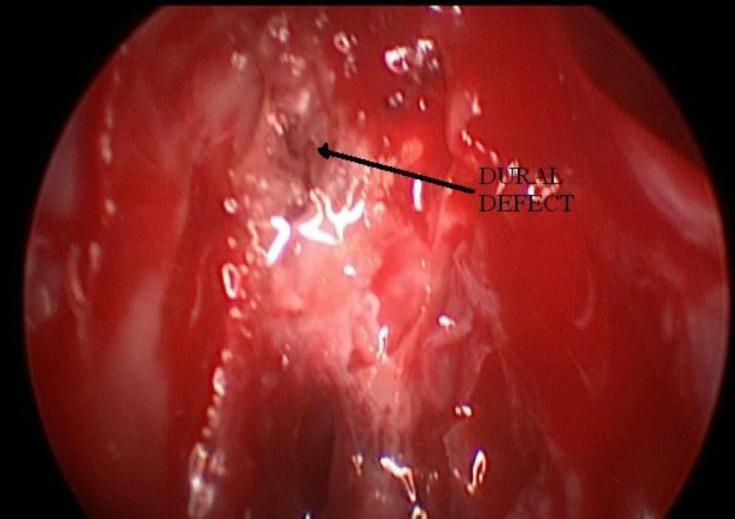
An intraoperative diagnostic nasal endoscopy image showing the skull base during a cerebrospinal fluid (CSF) rhinorrhea repair procedure. The view depicts highly vascular, erythematous (bright red) nasal mucosa and ethmoidal tissue. An arrow points to a centrally located 'Dural Defect', which appears as a small, irregular, dark brownish-black focal point within the surrounding inflamed tissue. White, translucent areas of possible CSF drainage or mucosal reflections are visible around the site of the leak. The image illustrates the localization of a dural breach, essential for subsequent endoscopic repair using fat or fascia lata grafts. This clinical photograph is used to demonstrate surgical anatomy and the visual presentation of skull base defects in neuro-otolaryngology.
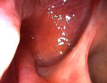
This endoscopic clinical photograph displays a postoperative view of the skull base 10 months following the repair of a clival cerebrospinal fluid (CSF) leak. The image highlights a successfully healed surgical site, showing the results of a multi-layer closure technique that utilized an onlay bovine pericardium graft, fibrin sealant, and a pedicled nasoseptal flap. The mucosal surface is notably smooth, continuous, and well-vascularized, appearing healthy with a pinkish-red hue. There is an absence of visible defects, graft displacement, dehiscence, or mucosal irregularities, indicating complete tissue integration and successful sealing of the initial skull base defect. No signs of active inflammation, crusting, or recurrent rhinorrhea are present, confirming stable long-term recovery within the otolaryngological and neurosurgical domain.
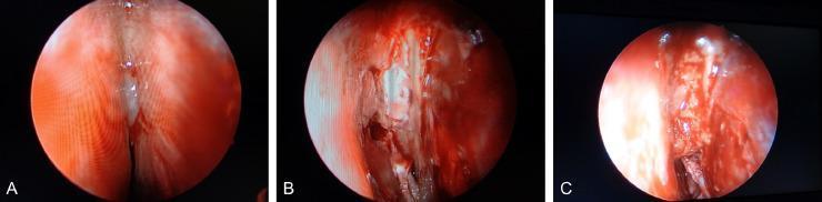
This composite of three intraoperative endoscopic images (A, B, C) documents the identification and surgical repair of a cerebrospinal fluid (CSF) rhinorrhea fistula within the nasal cavity. Image A shows the initial endoscopic view of the right olfactory fissure region, demonstrating mucosal swelling and a clear, watery discharge characteristic of a CSF leak. Image B reveals the underlying pathology after further exposure: a 1.2 cm bony defect in the ethmoid cribriform plate with visible dural exposure and active CSF leakage through the fistula. Image C demonstrates the multi-layered surgical closure, where the fistula has been packed with abdominal fat tissue and secured to provide a watertight seal. This clinical photograph is a primary educational resource for otorhinolaryngologists and neurosurgeons, illustrating the endoscopic management of spontaneous or traumatic skull base defects. Key concepts include intranasal anatomy, identification of dural fistulas, and the use of autologous fat grafts in endoscopic skull base reconstruction.
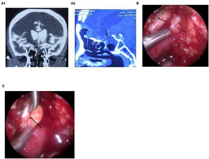
This composite educational graphic illustrates the diagnosis and endoscopic repair of a cerebrospinal fluid (CSF) leak. Panels A1 and A2 show Computed Tomography Cisternography (CTC) images in coronal and sagittal planes, respectively. Black arrows identify a skull base defect located at the right tuberculum sellae, adjacent to the sella turcica and sphenoid sinus. Panels B and C are intraoperative endoscopic photographs demonstrating a transnasal surgical approach. Image B captures the surgical site after saline injection to identify the precise site of the CSF leak (indicated by an arrow). Image C shows the 'bath-plug' repair technique, where a metallic surgical instrument is used to plug the defect with fascia lata (reddish-pink tissue, indicated by an arrow). This series demonstrates the clinical workflow from radiological localization to surgical intervention for managing refractory skull base fractures and associated rhinorrhea.
CSF Rhinorrhea
Definition
Classification
- A. Accidental (most common - ~80% of all CSF rhinorrhea)
-
- Immediate (within 48 hours of trauma)
-
- Delayed (onset days to weeks later, from meningeal herniation through fracture site)
-
- B. Surgical
-
- Complication of neurosurgical procedures: transsphenoidal hypophysectomy, frontal craniotomy, other skull base procedures
-
- Complication of rhinologic procedures: sinus surgery (FESS), septoplasty, other combined skull base procedures
-
- A. Elevated intracranial pressure (ICP)
- Hydrocephalus
- Intracranial mass (neoplasm eroding skull base)
- Benign intracranial hypertension (BIH) / pseudotumor cerebri
- B. Normal pressure
- Congenital skull base defects
- Erosive processes (osteomyelitis, cholesteatoma)
- Idiopathic ("spontaneous")
Note: Only 4% of all CSF leaks are truly nontraumatic; 16% follow intracranial/extracranial procedures. CSF rhinorrhea occurs in only 2-3% of serious head trauma, but skull base fractures carry a 12-30% risk of CSF fistula.
Pathophysiology
- Anatomical disruption - a defect in the arachnoid/dura, underlying bone, and paranasal sinus mucosa
- Pressure gradient - sufficient ICP to drive CSF through the defect
Clinical Presentation
- Unilateral watery nasal discharge - the classic presentation; clear, thin, watery
- Metallic or salty taste (CSF draining into nasopharynx)
- Positional variation - typically increases on leaning forward (the "reservoir sign" or "Dandy sign") or with Valsalva
- Halo sign - blood-stained discharge leaving a central red spot surrounded by a clear halo on gauze (low specificity)
- May be intermittent, particularly in nontraumatic/idiopathic cases
- Risk of bacterial meningitis - the most feared complication
Diagnosis
Biochemical Confirmation
| Test | Notes |
|---|---|
| β-2 transferrin | Gold standard - CSF-specific isoform not found in nasal secretions, tears, or serum; high sensitivity and specificity |
| β-trace protein (prostaglandin D-synthase) | Also highly specific; elevated in CSF; faster turnaround |
| Glucose testing | Low specificity - nasal secretions also contain glucose |
| "Halo sign" / ring test | Unreliable - poor sensitivity and specificity |
Localization (Imaging)
| Modality | Comments |
|---|---|
| High-resolution CT of skull base | First-line; best for identifying bony defects; thin-cut coronal sections optimal |
| CT cisternography | Requires LP for intrathecal contrast; excellent spatial resolution but needs active/large leak for reliable detection |
| MRI cisternography (MR cisternogram) | No LP needed; T2 CISS/FIESTA sequences; shows CSF as hyperintense signal tracking through defect; high spatial resolution; preferred when available |
| Radionuclide cisternography | Requires LP; poor sensitivity, poor spatial resolution - largely obsolete |
| Intrathecal fluorescein + nasal endoscopy | Confirms diagnosis AND localizes site intraoperatively; dilute concentration ESSENTIAL (serious neurological sequelae with high doses) |
Management
Conservative (Non-operative)
- Bed rest, head elevation (30°)
- Avoidance of nose blowing, Valsalva, straining
- Lumbar drainage - reduces CSF pressure and flow to allow natural healing
- Most traumatic leaks resolve within 7-10 days with conservative measures
- If CSF leak persists >7 days after conservative management or >5 days of lumbar drainage - surgical repair is indicated
- Prophylactic antibiotics - NOT routinely recommended (no confirmed benefit for preventing meningitis and risks selecting resistant organisms)
Surgical (Operative)
- Identify the leak site (pre-operatively with imaging + intraoperatively with fluorescein)
- Remove adjacent sinus mucosa to create a flat denuded bone surface for graft placement
- Close the defect with:
- Autologous grafts: fascia lata, free fat, free bone graft
- Allograft: acellular dermal allograft
- Xenogeneic: collagen dural substitutes
- Place a free mucosal overlay graft (middle turbinate or nasal floor mucosa)
- Secure with surgical sealant + resorbable packing ± nonresorbable sponge
- For high-flow leaks or large defects: vascularized pedicled mucosal flaps (e.g., nasoseptal flap) preferred
- Underlay technique - graft placed intradurally or epidurally under the defect
- Overlay technique - graft placed over denuded bone around defect
- "Bath-plug" technique - fat plug inserted into the defect, then overlay graft placed
- Nontraumatic CSF rhinorrhea (unlikely to resolve spontaneously)
- Traumatic leak that fails conservative measures (>7-10 days)
- Postoperative CSF rhinorrhea (most will require repair)
- Active meningoencephalocele or meningocele
- Brain tumor causing skull base erosion (after tumor management)
Management of Elevated ICP in Nontraumatic Rhinorrhea
- Acetazolamide (carbonic anhydrase inhibitor - reduces CSF production)
- Weight loss
- CSF diversion (lumboperitoneal or ventriculoperitoneal shunt) if medically refractory
- Failure to treat underlying elevated ICP significantly increases recurrence risk after repair
Intraoperative CSF Rhinorrhea (during FESS/sinus surgery)
- Repair at the time of surgery
- Remove bony partitions around defect to create flat surface
- Place free overlay nasal mucosal graft
- Secure with absorbable collagen packing ± nonabsorbable sponge
- Post-op: head CT and MRI to exclude intracranial bleeding/injury
- Neurosurgical and infectious disease consultations advised
- K.J. Lee's Essential Otolaryngology, p. 616
Endoscopic Images
Common Skull Base Defect Sites
| Site | Associated cause |
|---|---|
| Cribriform plate | Most common site; FESS injury, trauma |
| Ethmoid roof (fovea ethmoidalis) | FESS, trauma |
| Sphenoid sinus | Transsphenoidal surgery, spontaneous (IIH) |
| Frontal sinus | Frontal craniotomy, trauma |
| Tegmen tympani | Temporal bone fracture, arachnoid granulation |
Recent Evidence
- Cummings Otolaryngology Head and Neck Surgery, Ch. 48 (CSF Rhinorrhea and Anterior Meningoencephalocele)
- K.J. Lee's Essential Otolaryngology, 11th ed., p. 616
- Shambaugh Surgery of the Ear, 6th ed.