
Uterus : • Features • Blood supply • Nerve supply • Support ( Most asked ) • Lymphatic drainage ( Most asked ) Applied
Reading File
Now let me search the library for all relevant uterus sections in parallel.
Finding Sources
Finding Sources
The best anatomy source is Gray's Anatomy for Students. Let me read from that and also search for more specific uterus anatomy topics in parallel.
Reading File
Finding Sources
Finding Sources
Let me look for more dedicated uterus anatomy sections, especially support, blood supply, nerve supply, and lymphatics from Gray's Anatomy for Students and other sources.
Finding Sources
Finding Sources
Excellent! I found great sources. Let me now read the most comprehensive sections in parallel - Gray's, Berek & Novak, and the Color Atlas for vessels/nerves/lymphatics.
Reading File
Reading File
Reading File
Excellent data. Let me now read more from Gray's for the support/ligaments section, and from Berek & Novak for lymphatics and nerves, and fetch the key anatomical images.
Reading File
Reading File
Loading Image
I have rich data from multiple authoritative sources. Let me now get more on the support of the uterus (most asked) and lymphatics from Gray's and the Color Atlas.
Finding Sources
Finding Sources
Reading File
Reading File
Now I have comprehensive information from multiple textbooks. Let me also quickly get the Color Atlas's full support section and fetch the lymphatics image.
Reading File
Loading Image
I now have all the content needed for a comprehensive, well-illustrated answer. Let me compile the full response.
The Uterus - Complete Anatomy
Sources: Gray's Anatomy for Students, Berek & Novak's Gynecology, Color Atlas of Human Anatomy Vol. 2, Campbell-Walsh Urology
1. FEATURES
Position: The uterus is a thick-walled fibromuscular organ lying in the midline of the pelvis, between the urinary bladder (anteriorly) and the rectum (posteriorly). It is normally anteverted (angled forward relative to the vagina) and anteflexed (the body flexed forward at the isthmus).
Parts:
| Part | Details |
|---|---|
| Fundus | Rounded superior part, above the entry of the uterine tubes |
| Body (Corpus) | Main bulk; flattened anteroposteriorly; cavity is triangular (inverted triangle shape on AP view) |
| Isthmus | Junction between body and cervix (= lower uterine segment in pregnancy) |
| Cervix | Lower cylindrical part; 2-3 cm long; projects into the vagina as the portio vaginalis; external os (external opening), internal os (opens into endometrial cavity) |
| Cornu | Each upper lateral corner where the uterine tube inserts |
Layers of the Wall:
- Perimetrium - outer peritoneal/serous coat
- Myometrium - thick smooth muscle (1.5-2.5 cm thick); three sublayers - outer longitudinal (continuous with round ligament and tubal fibers), middle oblique/circular (carries blood vessels and nerves), inner longitudinal
- Endometrium - inner mucosa; undergoes cyclical change under hormonal influence
Size (normal adult): ~7.5 cm long × 5 cm wide × 3 cm thick; weighs ~60 g (nulliparous)
Squamocolumnar junction (transformation zone): Located at the cervix where stratified squamous epithelium (exocervix) meets mucus-secreting columnar epithelium (endocervical canal). This zone is the most vulnerable site for squamous neoplasia (cervical cancer).
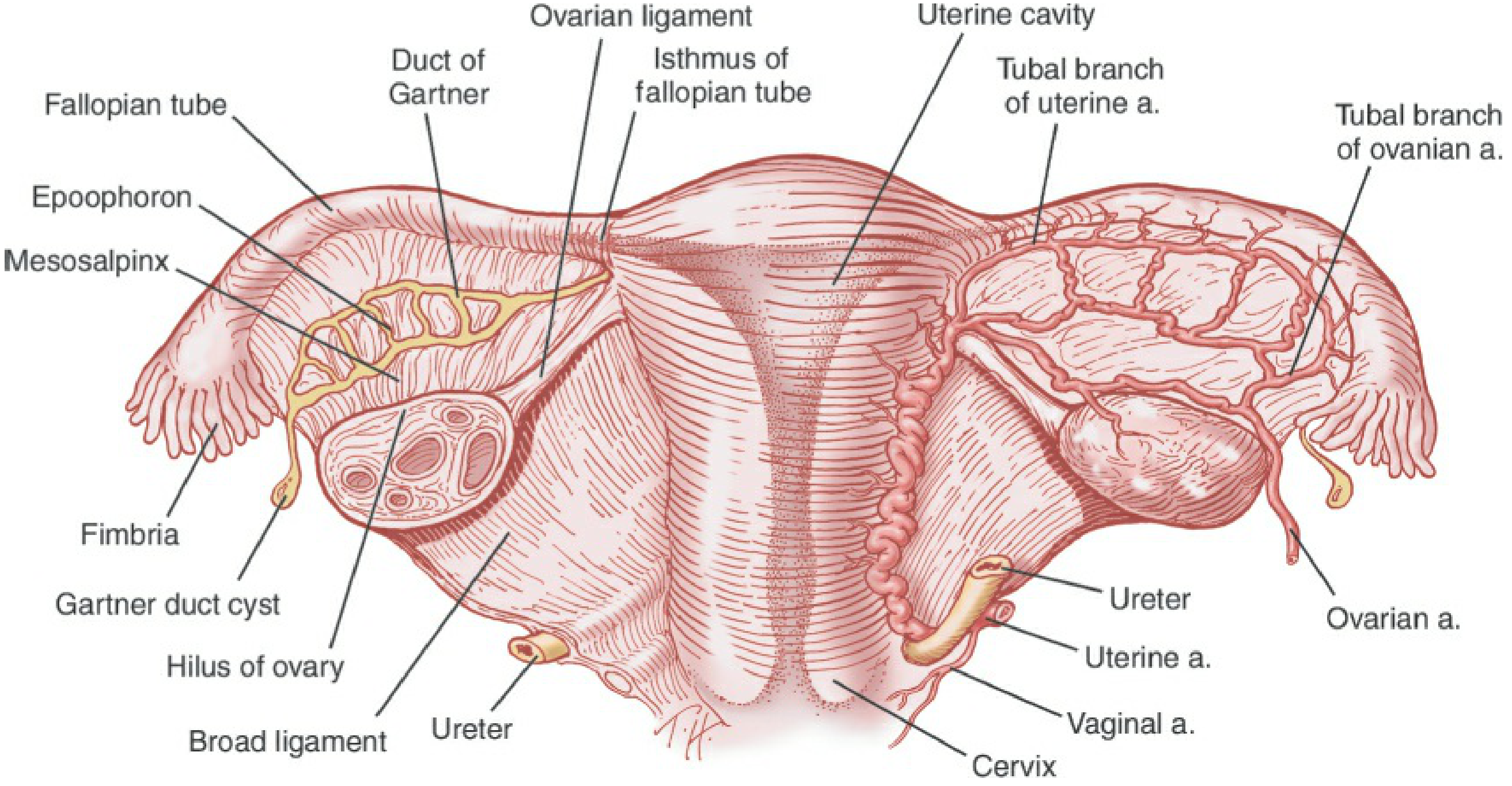
Fig. Uterus, fallopian tubes, and ovaries with associated vasculature - Berek & Novak's Gynecology
2. BLOOD SUPPLY
Arterial Supply
Main artery: Uterine artery (branch of the anterior trunk of the internal iliac artery)
- Courses medially and anteriorly in the base of the broad ligament
- Crosses OVER the ureter at ~2 cm lateral to the cervix ("water under the bridge" - ureter is the "water")
- Passes superiorly to the lateral vaginal fornix
- Reaches the cervix, then ascends along the lateral wall of the uterus in a tortuous course
- At the fundus, curves laterally and anastomoses with the ovarian artery
- Gives off: ascending main branch, descending vaginal artery (A4), tubal branch, and ovarian branch at the fundus
Supplementary arteries:
- Ovarian artery (from abdominal aorta) - anastomoses with uterine artery at the fundus; enlarges significantly in pregnancy; travels in the suspensory ligament of the ovary (infundibulopelvic ligament)
Clinical pearl: During hysterectomy, the uterine artery must be ligated without damaging the adjacent ureter. Failure to identify the ureter is the most common cause of ureteric injury in pelvic surgery.
Venous Drainage
- A network of valveless veins forms the uterine (venous) plexus around the body and cervix (in the parametrium)
- Drains via uterine veins → internal iliac veins
- Also connects with vaginal and ovarian plexuses
3. NERVE SUPPLY
Autonomic (the main innervation of the uterus)
- Via the inferior hypogastric plexus (pelvic plexus) and pelvic splanchnic nerves (S2-S4)
- These form a plexus lateral to the cervix with large ganglion cells - the uterovaginal plexus (Frankenhauser ganglion / Lee-Frankenhauser plexus)
Sympathetic: via hypogastric nerve (T10-L2) → inferior hypogastric plexus → uterus
- Causes uterine contraction and vasoconstriction
Parasympathetic: Pelvic splanchnic nerves (S2-S4) → inferior hypogastric plexus → uterus
- Causes uterine relaxation (in non-pregnant state) and vasodilation
Sensory (Pain) Fibers
- Pain from the body and fundus travels with sympathetic fibers → T10-T12, L1 (referred to umbilical and inguinal regions - explains labour pain felt in lower back and groin)
- Pain from the cervix travels with parasympathetic fibers → S2-S4 (referred to perineum, thighs, lower back)
Clinical: In spinal cord injuries below T10-T12, patients have an intact uterine nerve supply and experience labour pains; injuries above T10 abolish uterine pain sensation.
4. SUPPORT OF THE UTERUS ⭐ (Most Asked)
The uterus is supported by both active support (muscles) and passive support (ligaments/condensations of connective tissue). The pelvic floor muscles are the most important support - the ligaments mainly maintain position.
ACTIVE SUPPORT (Most Important)
Pelvic floor / levator ani muscles - the primary support; form the "pelvic diaphragm"
PASSIVE SUPPORT - Ligaments
A. CARDINAL LIGAMENT (Mackenrodt's Ligament / Transverse Cervical Ligament) ⭐ Most Important Clinical Ligament
- A condensation of connective tissue running from the cervix and upper vagina → lateral pelvic wall
- Contains the uterine artery and ureter
- Most important ligament for preventing uterine prolapse
- Weakness → cervical descent and uterovaginal prolapse
B. UTEROSACRAL LIGAMENTS (Rectouterine Ligaments / Sacrouterine Ligaments) ⭐ Most Important
- Dense subperitoneal connective tissue running from the posterior cervix → posterolateral pelvic wall / sacrum (S2-S4)
- Bound the rectouterine pouch (Pouch of Douglas)
- Maintain anteversion of the uterus by pulling the cervix posteriorly
- Carry autonomic nerve fibers (inferior hypogastric plexus)
- Palpable on per rectum examination in endometriosis (feel like nodular cords)
C. BROAD LIGAMENT
- A double fold of peritoneum extending from the lateral margin of the uterus to the lateral pelvic wall
- Not a true ligament - primarily contains vessels, nerves, and connective tissue
- Has subdivisions: mesometrium (covers uterus body), mesosalpinx (covers uterine tube), mesovarium (covers ovary)
- Provides lateral support; does not prevent prolapse
D. ROUND LIGAMENT OF THE UTERUS
- Arises from the uterine cornu (just anterior and below the uterine tube insertion)
- Passes through the inguinal canal → ends in labia majora
- Contains smooth muscle cells
- Maintains anteversion of the uterus
- Homologous to the gubernaculum testis in the male
- Carries the ilioinguinal nerve nearby
- Lymphatics from the fundus can follow this ligament → superficial inguinal nodes
E. PUBOCERVICAL LIGAMENT (Anterior Support)
- Condensation from cervix → posterior aspect of pubic symphysis
- Supports the bladder neck and anterior vaginal wall
F. OVARIAN LIGAMENT (Ligament of the Ovary)
- Connects the ovary to the uterine cornu; not a support ligament for the uterus per se
Summary Table: Support of the Uterus
| Ligament | Attachment | Primary Role |
|---|---|---|
| Cardinal (Mackenrodt's) | Cervix → lateral pelvic wall | MAIN: prevents prolapse |
| Uterosacral | Cervix → sacrum (S2-S4) | Maintains anteversion; 2nd most important |
| Round ligament | Uterine cornu → labia majora (via inguinal canal) | Anteversion; weaker support |
| Broad ligament | Lateral uterus → pelvic sidewall | Lateral stability (not anti-prolapse) |
| Levator ani (pelvic floor) | PRIMARY active support | Foundation support |
Applied: When the cardinal and uterosacral ligaments weaken (multiparity, menopause, chronic straining), the uterus descends - uterovaginal prolapse. The operation to repair this (Manchester repair / Fothergill's operation or pelvic floor repair) involves plication of these ligaments.
5. LYMPHATIC DRAINAGE ⭐ (Most Asked)
The lymphatic drainage of the uterus differs based on the region - this is critical for cancer staging and spread.
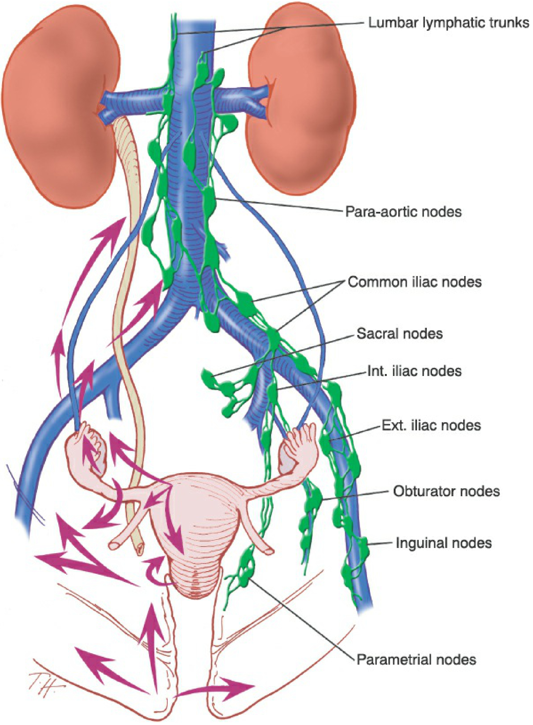
Fig. Lymphatic drainage of the female pelvis - Berek & Novak's Gynecology
Region-by-Region Drainage
| Region | Primary Lymph Nodes | Route |
|---|---|---|
| Fundus | Para-aortic (lateral aortic) nodes | Along ovarian vessels (infundibulopelvic ligament) / suspensory ligament of ovary |
| Fundus (minor) | Superficial inguinal nodes | Along round ligament through inguinal canal |
| Body (corpus) | External iliac nodes | Via broad ligament |
| Lower uterine body | External and internal iliac nodes | Same as cervix |
| Cervix | External iliac, internal iliac, and sacral nodes (via uterosacral ligaments) | Lateral parametrium → external iliac; posterior → sacral; via obturator nodes |
| Cervix (also) | Obturator nodes (a subgroup of external iliac) | Along lateral parametrium |
All nodes ultimately drain to:
Common iliac nodes → para-aortic nodes → lumbar lymphatic trunk → cisterna chyli → thoracic duct
Key Clinical Points - Lymphatic Spread
Endometrial (uterine body) cancer: Spreads to external iliac, internal iliac, and obturator nodes first; then para-aortic nodes (especially for fundal tumors which drain with ovarian vessels)
Cervical cancer: Spreads to obturator and external iliac nodes first (the "primary" nodes). Sentinel node concept is used in staging. Posterior spread is to sacral nodes via uterosacral ligaments.
Round ligament route (fundus → inguinal nodes): Explains rare inguinal node involvement in endometrial cancer and why fundal tumors can spread to inguinal nodes.
6. APPLIED ANATOMY ⭐
A. Uterine Artery & Ureter Relationship
- The uterine artery crosses over the ureter ("water under the bridge") about 1.5-2 cm lateral to the cervix at the base of the broad ligament
- Critical in hysterectomy: Ureter is at risk when ligating the uterine artery
- Ureter can also be injured when clamping the cardinal ligament
B. Uterine Artery Embolization (UAE)
- An interventional radiology procedure where small particles are injected into both uterine arteries via the femoral approach
- Blocks blood supply to fibroids and adenomyosis, causing infarction and shrinkage
- Also used in postpartum hemorrhage
C. Hysterectomy Types
- Total abdominal hysterectomy (TAH): Removes body, fundus, and cervix
- TAH + bilateral salpingo-oophorectomy (BSO): Also removes tubes and ovaries
- Subtotal/supracervical: Cervix left in situ (rare)
- Performed via Pfannenstiel (transverse suprapubic) incision
D. Uterine Prolapse
- Caused by weakening of the cardinal and uterosacral ligaments and pelvic floor
- Degrees:
- 1st degree - cervix descends into vagina
- 2nd degree - cervix at vaginal introitus
- 3rd degree (procidentia) - entire uterus outside the vagina
- Risk factors: multiparity, prolonged labour, obesity, chronic straining, menopause (estrogen loss)
- Treatment: pelvic floor exercises, pessary, surgical repair (Manchester operation, pelvic floor repair, or vaginal hysterectomy)
E. Referred Pain in Dysmenorrhoea / Labour
- Uterine body pain transmitted via T10-L1 sympathetic fibers → referred to lower back, umbilical region, and inner thighs
- Cervical pain via S2-S4 → referred to perineum, inner thighs, lower sacrum
F. Ectopic / Abnormal Implantation
- Normal implantation is in the body of the uterus (upper posterior wall preferred)
- Placenta praevia: implantation in the lower uterine segment / isthmus (over the internal os)
- Ectopic pregnancy: implantation outside uterus (most commonly ampulla of the tube)
G. Uterine Malignancy and Lymph Node Dissection
- Cervical cancer surgery (Wertheim's radical hysterectomy) removes the uterus + parametrium + upper vagina + pelvic lymph nodes (external iliac, internal iliac, obturator)
- Para-aortic node sampling is done for advanced disease or fundal tumors
Quick Recall Mnemonics:
- "Water under the bridge" = Ureter (water) passes UNDER the uterine artery (bridge) at the lateral cervix
- Ligament importance order: Cardinal > Uterosacral > Round > Broad (for prolapse prevention)
- Lymphatics by region: Fundus → para-aortic; Body → external iliac; Cervix → external iliac + internal iliac + sacral nodes; Fundus (minor) → inguinal (via round ligament)
- Nerve supply levels: Body/fundus pain → T10-L1 (labour pain felt in back); Cervical pain → S2-S4
This is a shared conversation. Sign in to Orris to start your own chat.