
Obstructive jaundice patient for whipple surgery 20 mark answer
Reading File
Now let me search the medical library for comprehensive content on this topic.
Finding Sources
Finding Sources
Finding Sources
Reading File
Finding Sources
Finding Sources
Reading File
Reading File
Reading File
Finding Sources
Finding Sources
Finding Sources
Reading File
Reading File
Reading File
Reading File
Reading File
Finding Sources
Reading File
I now have comprehensive material from multiple textbooks. Let me compile the full 20-mark answer.
Searching Images
Whipple pancreaticoduodenectomy surgical anatomy diagram reconstruction
Loading Image
I now have all the material needed for a comprehensive 20-mark answer. Here it is:
Obstructive Jaundice Patient for Whipple Surgery
1. Introduction
Obstructive (post-hepatic/surgical) jaundice results from impaired flow of conjugated bilirubin through the biliary tree into the duodenum. When caused by a periampullary malignancy - carcinoma of the pancreatic head, distal cholangiocarcinoma, ampullary carcinoma, or duodenal carcinoma - the definitive curative treatment is pancreaticoduodenectomy (Whipple procedure). These patients require systematic preoperative evaluation, optimization, and careful surgical planning.
2. Pathophysiology of Obstructive Jaundice Relevant to Surgery
Chronic biliary obstruction causes a cascade of systemic effects that directly impair surgical outcomes:
| System | Effect |
|---|---|
| Coagulation | Fat malabsorption - vitamin K deficiency - reduced synthesis of factors II, VII, IX, X - prolonged PT |
| Renal | Hepatorenal syndrome risk; bile salts impair renal tubular function; endotoxaemia |
| Immune | Endotoxaemia from bacterial translocation; impaired Kupffer cell function |
| Nutritional | Anorexia, weight loss, hypoalbuminaemia |
| Cardiovascular | Bradycardia (bile salt effect on SA node); hypotension |
| Wound healing | Impaired due to hypoalbuminaemia and malnutrition |
- Current Surgical Therapy 14e (obstructive jaundice chapter): cholestasis impairs clotting and immunologic responses.
- Pye's Surgical Handicraft: coagulation defects in jaundice involve prothrombin, factor V, factor VII; a good response to IV vitamin K confirms posthepatic (surgical) jaundice.
3. Causes Requiring Whipple Surgery
Malignant periampullary causes:
- Carcinoma of the head of pancreas (most common)
- Distal cholangiocarcinoma (below the cystic duct junction)
- Ampullary carcinoma
- Duodenal carcinoma
Benign causes occasionally requiring Whipple:
- Chronic pancreatitis with head mass lesion
- Pseudotumoral chronic pancreatitis
4. Clinical Presentation
- Progressive, painless jaundice - classic feature of malignancy (fluctuating jaundice suggests stones)
- Dark urine, clay-coloured stools, pruritus, fatigue
- Courvoisier's sign - palpable, non-tender, distended gallbladder (total bilirubin >10 mg/dL is highly suggestive of malignancy)
- Weight loss, anorexia, nausea
- Fever if cholangitis is present
5. Preoperative Investigations
Biochemical
- LFTs: elevated bilirubin (predominantly direct), ALP (3-4x elevation in 75% of cholestasis), GGT, 5'-nucleotidase
- PT/INR, aPTT (assess coagulopathy)
- Serum albumin (nutritional status)
- Blood sugar (pre-existing or new-onset diabetes)
- Urea, creatinine (renal function baseline)
- FBC (anaemia, leucocytosis)
- Tumour markers: CA 19-9 (pancreatic/biliary malignancy), CEA
- Serum IgG4 (if autoimmune pancreatitis suspected)
Imaging
- Abdominal ultrasound - first-line; shows biliary dilation, level of obstruction, distended gallbladder
- CT (triple-phase: arterial, portal venous, pancreatic phase) - defines tumour extent, vascular involvement (SMA, SMV, PV, coeliac axis), lymphadenopathy, and distant metastases. Key for resectability assessment
- MRCP - excellent non-invasive imaging of biliary and pancreatic ducts; sensitivity 95% for biliary obstruction; differentiates benign from malignant strictures
- EUS (endoscopic ultrasound) - detects tumours at hilum, ampullary region, pancreatic head; guides fine-needle aspiration (FNA); sensitivity 84-91%, specificity 71-100% for distal biliary strictures
- ERCP - therapeutic (stenting) rather than primarily diagnostic; useful in cholangitis or severe hepatic congestion
- Chest CT - excludes pulmonary/mediastinal metastases
- Diagnostic laparoscopy - selectively used in high CA 19-9 or equivocal lesions to exclude occult metastases before laparotomy
Resectability Assessment (CT Criteria)
- Resectable: no arterial contact, <180° venous contact with SMV/PV
- Borderline resectable: >180° venous contact, <180° arterial contact
- Locally advanced/unresectable: >180° arterial contact (SMA/coeliac), complete venous occlusion
6. Preoperative Optimisation
Correction of Coagulopathy
- IV Vitamin K (10 mg daily for 3 days) - corrects PT if hepatocellular function is intact (confirming surgical jaundice)
- Fresh frozen plasma if urgent correction needed
- Check response: normalisation of PT indicates adequate hepatocyte synthetic function
Nutritional Optimisation
- High-protein, high-calorie diet
- Consider nasojejunal or parenteral nutrition if severely malnourished
- Correct hypoalbuminaemia
Renal Protection
- Adequate hydration/IV fluids preoperatively
- Mannitol infusion during surgery (renal protection against hepatorenal syndrome)
- Avoid nephrotoxic drugs
Preoperative Biliary Drainage
- Controversial - routine preoperative biliary stenting is NOT recommended
- Randomised controlled trial data (Maingot's; Current Surgical Therapy 14e) shows routine preoperative stenting in patients undergoing pancreaticoduodenectomy is associated with increased perioperative morbidity, especially infectious complications (bile contamination)
- Selective indications for biliary drainage (ERCP/stent or PTC):
- Cholangitis - urgent drainage needed
- Severe hepatic congestion/derangement
- Significant delay until surgery (e.g., awaiting neoadjuvant therapy)
- Bilirubin very high (>15 mg/dL) with planned delay >2 weeks
- Metallic stents are preferred over plastic if stenting is done before pancreaticoduodenectomy
Other
- Antibiotic prophylaxis (preoperative, broad-spectrum)
- DVT prophylaxis (subcutaneous heparin before induction)
- Cardiac and pulmonary evaluation (ECG, echo, PFTs as indicated)
- Optimise blood sugar
- Cross-match blood products
7. The Whipple Procedure (Pancreaticoduodenectomy)
Anatomy Removed
- Head, neck, and uncinate process of the pancreas
- Duodenum (entire)
- Distal stomach (antrum) in classic Whipple; preserved in pylorus-preserving variant (PPPD)
- Gallbladder
- Distal common bile duct
- Regional lymph nodes (aiming for ≥12 nodes)
Surgical Steps (Open Approach)
Step 1 - Exposure and Laparoscopy
Midline or bilateral subcostal incision. Diagnostic laparoscopy first (selective) to rule out occult metastases. Explore peritoneal cavity and liver.
Step 2 - Kocherisation
Wide Kocher manoeuvre - mobilise duodenum and pancreatic head off the inferior vena cava and aorta to assess posterior resectability.
Step 3 - Hepatoduodenal Ligament Dissection
- Identify and divide the gastroduodenal artery (GDA) after confirming adequate hepatic arterial perfusion
- Skeletonise the portal vein (PV) and superior mesenteric vein (SMV) tunnel behind the pancreatic neck
- Cholecystectomy (top-down); divide CBD above cystic duct junction; bile cultures obtained
Step 4 - Stomach/Pylorus Division
- Classic Whipple: antrectomy (gastrojejunostomy reconstruction later)
- Pylorus-preserving (PPPD): divide duodenum 2-3 cm distal to pylorus (duodenojejunostomy later)
Step 5 - Pancreatic Neck Transection
Divide pancreatic neck over the SMV/PV tunnel using electrocautery or scalpel.
Step 6 - Division of Proximal Jejunum
Divide jejunum ~15 cm distal to ligament of Treitz; bring through the transverse mesocolon defect.
Step 7 - Uncinate Process Dissection
Most demanding step - careful dissection of uncinate from the SMA (posterior to the SMV). Multiple small branches from the SMA must be ligated individually (inferior pancreaticoduodenal arteries).
Step 8 - Specimen Removal
En-bloc removal of pancreatic head, duodenum, distal CBD, gallbladder, and antrum/pylorus.
Reconstruction (Child's Sequence)
Three anastomoses are performed in sequence:
- Pancreaticojejunostomy (PJ) - pancreatic remnant anastomosed end-to-side to jejunum (duct-to-mucosa technique preferred); most critical anastomosis
- Hepaticojejunostomy/Choledochojejunostomy (CJ) - bile duct anastomosed end-to-side to jejunum, 10-15 cm downstream from PJ
- Gastrojejunostomy (GJ) or Duodenojejunostomy - antrum/pylorus anastomosed further downstream, completing reconstruction
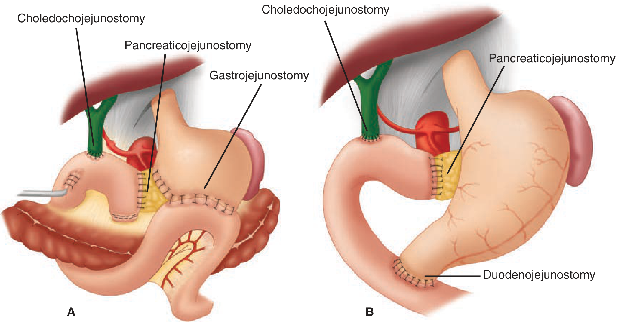
A: Classic Whipple with gastrojejunostomy. B: Pylorus-preserving pancreaticoduodenectomy (PPPD) with duodenojejunostomy. Both include pancreaticojejunostomy and choledochojejunostomy. Source: Schwartz's Principles of Surgery.
8. Postoperative Complications (ISGPS-Defined)
Mortality at high-volume centres: <3-5%. Morbidity: 40-60%.
Procedure-Specific Complications
1. Clinically Relevant Post-operative Pancreatic Fistula (CR-POPF)
- Most feared complication; incidence ~15%; responsible for 36% of mortality after PD
- Defined as: drain amylase >3x upper limit of normal on/after POD 3 + clinical impact
- Risk factors (Fistula Risk Score): soft gland texture, small duct (<3 mm), pathology other than pancreatic cancer/pancreatitis, high intraoperative blood loss
- Management: prolonged drainage, octreotide, nil by mouth, TPN; interventional radiology drainage; rarely re-operation
2. Delayed Gastric Emptying (DGE)
- Inability to tolerate oral intake by POD 7 or nasogastric tube in situ beyond POD 3
- Grade A/B/C based on clinical impact
- Management: nasogastric decompression, prokinetics (metoclopramide, erythromycin), parenteral nutrition
3. Post-pancreatectomy Haemorrhage (PPH)
- Early (<24h): surgical bleeding from anastomosis or vessels
- Late (>24h): often sentinel bleed from pseudoaneurysm of GDA stump (alarming)
- Management: early - re-operation; late - CT angiography and interventional radiology embolisation
4. Bile Leak
- From hepaticojejunostomy or cystic duct stump
- Management: ERCP and stenting, percutaneous drainage
5. Chyle Leak
- Lymphorrhea from lymphatic channels
- Management: low-fat/medium-chain triglyceride diet, octreotide
General Complications
- Surgical site infection (deep/superficial)
- Pneumonia
- DVT/PE
- Acute kidney injury
- Hepatic failure (particularly in severely jaundiced patients)
- Wound dehiscence
9. Perioperative Care
- ICU/HDU for at least 24-48 hours
- Arterial line - continuous BP monitoring intraoperatively
- Goal-directed fluid resuscitation - avoid over- or under-hydration
- Central venous access - avoid left neck if venous resection anticipated (left IJV may be needed as conduit)
- Drains - closed-suction abdominal drains placed near the pancreatic and biliary anastomoses; output monitored for amylase on POD 3 to detect fistula
- Nasogastric tube - early removal encouraged in PPPD
- Early enteral nutrition preferred when tolerated
- DVT prophylaxis - LMWH + compression stockings
- Perioperative antibiotics - redosed intraoperatively at appropriate intervals
- Blood glucose monitoring - newly or worsened diabetes common
10. Prognosis
| Pathology | 5-Year Survival After R0 Resection |
|---|---|
| Ampullary carcinoma | 40-50% (best prognosis) |
| Distal cholangiocarcinoma | 25-35% |
| Pancreatic adenocarcinoma | 15-25% |
| Duodenal carcinoma | 40-60% |
- R0 (margin-negative) resection is the single most important prognostic factor
- Adjuvant chemotherapy (gemcitabine + capecitabine; or FOLFIRINOX) is standard after PD for pancreatic cancer
- Neoadjuvant therapy is increasingly preferred to improve R0 rates and select against rapid progressors
Summary Table
| Domain | Key Point |
|---|---|
| Cause | Periampullary malignancy (PaHoP most common) |
| Presentation | Painless progressive jaundice, Courvoisier's sign |
| Investigation | CT triple-phase, MRCP, EUS-FNA, tumour markers |
| Optimisation | Vitamin K, nutrition, renal protection |
| Biliary drainage | Selective, not routine |
| Operation | PD with Child's reconstruction (PJ - CJ - GJ/DJ) |
| Critical step | Uncinate dissection from SMA |
| Commonest complication | Pancreatic fistula (CR-POPF) |
| Mortality | <5% at high-volume centres |
Sources: Current Surgical Therapy 14e; Maingot's Abdominal Operations; Fischer's Mastery of Surgery 8e; Schwartz's Principles of Surgery 11e; Pye's Surgical Handicraft 22e.
Give answer for an anaesthesia exam going student
Reading File
Finding Sources
Finding Sources
Finding Sources
Finding Sources
Finding Sources
Reading File
Reading File
Reading File
Finding Sources
Finding Sources
Finding Sources
Reading File
Reading File
Reading File
Finding Sources
Reading File
Reading File
Reading File
Finding Sources
I now have all the anaesthesia-specific content needed. Here is the complete 20-mark answer reframed entirely from an anaesthesia perspective:
Anaesthetic Management of a Patient with Obstructive Jaundice for Whipple Surgery (Pancreaticoduodenectomy)
1. Introduction
Obstructive jaundice presents the anaesthetist with a patient who has multi-system physiological derangements secondary to cholestasis and biliary obstruction. When planned for a Whipple procedure (pancreaticoduodenectomy) - one of the most complex and lengthy abdominal operations - a thorough anaesthetic assessment and preparation are essential to minimise the high morbidity associated with this combination.
2. Pathophysiological Effects of Obstructive Jaundice Relevant to Anaesthesia
This is the cornerstone of understanding every anaesthetic decision:
| System | Derangement | Anaesthetic Implication |
|---|---|---|
| Coagulation | Vitamin K deficiency (fat malabsorption) - reduced factors II, VII, IX, X - prolonged PT/INR | Risk of epidural haematoma; massive intraoperative haemorrhage |
| Renal | Bile salts + endotoxaemia impair tubular function; hepatorenal syndrome (HRS) risk | Avoid nephrotoxins; maintain euvolaemia; mannitol diuresis |
| Cardiovascular | Bile salt-mediated bradycardia; peripheral vasodilation; blunted response to vasopressors | Intraoperative haemodynamic instability; vasopressor resistance |
| Immune/Sepsis | Endotoxaemia from bacterial translocation via portal system; cholangitis | Increased risk of septic shock; antibiotic cover essential |
| Pharmacokinetics | Hypoalbuminaemia - reduced protein binding - increased free drug fraction; reduced hepatic metabolism | Drug effects are unpredictable and prolonged |
| GI | Gastroparesis, delayed gastric emptying | Full stomach precautions; RSI may be needed |
| Haematological | Anaemia from haemolysis and poor nutrition | Pre-op cross-match; likely transfusion requirement |
| Metabolic | Hyponatraemia, hypokalemia, hypoglycaemia, hypoalbuminaemia | Electrolyte correction essential preoperatively |
3. Preoperative Anaesthetic Assessment
History
- Duration and depth of jaundice (total bilirubin level)
- Presence of pruritus, cholangitis (fever, rigors - Charcot's triad)
- Comorbidities: diabetes, cardiovascular disease, renal disease
- Drug history (NSAIDs, anticoagulants, hepatotoxins)
- Prior surgeries and anaesthetic history
Examination
- Jaundice, scratch marks (pruritus)
- Signs of malnutrition (muscle wasting, cachexia)
- Cardiovascular: bradycardia, peripheral vasodilation, volume status
- Ascites (not common in obstructive, unlike cirrhotic - but may be present with malignancy)
- Airway assessment (routine Mallampati, neck mobility)
Investigations
Haematological:
- FBC - anaemia, leucocytosis (cholangitis/infection)
- PT/INR, aPTT - critical for epidural decision and bleeding risk
- Platelet count
- Group and cross-match (2-4 units RBC, FFP, platelets)
Biochemical:
- Bilirubin (total and direct), ALT, AST, ALP, GGT
- Serum albumin (below 3.5 g/dL = significant nutritional deficit)
- Urea, creatinine, electrolytes (Na+, K+, Mg2+) - baseline renal function
- Blood glucose (diabetes or new-onset in pancreatic disease)
- Ca 19-9, CEA (for oncological context)
Cardiac:
- ECG (bradycardia, QT prolongation)
- Echocardiography if significant cardiac history or poor functional capacity
Respiratory:
- CXR, pulse oximetry
- PFTs if clinically indicated
- ABG if hypoxaemia suspected
Imaging:
- CT/MRI reviewed for vascular anatomy (crucial for expected blood loss and vessel injury planning)
Risk Stratification
- Child-Pugh score (if hepatocellular component suspected): Class A acceptable, Class C contraindicates elective surgery
- MELD score: MELD <11 = low postoperative mortality; MELD ≥20 = high risk
- SORT/ASA score: Most patients are ASA III or IV
4. Preoperative Optimisation
Correction of Coagulopathy
- IV Vitamin K 10 mg daily x 3 days - corrects PT if hepatocyte synthesis intact (this also confirms the jaundice is obstructive, not hepatocellular - a positive response to Vitamin K = surgical jaundice)
- FFP/cryoprecipitate if urgent surgery or non-responsive to Vitamin K
- Target INR <1.5 before epidural placement
- Platelet transfusion if count <80,000/μL (for neuraxial anaesthesia)
Renal Protection
- Adequate IV hydration preoperatively
- Mannitol 0.5 g/kg IV given perioperatively - osmotic diuresis, free radical scavenging, renal protection
- Avoid NSAIDs, aminoglycosides, contrast agents
- Baseline creatinine documented; urine output targets set
Nutritional
- High-protein supplementation
- TPN if severely malnourished (albumin <2.5 g/dL) - start 7-10 days preoperatively
- Correct electrolytes (K+, Mg2+, glucose)
Biliary Drainage (Anaesthetic Perspective)
- If biliary stenting is performed: contaminated bile = higher risk of infectious complications intraoperatively
- Metal stents preferred over plastic if decompression is done before Whipple
- Routine preoperative stenting is NOT recommended - increases infection risk without improving mortality (Maingot's; Current Surgical Therapy)
- Exception: cholangitis, very high bilirubin with planned delay, or need for neoadjuvant therapy
Blood Bank Preparation
- Cross-match 4 units packed RBC, 4 units FFP, 1 adult dose platelets
- Cell salvage (autologous blood recovery) may be used if no malignancy spillage risk
- Discuss with surgeon expected blood loss (can be 500-2000 mL)
5. Choice of Anaesthetic Technique
General Anaesthesia + Thoracic Epidural: Gold Standard
Combined general and epidural anaesthesia (TEA) is the technique of choice for Whipple procedure.
Benefits of TEA (T6-T8 level):
- Excellent intraoperative and postoperative analgesia
- Reduces stress response and sympathetic activation (preserves hepatic blood flow)
- Decreases intraoperative opioid requirements (less hepatic metabolism burden)
- Reduces postoperative pulmonary complications (especially important in long procedure)
- Facilitates early extubation and ambulation (ERAS protocol)
- Associated with reduced postoperative ileus (sympathetic block promotes gut motility)
Contraindications to epidural in jaundiced patients:
- Uncorrected coagulopathy (INR >1.5, platelets <80,000)
- Patient refusal or sepsis/bacteraemia
- If contraindicated: TAP (transversus abdominis plane) block or QL (quadratus lumborum) block is an effective alternative for abdominal surgery
6. Induction of Anaesthesia
Premedication
- Anxiolytic (oral midazolam) - use cautiously and in reduced dose (prolonged effect due to reduced protein binding and impaired metabolism in jaundiced patients)
- H2 blocker/PPI + metoclopramide (aspiration prophylaxis)
- Antifibrinolytics: Tranexamic acid 1 g IV at induction if major bleeding expected
Induction Agents
- Propofol (preferred) - highly lipophilic, high extraction ratio; clearance relatively preserved in jaundice without cirrhosis; use reduced dose due to hypoalbuminaemia (increased free fraction)
- Thiopentone - alternative; pharmacodynamic effects more pronounced
- Ketamine - useful if haemodynamically compromised (but avoid in raised ICP/tachycardia)
- Etomidate - preferred if cardiovascular instability (minimal cardiac depression)
Rapid Sequence Induction (RSI)
- Consider RSI if gastric emptying is delayed (periampullary malignancy) or if ascites is present
- Pre-oxygenation with 100% O2 for 3 minutes
- Cricoid pressure while airway secured
Neuromuscular Blockade
- Atracurium or cisatracurium: DRUGS OF CHOICE in jaundice/liver disease
- Hoffman elimination + ester hydrolysis = organ-independent elimination
- Duration unaffected by liver disease
- Laudanosine (metabolite) is hepatically cleared but neurotoxicity not clinically significant
- Avoid vecuronium and rocuronium as sole agents - prolonged duration due to reduced hepatic clearance and increased volume of distribution
- Avoid succinylcholine if suspecting plasma cholinesterase deficiency (reduced in liver disease)
Intubation
- Rapid sequence with RSI if risk factors present
- Standard orotracheal intubation otherwise
- Thoracic epidural placed prior to induction (or post-induction in anaesthetised patient at T6-T8)
7. Intraoperative Monitoring
Standard:
- ECG (5-lead), SpO2, ETCO2, temperature
- Urine output (Foley catheter - target ≥0.5 mL/kg/h)
Advanced (mandatory for Whipple):
- Arterial line (radial artery) - beat-to-beat BP monitoring; frequent ABG and blood sampling; assessment of pulse pressure variation (PPV) for fluid responsiveness
- Central venous catheter - CVP guidance; vasoactive drug infusion; avoid left internal jugular vein if venous resection/reconstruction anticipated (IJV may be needed as vascular conduit)
- Cardiac output monitoring (FloTrac/Vigileo, LIDCO, oesophageal Doppler) - goal-directed fluid therapy in long procedures
- BIS/depth of anaesthesia monitor - avoid awareness (particularly important with reduced drug requirements in jaundice)
- Blood glucose monitoring - hourly; target 6-10 mmol/L
- Temperature monitoring - active warming (Bair Hugger); hypothermia worsens coagulopathy
- Neurological (if epidural) - test dose before activation; monitor for local anaesthetic toxicity
8. Maintenance of Anaesthesia
Volatile Agents
- Isoflurane or Sevoflurane: agents of choice in patients with liver disease
- At 1 MAC: minimal reduction of hepatic blood flow
- Maintain hepatic arterial buffer response
- Sevoflurane: dose-dependent reduction does NOT occur at higher MAC (safest)
- Isoflurane: minimal TFA (trifluoroacetate) production (0.2% metabolised vs 20% halothane)
- Avoid Halothane: greatest cardiovascular depression, maximal hepatic blood flow reduction, risk of halothane hepatitis
- Desflurane: 30% reduction in hepatic blood flow at 1 MAC - less preferred
- Nitrous oxide: avoid due to bowel distension and impaired gut healing
Key Intraoperative Goals
- Maintain normotension (MAP >65 mmHg): hypotension reduces hepatic blood flow and precipitates HRS
- Avoid excessive sympathetic activation (light anaesthesia, pain, laryngospasm)
- Avoid high mean airway pressures: reduces hepatic venous outflow
- Normoventilation (avoid hypercapnia and hyperventilation)
Opioids
- Fentanyl (intraoperative): short-acting; redistribution governs single-dose elimination
- Remifentanil infusion: ideal in jaundice - eliminated by blood and tissue esterases, completely independent of hepatic function regardless of infusion duration
- Morphine and meperidine: avoid or use with extreme caution - prolonged elimination in liver disease; normeperidine accumulation = neurotoxicity
- Reduce all opioid doses due to pharmacodynamic sensitivity (increased CNS sensitivity, risk of precipitating encephalopathy)
Fluid Management (Goal-Directed)
- Balanced crystalloids (Hartmann's/Plasmalyte) as maintenance
- Albumin infusions if serum albumin <25 g/L (especially after large fluid shifts intraoperatively)
- Blood products guided by POC coagulation testing (TEG/ROTEM preferred over standard PT/INR in the operating room - gives real-time assessment of coagulation status)
- Avoid overhydration: worsens bowel oedema and anastomotic healing
- Avoid underhydration: precipitates HRS, AKI
- Target CVP 5-8 mmHg during resection phase, allow higher during reconstruction
- Mannitol 0.5 g/kg given at start of surgery - renal protection from hepatorenal syndrome risk
- Cell salvage device on standby
Vasopressors
- Jaundiced patients have reduced response to vasoconstrictors (vasoplegia from nitric oxide, prostacyclin)
- Noradrenaline (norepinephrine): first-line vasopressor
- Vasopressin: useful adjunct, promotes renal perfusion
- Note: catecholamines may paradoxically reduce hepatic blood flow via hepatic arterial vasoconstriction
9. Regional Anaesthesia Considerations
Thoracic Epidural Analgesia (TEA) - T6-T8
- Placed pre-induction or post-induction
- Test dose: 3 mL lignocaine 2% with adrenaline 1:200,000 (checks for IV or intrathecal placement)
- Maintenance: bupivacaine 0.1-0.125% + fentanyl 2 mcg/mL at 6-12 mL/hr OR PCEA (patient-controlled epidural analgesia)
- Monitor: sensory block level, BP, HR, motor block in lower limbs
- Prerequisite: INR <1.5, platelets >80,000 before placement
Alternatives if Epidural Contraindicated
- TAP block (transversus abdominis plane): bilateral ultrasound-guided; effective for anterior abdominal wall pain (T10-L1)
- Quadratus Lumborum (QL) block: wider coverage (T7-L1), increasingly used for hepatopancreatic surgery
- IV ketamine infusion (sub-anaesthetic dose 0.1-0.3 mg/kg/h): opioid-sparing, prevents central sensitisation
- Multimodal analgesia: paracetamol (caution in hepatic impairment; reduce dose), avoid NSAIDs (renal risk + platelet dysfunction)
10. Extubation and Recovery
- Criteria for extubation on table: haemodynamically stable, normothermic, coagulation corrected, responsive, adequate respiratory effort, ETCo2 normal
- If prolonged surgery (>6 hours), massive transfusion, or haemodynamic instability - planned elective ventilation in ICU
- Reverse neuromuscular blockade: neostigmine + glycopyrrolate (or sugammadex if rocuronium used)
- Adequate analgesia confirmed before extubation (check epidural function)
- ICU/HDU admission: all patients post-Whipple require at least 24-48 hours high-dependency monitoring
11. Postoperative Anaesthetic Management
Pain
- Continue TEA for 3-5 days postoperatively (PCEA most effective)
- Step down to oral multimodal analgesia (paracetamol ± tramadol ± NSAIDs - NSAIDs cautiously only once renal function confirmed stable)
- Avoid excessive opioids: risk of respiratory depression, ileus, encephalopathy
Postoperative Renal Protection
- Maintain urine output ≥0.5 mL/kg/h
- Daily creatinine monitoring
- Avoid nephrotoxins (aminoglycosides, NSAIDs)
- Consider mannitol or frusemide if oliguria develops
Glucose Management
- Postoperative hyperglycaemia common (pancreatic head resection disrupts insulin secretion)
- Insulin sliding scale or infusion; target 6-10 mmol/L
- Risk of hypoglycaemia after total pancreatectomy (absent glucagon)
ICU Monitoring
- Hourly urine output, 4-hourly blood glucose
- Daily LFTs, coagulation profile, electrolytes
- Drain output (amylase on POD 3 for CR-POPF screening)
- DVT prophylaxis: LMWH + compression stockings once haemostasis confirmed
12. Specific Anaesthetic Risks in This Setting
| Complication | Mechanism | Prevention/Management |
|---|---|---|
| Hepatorenal Syndrome | Renal vasoconstriction from bile salts, endotoxaemia | Euvolaemia, mannitol, noradrenaline, avoid nephrotoxins |
| Coagulopathy/Massive Haemorrhage | Vitamin K deficiency, dilutional, fibrinolysis | TEG-guided transfusion, FFP, tranexamic acid, cell salvage |
| Drug toxicity | Unpredictable pharmacokinetics | Dose reduction, titrate to effect, use organ-independent drugs |
| Postoperative jaundice | Halothane hepatitis, ischaemic hepatitis, anastomotic bile leak | Use isoflurane/sevoflurane, maintain hepatic blood flow |
| Sepsis/Cholangitis | Bile contamination, stent-related infection | Broad-spectrum antibiotics, biliary cultures intraoperatively |
| Aspiration | Gastroparesis, ascites | RSI, pre-op antacids, NG tube if needed |
| Hypoglycaemia (post-op) | Reduced glycogen stores + insulin dysregulation | Glucose monitoring, dextrose infusion |
13. ERAS Protocol for Whipple Surgery (Anaesthetic Components)
The Enhanced Recovery After Surgery (ERAS) pathway for pancreaticoduodenectomy includes key anaesthetic elements:
- No prolonged fasting (clear fluids up to 2 hours; carbohydrate loading 6 hours before)
- Avoid routine bowel preparation
- Thoracic epidural placement
- Goal-directed fluid therapy (avoid over-hydration)
- Opioid-minimising multimodal analgesia
- Early extubation (avoid prolonged ventilation)
- Postoperative nausea and vomiting (PONV) prophylaxis (ondansetron + dexamethasone - caution with dexamethasone in diabetics)
- Early nasogastric removal and early oral intake
- DVT prophylaxis from day 0
Summary for Exam
| Phase | Key Anaesthetic Points |
|---|---|
| Preop | Vitamin K correction, albumin, renal baseline, coag correction, epidural eligibility |
| Drug choice | Atracurium/cisatracurium (NMB), isoflurane/sevoflurane (volatile), remifentanil (opioid), propofol (induction) |
| Technique | GA + thoracic epidural (T6-T8); or TAP/QL block if epidural contraindicated |
| Monitoring | Art line, CVP/CVC, urinary catheter, BIS, temperature, CO monitoring |
| Fluid | Goal-directed; balanced crystalloid + albumin; TEG-guided blood products; mannitol for renal protection |
| Pharmacology | Reduced doses; organ-independent drugs preferred; avoid prolonged opioids |
| Postop | ICU/HDU, epidural analgesia 3-5 days, glucose monitoring, renal monitoring |
Sources: Morgan & Mikhail's Clinical Anesthesiology 7e; Barash, Cullen & Stoelting's Clinical Anesthesia 9e (Chapter 46 - The Liver: Surgery and Anesthesia); Miller's Anesthesia 10e; Current Surgical Therapy 14e; Maingot's Abdominal Operations.
This is a shared conversation. Sign in to Orris to start your own chat.