
from Guyton and Hall textbook of medical physiology, 4th South Asia edition write a 5 mark short note on .Properties of Cardiac Muscle,Ionic Basis of Autorhythmicity. Why Heart Muscle Cannot be Tetanized.
Reading File
Finding Sources
Finding Sources
Finding Sources
Finding Sources
Finding Sources
Finding Sources
Reading File
Finding Sources
Reading File
Finding Sources
Reading File
Finding Sources
Reading File
Reading File
Reading File
Finding Sources
Reading File
Reading File
Reading File
Loading Image
Loading Image
Loading Image
Loading Image
I now have all the content needed from Guyton and Hall. Here is the complete 5-mark short note:
Short Note: Properties of Cardiac Muscle, Ionic Basis of Autorhythmicity, and Why the Heart Cannot be Tetanized
Source: Guyton and Hall Textbook of Medical Physiology, 4th South Asia Edition (ISBN 9780443111013)
1. Properties of Cardiac Muscle
Cardiac muscle shares features with skeletal muscle but has several unique properties that make it suited for continuous, rhythmic pumping.
Structural Syncytium
Cardiac muscle fibers are arranged in a lattice, dividing and recombining. The cell membranes at the intercalated discs form gap junctions (permeable communicating junctions) that allow rapid diffusion of ions along the longitudinal axes of muscle fibers. This means action potentials travel with ease from one cardiac cell to the next. The heart is therefore a syncytium - two functional syncytia, one atrial and one ventricular, separated by fibrous tissue. An impulse initiated in any part of either syncytium spreads across the entire muscle mass on an "all or nothing" basis.
Striation and Contractile Apparatus
Cardiac muscle is striated, containing actin and myosin filaments arranged side by side, identical to skeletal muscle. Contraction follows the sliding-filament mechanism. The left ventricle is organized into complex helical fiber layers (subepicardial and subendocardial layers running in opposite directions) that generate a wringing or twisting motion (torsion) during systole, aiding ventricular ejection.
Action Potential with Plateau
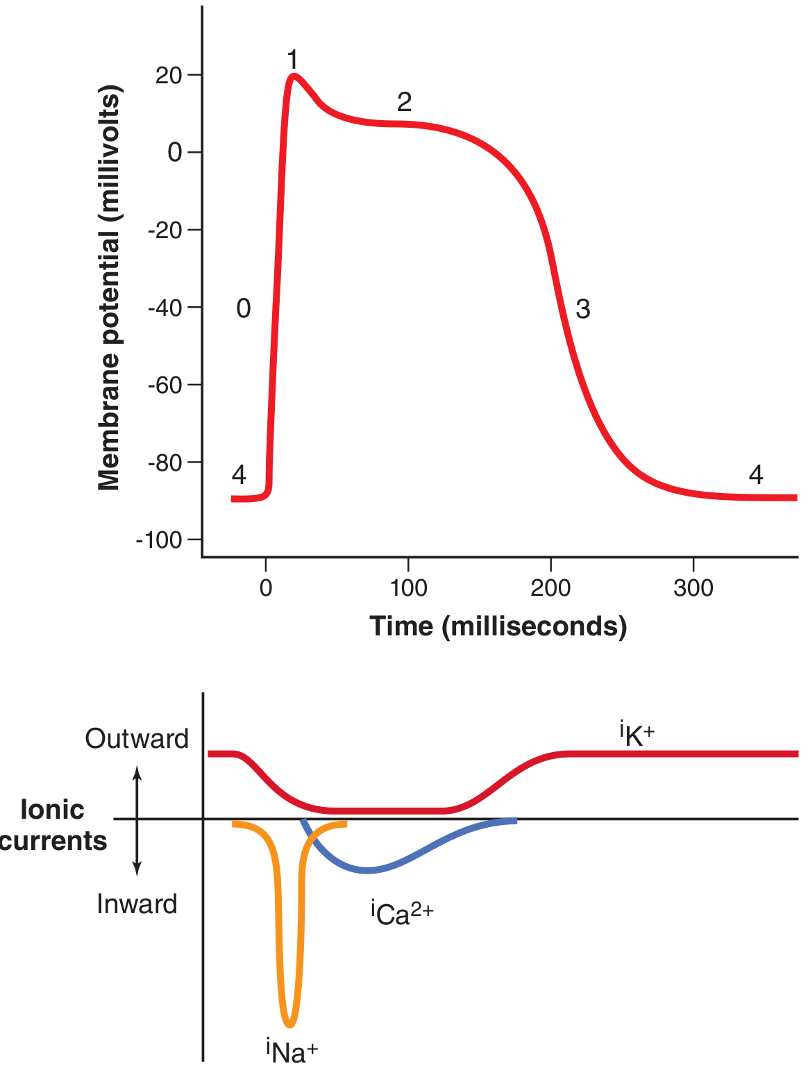
The ventricular action potential averages about 105 mV (resting: -85 mV, peak: +20 mV). Unlike skeletal muscle, the cardiac action potential has a prolonged plateau lasting about 0.2 seconds. This plateau is due to:
- Opening of L-type (slow) calcium channels, allowing sustained inflow of Ca²+ and Na+
- A simultaneous 5-fold decrease in K+ permeability, reducing outward K+ current
This makes the contraction of cardiac muscle last up to 15 times longer than skeletal muscle.
Phases of the Cardiac Action Potential (Fig. 9.5):
| Phase | Event | Ionic Basis |
|---|---|---|
| 0 | Rapid depolarization | Fast Na+ channels open; Na+ rushes in; membrane reaches +20 mV |
| 1 | Initial repolarization | Fast Na+ channels close; K+ exits |
| 2 | Plateau | L-type Ca²+ channels open (Ca²+ and Na+ enter); K+ channels close |
| 3 | Rapid repolarization | Ca²+ channels close; slow K+ channels open |
| 4 | Resting potential | -80 to -90 mV |
Excitation-Contraction Coupling
When the action potential spreads along the T tubules (which are 5x wider than those in skeletal muscle), it triggers L-type Ca²+ channels in the T tubule membrane. This Ca²+ entry activates ryanodine receptor channels in the sarcoplasmic reticulum (SR) membrane (calcium-induced calcium release), releasing a much larger Ca²+ store from the SR. The free intracellular Ca²+ then binds troponin to initiate cross-bridge cycling and contraction. Cardiac muscle depends on extracellular Ca²+ more than skeletal muscle because its SR is less well developed.
2. Ionic Basis of Autorhythmicity
Autorhythmicity (self-excitation) is the ability of certain cardiac fibers to initiate action potentials spontaneously and rhythmically without external neural input. It is a property of the SA node, AV node, and Purkinje fibers.
Why the SA Node Drives the Heart
The SA node (sinoatrial node) is a small strip of specialized cardiac muscle (3 mm x 15 mm x 1 mm) in the superior posterolateral wall of the right atrium. Its fibers have almost no contractile filaments but fire intrinsically at 70-80 beats/min - faster than the AV node (40-60/min) or Purkinje fibers (15-40/min). Since the SA node fires first each time, it suppresses spontaneous discharge in the lower centers before they reach threshold. The SA node is therefore the dominant pacemaker of the normal heart.
Ionic Mechanism - Why Sinus Nodal Fibers are "Leaky"
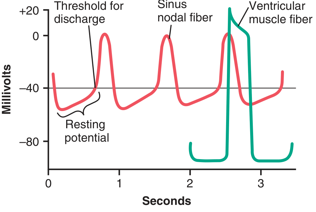
The resting potential of SA nodal fibers is only -55 to -60 mV (compared to -85 to -90 mV in ventricular muscle). At this less-negative potential, fast Na+ channels are already inactivated (their inactivation gates close when potential is less negative than -55 mV for more than a few milliseconds). Only slow L-type Ca²+ channels can open to generate the action potential - so the SA nodal action potential is slower to develop and return than the ventricular action potential.
The "Funny" (If) Current - Pacemaker Depolarization
Between heartbeats, Na+ ions from the extracellular fluid continuously leak inward through non-specific cation channels (HCN channels, carrying the "funny" current, I_f). This slow inward leak of positive Na+ (and some Ca²+) causes the resting membrane potential to drift progressively upward (less negative) - called the pacemaker potential or prepotential.
Threshold and Firing:
When this spontaneous depolarization reaches the threshold of about -40 mV, L-type Ca²+ channels are activated - generating the upstroke of the SA node action potential. After the action potential, repolarization occurs as Ca²+ channels close and K+ channels open. The resulting hyperpolarization is then followed by the same slow upward drift as K+ channels progressively close again and inward Na+/Ca²+ leakage resumes. This self-regenerating cycle continues indefinitely.
Summary of the Ionic Cycle of Autorhythmicity:
- End of action potential: K+ channels open → hyperpolarization to ~-60 mV
- K+ channels gradually close → inward Na+ leak (If/funny current) begins
- Resting potential slowly drifts upward toward threshold (-40 mV)
- Threshold reached → L-type Ca²+ channels open → action potential fires
- Cycle repeats automatically
3. Why the Heart Cannot be Tetanized
Tetanus in skeletal muscle occurs when a second stimulus is delivered before the muscle has finished relaxing from the first contraction, causing summation of contractions into a sustained, fused state. This is possible in skeletal muscle because its refractory period is very short (a few milliseconds) compared to its contraction duration.
Cardiac muscle cannot be tetanized because its refractory period is almost as long as the contraction itself.
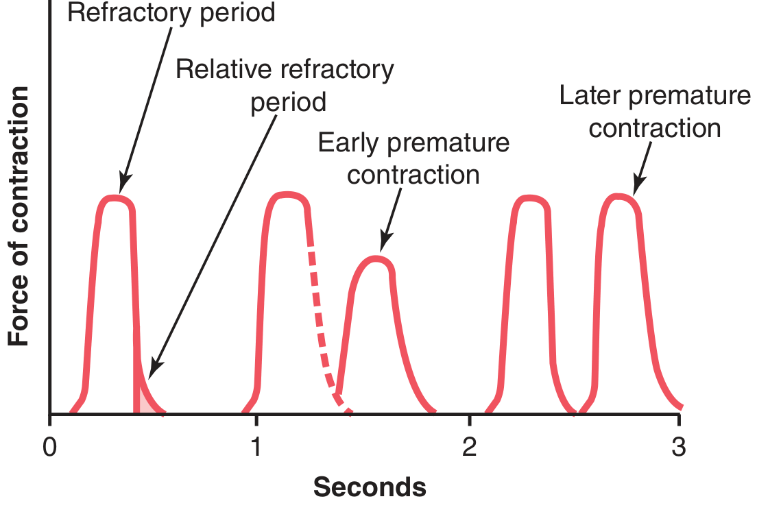
The normal absolute refractory period of ventricular muscle is 0.25 to 0.30 seconds - approximately equal to the duration of the plateau action potential and the period of ventricular contraction. During this entire period, no new stimulus, however strong, can re-excite the muscle. A second stimulus delivered during this window simply cannot produce another action potential.
There is also a relative refractory period of about 0.05 seconds immediately after the absolute refractory period, during which the muscle can only be excited by an unusually strong stimulus, producing a weaker-than-normal contraction (the "early premature contraction" shown below).
Why this Matters:
The prolonged refractory period is a direct consequence of the prolonged plateau of the cardiac action potential (maintained by L-type Ca²+ channels). Because the muscle cannot respond to a new stimulus until it has nearly fully relaxed and repolarized, wave summation and tetanus are physically impossible in cardiac muscle under normal physiological conditions.
This is of immense physiological importance - tetanization of the heart would stop its pumping action entirely (no filling between contractions), which would be immediately fatal. The long refractory period thus acts as a built-in protection against cardiac tetanus.
Key Comparison:
| Feature | Skeletal Muscle | Cardiac Muscle |
|---|---|---|
| Refractory period | ~2 ms (short) | 250-300 ms (long) |
| Contraction duration | ~20-100 ms | ~300 ms |
| Plateau in AP | Absent | Present (0.2 sec) |
| Tetanization | Possible | Impossible |
| Wave summation | Occurs | Does not occur |
Guyton and Hall Textbook of Medical Physiology, 4th South Asia Edition, Chapter 9 (pp. 122-126) and Chapter 10 (pp. 135-137)
This is a shared conversation. Sign in to Orris to start your own chat.