
Blister or wound in right side of the tounge near last tooth
tongue ulcer lateral border aphthous traumatic

This clinical photograph displays a black-and-white lateral view of a human oral cavity, focusing on a localized lesion on the tongue. A single, discrete ulcer is visible on the lateral border of the tongue. The lesion, identified as aphtha minor, is round-to-oval in shape with well-defined margins. It features a slightly depressed central base and is surrounded by a subtly raised or inflamed border. The ulcer appears relatively small, consistent with minor aphthous stomatitis (typically 3 to 10 millimeters in diameter). Other visible anatomical structures include the upper and lower teeth, the lips, the philtrum, and the inner cheek mucosa. The image serves as a clinical example of Recurrent Aphthous Stomatitis (RAS), specifically the minor type, which characteristically occurs on non-keratinized mobile mucosa such as the lateral tongue. This visual is relevant for medical education in oral pathology, dermatology, and general clinical diagnostics.
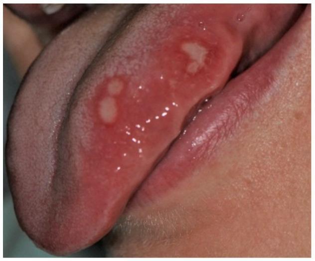
This clinical photograph shows a close-up view of the oral cavity focusing on the left lateral margin of the tongue. The image demonstrates minor aphthae, appearing as four discrete, oval-shaped ulcerations. Each ulcer is characterized by a central yellowish-whitish fibrinopurulent membrane that appears removable. A well-defined, prominent erythematous halo surrounds each lesion, signifying localized inflammation of the surrounding non-keratinized mucosa. The lesions are located on the ventral-lateral aspect of the tongue, a common site for recurrent aphthous stomatitis (RAS). This visual is representative of pediatric oral pathology, specifically illustrating the classic clinical presentation of minor aphthous ulcers in an adolescent patient. The morphology—small size, oval shape, and inflammatory border—is a key diagnostic feature for distinguishing these from other oral mucosal lesions like traumatic ulcers or herpetic stomatitis.
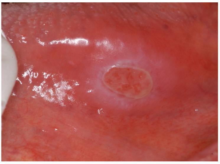
This clinical photograph displays a solitary, well-demarcated oral eosinophilic ulcer, also known as a Traumatic Ulcerative Granuloma with Stromal Eosinophilia (TUGSE). The lesion is located on the lateral border of the tongue, extending toward the ventral surface. The ulcer is characterized by an oval-shaped, erythematous central bed showing irregular texture and granulated tissue. A diagnostic hallmark visible here is the prominent, raised, indurated border with a pale or whitish fibrinous appearance, which clearly separates the ulcer from the surrounding inflamed (erythematous) mucosa. The surrounding tongue tissue appears smooth and lacks the typical papillary texture, likely due to local inflammation. This image is representative of an inflammatory reactive lesion often triggered by trauma. It is a key educational resource for distinguishing benign, self-limiting eosinophilic ulcers from more aggressive conditions like oral squamous cell carcinoma or major aphthous ulcers in clinical dentistry and oral pathology.
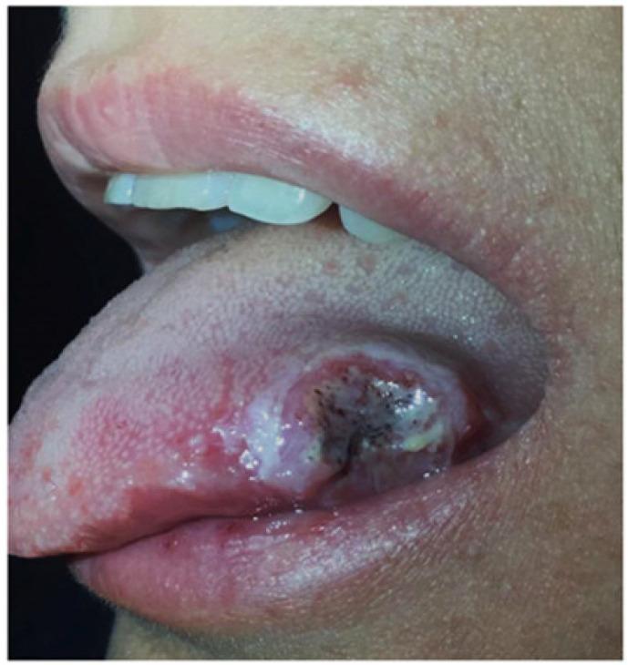
This clinical photograph displays a large, solitary aphthous ulcer on the lateral border and dorsal surface of the tongue in a patient with Crohn's disease. The lesion is characterized by a central necrotic base covered with a yellowish-white fibrinopurulent pseudomembrane and dark, irregular punctate areas. The ulcer has well-defined, raised, and intensely erythematous borders, forming a classic inflammatory halo. The surrounding lingual mucosa exhibits significant erythema and localized swelling, indicating secondary inflammation. Such oral manifestations are recognized as extraintestinal manifestations of inflammatory bowel disease (IBD), specifically Crohn's disease, and may correlate with systemic disease activity. This image serves as an educational example of aphthous stomatitis for gastroenterology and oral pathology, highlighting the importance of intraoral examination in the diagnosis and management of systemic inflammatory conditions.
Blister or Ulcer on the Right Side of the Tongue (Near Last Tooth)
What Is It Most Likely?
1. Traumatic Ulcer (Most Common at This Site)
- Appears exactly where the tooth edge contacts the tongue
- Painful, shallow, with a whitish/yellow base and red rim
- Usually single
- Heals in 7-14 days if the trauma source is removed
- Have a dentist smooth the rough tooth edge or adjust any sharp filling/crown
- Avoid pressing the tongue against the tooth
- Salt water rinses (1 tsp salt in warm water) 3-4 times a day
2. Aphthous Ulcer (Canker Sore)
- Round/oval ulcer with a yellowish-white center and a red halo (erythematous ring)
- Painful, especially on eating or speaking
- No blister phase before the ulcer appears
- Heals in 7-10 days without scarring (minor type)
- Triggered by stress, local trauma, certain foods (acidic), hormonal changes
- For mild ulcers: chlorhexidine mouthwash, topical analgesics (benzocaine gel), warm salt water rinses
- For more painful ulcers: topical steroid gel (e.g., triamcinolone acetonide in orabase) - applied directly to the ulcer after meals and at night
- Avoid spicy, acidic, or hard foods while healing
- Most heal on their own within 10 days
3. Herpetic Lesion (Cold Sore / HSV)
- Usually begins with a tingling/burning feeling
- Small fluid-filled blister(s) that rupture and leave shallow ulcers
- Can appear on the tongue, palate, or lips
- More common in immunocompromised individuals for intraoral HSV
| Feature | Herpes (HSV) | Aphthous Ulcer |
|---|---|---|
| Blister phase | YES | No |
| Location | Keratinized or non-keratinized mucosa | Non-keratinized only |
| Duration | 7-14 days | 7-10 days |
| Treatment | Antiviral (acyclovir) | Topical steroid |
4. When to Be Concerned (See a Doctor/Dentist)
- The ulcer does not heal in 2-3 weeks
- It is painless (a painless ulcer on the tongue can be a sign of oral cancer)
- It is hard, raised, or indurated around the edges
- It is getting bigger over time
- You use tobacco or alcohol heavily
- You have other symptoms: weight loss, difficulty swallowing, ear pain
Immediate Self-Care Tips
- Salt water rinse - 1/2 to 1 tsp salt dissolved in a glass of warm water, gargle/rinse 3-4 times/day
- Avoid irritants - spicy, sour, hot, or crunchy foods
- OTC topical gel - benzocaine (Orajel) or triamcinolone gel (available at pharmacy) for pain and faster healing
- Chlorhexidine mouthwash - helps prevent secondary infection
- See a dentist - to check if the last tooth has a sharp edge causing repeated trauma
Bottom Line
Disclaimer: This is educational information only. Please see a healthcare professional for a proper diagnosis and treatment.