
1) Medial maxillectomy- indications, steps, limitations, and complications 2) moure’s lateral rhinotomy, both in detail according to standard ent textbook and in simplified manner
I'll start by reading the medical library skill, then search for both topics in parallel.
Reading File
Now let me search for both topics in parallel across the medical library.
Finding Sources
Finding Sources
Finding Sources
Finding Sources
Excellent findings. Now let me read the key sections in parallel from multiple textbooks.
Reading File
Reading File
Reading File
Good. Now let me read more sections - the Scott-Brown's detailed content on lateral rhinotomy/medial maxillectomy, and the Cummings content on complications and limitations.
Reading File
Reading File
Reading File
Now let me fetch the key images and also read any more content about complications and the Cummings lateral rhinotomy figure section.
Reading File
Finding Sources
Loading Image
Now let me get the lateral rhinotomy diagram (Fig 176.21) and fetch more details on complications/limitations.
Loading Image
Finding Sources
Loading Image
I have excellent material from multiple authoritative textbooks plus the key images. Let me now compile the comprehensive answer.
1. MEDIAL MAXILLECTOMY
Indications
Medial maxillectomy is used for lesions involving up to the entire lateral nasal wall, but NOT extending to:
- The orbit
- Anterior cranial fossa
- Lateral maxilla (beyond the V2 plane)
- Alveolus/hard palate
A complete medial maxillectomy encompasses the middle turbinate, inferior turbinate, and the contents within the ethmoid and maxillary sinuses.
Specific indications include:
- Benign and low-grade malignant sinonasal neoplasms (inverted papilloma, low-grade adenocarcinoma, esthesioneuroblastoma - limited)
- Tumors of the lateral nasal wall and medial maxillary sinus
- Recurrent or extensive inverted papillomas with broad attachment to the lateral nasal wall
- As part of endoscopic sinonasal cancer surgery (endoscopic medial maxillectomy)
The endoscopic modified medial maxillectomy is specifically indicated for benign sinonasal neoplasms, requiring complete inferior turbinectomy and widening of the antral ostium. It is also used when mucociliary clearance is ineffective.
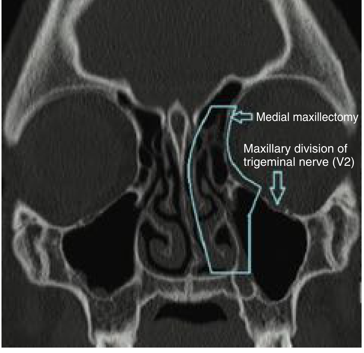
The lateral margin of the resection does not cross the plane of the maxillary division of the trigeminal nerve (V2) in the orbital floor - this is the key anatomical limit of a medial maxillectomy.
Steps (Open Approach via Lateral Rhinotomy)
(Cummings Otolaryngology, Scott-Brown's)
1. Incision - Lateral Rhinotomy
- An extended lateral rhinotomy incision is made
- After the incision, an upper cheek flap is raised
2. Anterior Maxillary Sinus Exposure
- The anterior wall of the maxillary sinus is burred out to allow exposure into the nasal and maxillary sinus cavities
3. Orbital Periosteum Elevation
- The medial wall periosteum is incised and elevated off the orbital rim and lamina papyracea
- The medial canthal ligament is transected and tagged with a suture for later reattachment to the nasal bone
4. Lacrimal System Management
- The lacrimal sac and duct are elevated
- The duct is transected flush against the orbital rim
- Further elevation is performed posteriorly
- The anterior and posterior ethmoid arteries are ligated
5. Osteotomies (Three Cuts)
- Cut 1 (inferior): A curved osteotome frees the medial wall of the maxillary sinus in a plane parallel to the nasal cavity floor, all the way back to the posterior sinus margin
- Cut 2 (superior): A similar cut made superiorly, back to the posteromedial orbital wall
- Cut 3 (outfracture): A cut outfractures the lamina papyracea from the maxilla, nasal bone, and orbital surface of the frontal bone
6. Specimen Delivery
- A bimanual rocking motion is performed to fracture the posterior ethmoid cells
- Angled scissors then transect the remaining posterior attachments near the choanae
7. Closure
- The nasolacrimal duct is stented
- The medial canthal ligament is sutured back to the nasal bone
- Meticulous two-layered closure is performed for soft tissue and skin
(Scott-Brown's adds: the resection can be extended into the sphenoid, frontal sinuses, or pterygopalatine fossa; packing is rarely required after resection)
Boundaries of Resection (Endoscopic Medial Maxillectomy)
(Cummings Fig. 94.28 - CT coronal shown below)
The anatomic segments resected are:
- Medial maxillary wall (to the palate)
- Uncinate process
- Inferior turbinate (with its attachment)
- Orbital lamina (if involved)
- Posterior margin can extend to the nasopharynx
- Lateral margin = V2 plane (not crossed)
Limitations
- Cannot be used when the lesion extends to the orbit, anterior cranial fossa, lateral maxilla, or alveolus - these require subtotal/total maxillectomy or craniofacial resection
- The lateral margin does not cross V2 in the orbital floor - extension beyond this requires a wider resection
- Involvement of the cribriform plate mandates a craniofacial resection approach
- Lesions with skin involvement may not be adequately addressed by medial maxillectomy alone
- Limited access to the anterolateral maxillary sinus - Denker's extension (endoscopic) or Weber-Ferguson approach needed
- Postoperative crusting is a known functional limitation - the modified technique preserving the inferior turbinate reduces this
Complications
(Cummings, Scott-Brown's, Miller's Anesthesia)
Intraoperative
- Bleeding - from anterior/posterior ethmoid arteries; internal maxillary artery branches
- Orbital entry - inadvertent penetration of orbital periosteum; ecchymosis/hematoma
- CSF leak - if cribriform plate is approached or violated
- Nasolacrimal duct injury - transection is deliberate but duct stenting reduces epiphora; inadvertent damage without stenting leads to dacryocystitis
Early Postoperative
- Epiphora - from disrupted nasolacrimal drainage (common if duct not stented)
- Orbital hematoma - requires urgent decompression
- Wound dehiscence/infection
- Crusting and synechiae formation
Late/Long-term
- Ectropion - due to lower eyelid involvement of the incision (if subciliary extension used); the modified lateral rhinotomy variation avoids this
- Medial canthal web/dystopia - if medial canthal ligament reattachment is suboptimal
- Epiphora (chronic) - dacryocystitis, stenosis
- Cosmetic deformity - asymmetry, notching of alar base
- Hypoesthesia in V2 distribution - infraorbital nerve damage
- Empty nose syndrome / hyposmia - from turbinate removal
- Diplopia - rare; from orbital floor manipulation
- Recurrence of tumor due to incomplete margins (especially inverted papilloma)
2. MOURE'S LATERAL RHINOTOMY
Historical Note
(Scott-Brown's Otorhinolaryngology)
Although this approach is usually attributed to Moure, it was actually originally described by Michaux some 50 years earlier in 1854. Moure's name is conventionally attached to it in ENT literature. The approach gives excellent exposure of both the nasal cavities and medial maxilla with a cosmetically acceptable incision in the lateral nasal crease.
Standard Textbook Description
Incision Design - The Three Landmark Points (Cummings Fig. 176.21)
The basic lateral rhinotomy connects three surface landmarks:
- Point 1 - halfway between the nasion and the medial canthus
- Point 2 - where the alar crease begins
- Point 3 - at the base of the columella
The incision runs along the lateral nasal crease from the medial canthal level, along the junction of the nasal sidewall, around the alar groove, to the columella base. A further inferior extension toward the philtrum/upper lip can be added.
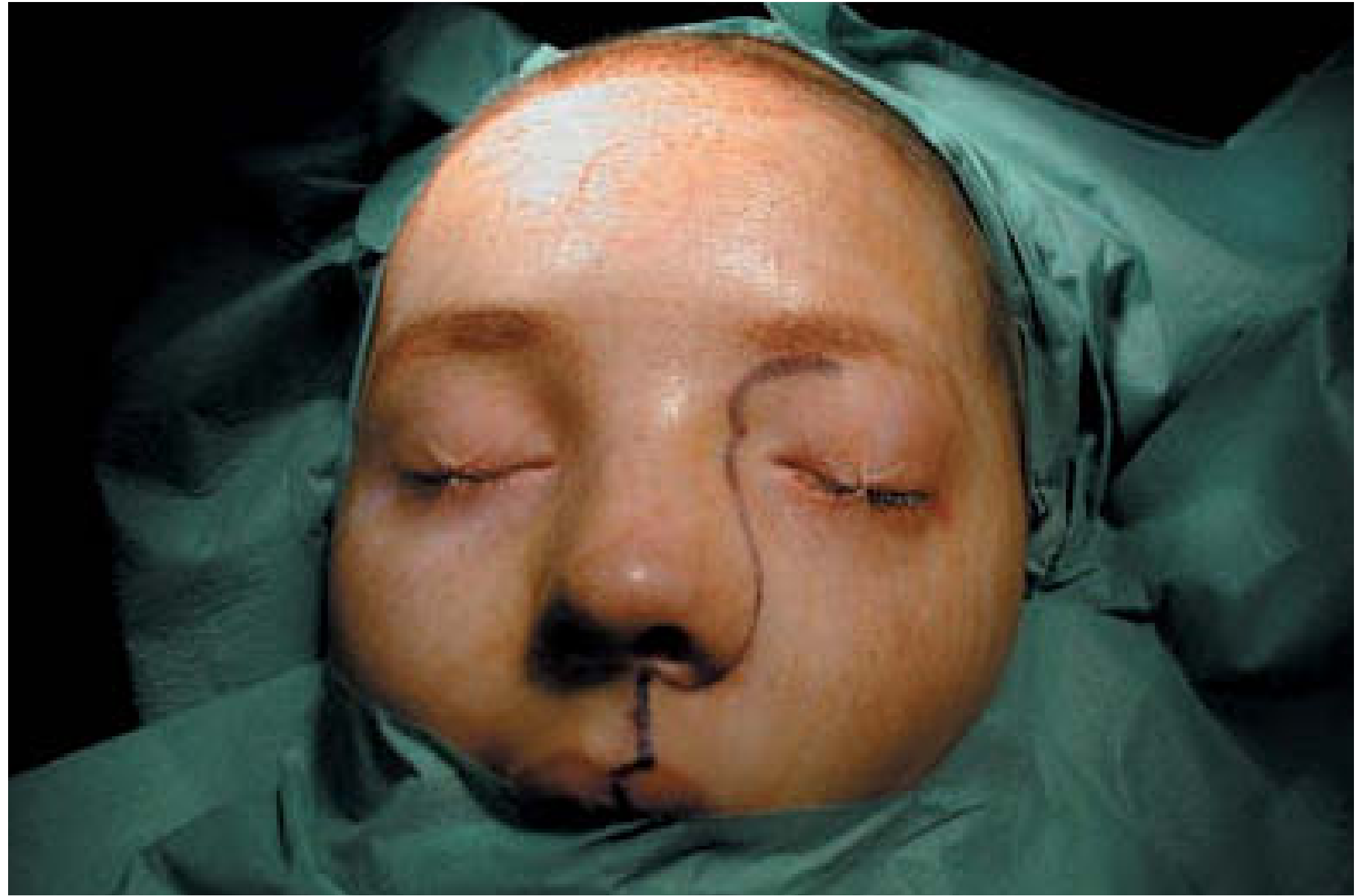
Standard (Cummings): The incision begins inferiorly at the philtrum above the lip, traverses across the nasal cavity floor and around the lateral nasal ala, then travels up along the junction of the nasal sidewall to the level of the medial canthus.
Modified variation (Cummings): Enters and exits the nasal vestibule floor at a 45-degree angle, hugs the lateral alar groove, and travels up the medial aspect of the sidewall - this modification avoids potential distortion of facial contour, ectropion, and cosmetic deformity.
Extensions of the Lateral Rhinotomy
(Cummings Fig. 176.21 - diagram below)
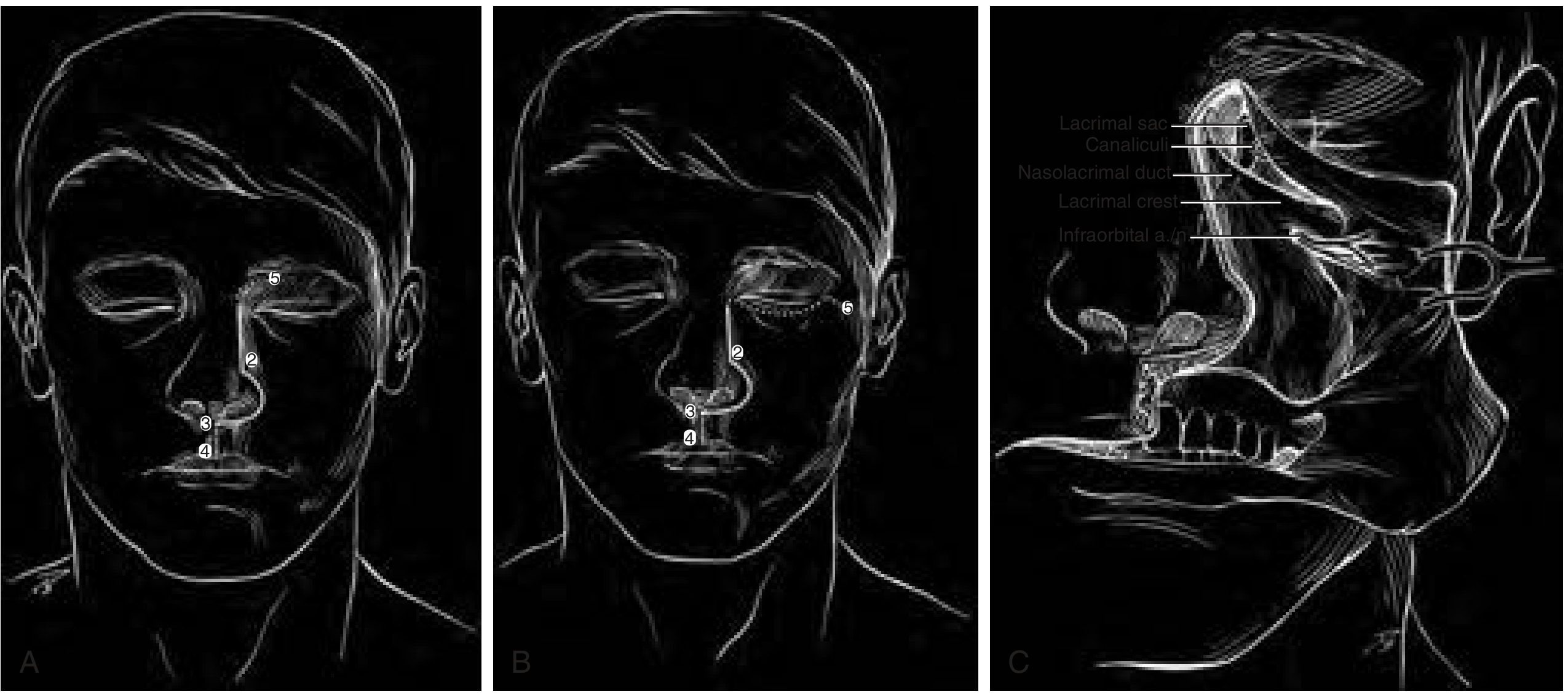
| Extension | Direction | Gives Access To |
|---|---|---|
| Lynch (cephalad) | Points 1 to 5 (up to medial eyebrow) | Medial orbital wall, medial canthal ligaments, lacrimal duct |
| Subciliary extension | 90° laterally across lower eyelid crease | Orbital floor, zygoma |
| Lip-split (Weber-Ferguson) | Points 3 to 4 (philtrum down to lip) | Inferior access to palate, total maxillectomy |
| Weber-Ferguson with subciliary | Combined | Full maxillectomy including orbital floor (subtotal/total) |
Surgical Steps of Lateral Rhinotomy
(Scott-Brown's + Cummings)
- Marking: Three landmark points are identified and the incision is drawn in the natural lateral nasal crease
- Incision: Full-thickness incision from just above the medial canthus level, along the lateral nasal wall, around the alar groove, to the columella base (or into the floor of the nasal vestibule for the modified approach)
- Flap elevation: An upper cheek flap is raised in the subperiosteal plane over the maxilla
- Orbital periosteum elevation: As for an external ethmoidectomy; extended laterally over the maxilla to the infraorbital nerve
- Lacrimal system: The lacrimal sac and duct are exposed by removing the overlying bone; the sac is divided low down, and the anterior ethmoidal artery is clipped and divided
- Trochlea: The insertion of the trochlea is freed by sharp dissection
- Bony window: Extended as required to access nasal cavities, ethmoids, antrum, with wide exposure - can be extended into the sphenoid, frontal sinuses, or pterygopalatine fossa
- Resection: Mucosa of the nose, antrum and ethmoids is resected along with the entire lateral nasal wall en bloc
- Closure: Meticulous layered closure; medial canthal ligament reattached; nasolacrimal duct stented
Subsequent partial maxillectomy via this approach allows access to deeper structures: paranasal sinuses, nasopharynx, and pterygopalatine fossa.
Indications for Lateral Rhinotomy
- Invasive sinonasal tumors involving anterior nasal cavity, facial skin, dentulo-alveolar, palatal structures, and maxilla
- Medial maxillectomy for lesions of the lateral nasal wall
- Tumors of the ethmoid sinuses and nasal cavity
- Access to nasopharynx, pterygopalatine fossa, and anterior sphenoid
- Combined with craniofacial resection for en bloc resection of extensive tumors
- Angiofibroma - surgical resection requiring adequate exposure (alongside midfacial degloving)
- Nasal glioma (extracranial lesions)
Simplified Explanation
Think of the lateral rhinotomy as drawing a line along the natural shadow crease that runs beside your nose - from just below the inner corner of your eye, down to the base of your nostril, and sometimes down to the upper lip. Because it follows a natural crease, the scar heals very well cosmetically.
Why is it done?
To open up a "door" on the side of the nose so the surgeon can reach tumors deep inside the nasal cavity or the sinuses behind the face.
The key steps in simple terms:
- Cut along the nasal crease (the "door" incision)
- Lift the soft tissues of the cheek off the underlying bone (like peeling back a flap)
- Move the tear-sac (lacrimal) out of the way and ligate the blood vessels
- Use osteotomes to chisel out the bone of the medial sinus wall (medial maxillectomy)
- Remove the tumor along with the lateral nasal wall en bloc
- Stitch everything back in layers, reattach the inner eyelid ligament, stent the tear duct
The extensions - think of it like a zipper:
- Zip it upward toward the eyebrow (Lynch extension) = access to the orbit
- Zip it across the lower eyelid (subciliary extension) = access to orbital floor
- Zip it down through the lip (Weber-Ferguson) = access to the palate for total maxillectomy
What it can and cannot reach:
- ✅ Lateral nasal wall, ethmoids, maxillary sinus medially, nasopharynx, pterygopalatine fossa
- ❌ Lesions involving the orbit proper, hard palate, lateral maxilla, or skull base (need bigger operations)
Sources
- Cummings Otolaryngology Head and Neck Surgery - Chapters 94 (Sinonasal Malignancies) and 176 (Skull Base Approaches)
- Scott-Brown's Otorhinolaryngology Head & Neck Surgery - Chapter 7 (Sinonasal Tumours)
- K.J. Lee's Essential Otolaryngology - Sinonasal Procedures
This is a shared conversation. Sign in to Orris to start your own chat.