
Definition Epidemiology Etiology Clinical picture Diagnosis Treatment Vaccination 51 to last Rabies
rabies virus negri bodies histology brain
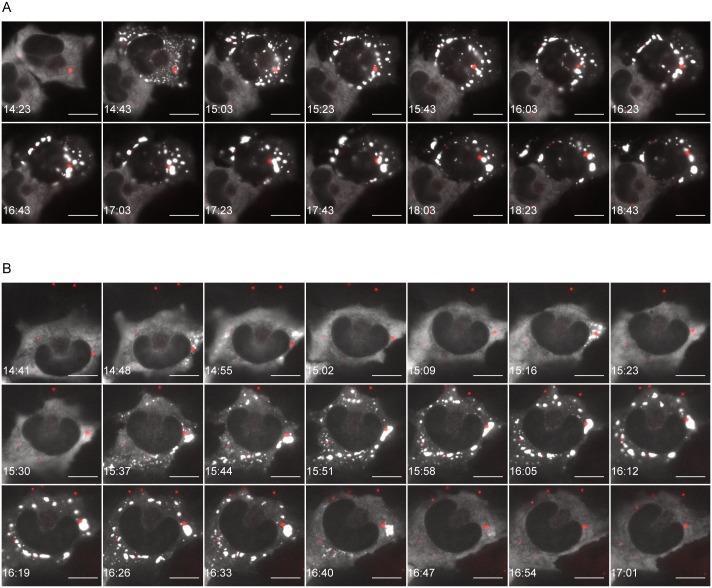
Time-lapse fluorescence microscopy of U373-MG cells infected with Rabies virus (RABV), illustrating the dynamics of stress granules (SGs) and Negri bodies (NBs). Panels A and B display two distinct temporal patterns of SG formation. The white signals represent G3BP-eGFP, a marker for SGs, while the red signals indicate P-mCherry, marking viral NBs. In cell A, a 'persistent pattern' is observed: initially diffuse cytoplasmic G3BP-eGFP (white) coalesces into bright punctate granules that increase in size through fusion and persist over several hours. In cell B, a 'transient pattern' is shown: G3BP-eGFP initially forms a peripheral ring-like structure near the cell membrane (15:30–16:26) before subsequently breaking up and losing signal intensity. Throughout both sequences, the red P-mCherry signals (NBs) appear as stable, distinct puncta, often in close proximity to but separate from the SGs. Scale bars represent 15 μm. This visualization demonstrates the liquid-droplet behavior and phase-transition properties of cytoplasmic RNA granules during viral infection.
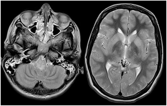
Two axial T2-weighted MRI scans of the brain demonstrating neuroimaging findings in a patient with Duvenhage virus (rabies) infection. The left image shows a cross-section of the posterior fossa, featuring the medulla oblongata, pons, and cerebellum. A distinct area of hyperintensity (increased signal) is indicated by an arrow in the posterior aspect of the medulla oblongata. The right image shows a higher axial section through the cerebral hemispheres, including the lateral ventricles and deep gray matter. Multiple white arrows highlight diffuse, bilateral areas of increased signal intensity within the basal ganglia. The ventricles appear fluid-filled and dark, while the surrounding brain parenchyma shows heterogeneous signals. These findings are consistent with viral encephalitis, specifically highlighting the predilection of rabies viruses for brainstem and deep gray matter structures. These diagnostic images illustrate the progression of infectious encephalitis and provide clinical evidence of brainstem and subcortical involvement.
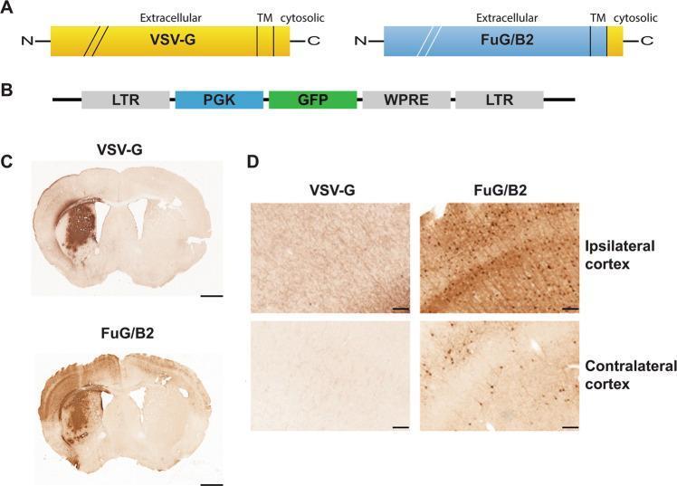
This composite educational image illustrates a comparative study of retrograde gene transport in the mouse brain using different lentiviral vector envelopes. Panel A displays diagrams of the VSV-G and FuG/B2 (rabies virus glycoprotein-derived) envelopes. Panel B shows the SIN-PGK-GFP-WPRE lentiviral vector construct used to express green fluorescent protein. Panel C provides coronal brain sections highlighting the primary transduction sites in the striatum. The VSV-G group shows localized staining at the injection site, whereas the FuG/B2 group shows broader diffusion. Panel D consists of high-magnification photomicrographs comparing retrograde transport in cortical regions. In the VSV-G group, the ipsilateral and contralateral cortex show only minimal GFP-positive fibers. Conversely, the FuG/B2 group demonstrates numerous GFP-positive neuronal cell bodies in both the ipsilateral and contralateral cortex, confirming the superior retrograde transport capabilities of the FuG/B2 pseudotyped vector for targeting distant connected brain regions. This visual material is used to teach neuroanatomical tracing and gene therapy delivery strategies.
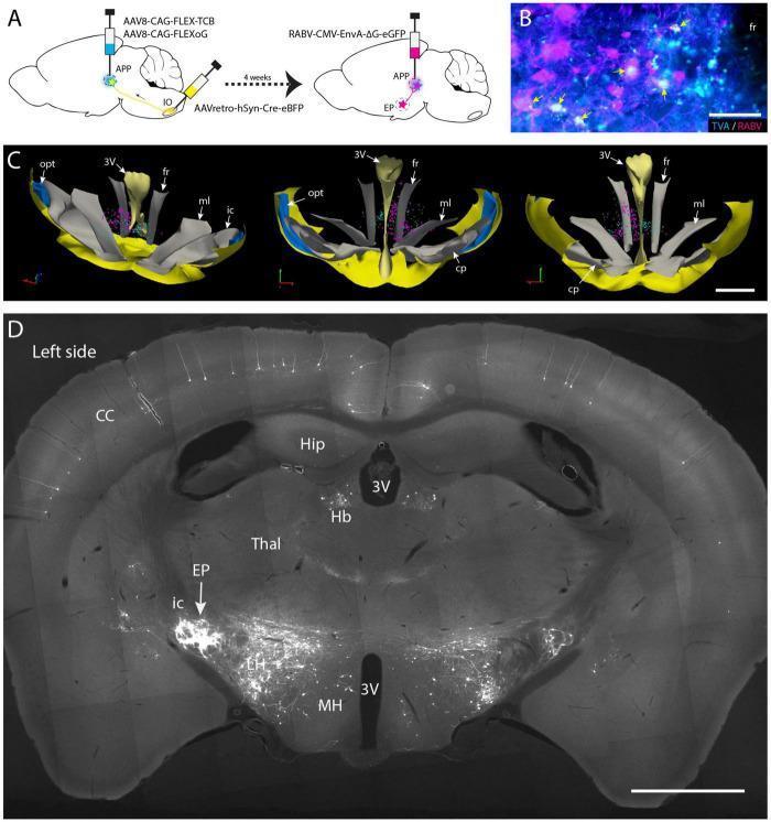
This composite educational graphic illustrates a neuroanatomical study using conditional monosynaptic rabies virus (RABV) tracing in a mouse brain. Panel A provides a schematic of the viral strategy, involving helper virus injections into the APP and AAVretro-Cre into the inferior olive (IO), followed by EnvA-pseudotyped RABV. Panel B is a fluorescence micrograph showing double-labeled 'starter cells' in the fasciculus retroflexus (fr) area, co-expressing TVA (cyan) and RABV (magenta). Panel C presents a 3D reconstruction from sequential brain sections, visualizing the spatial distribution of starter cells (magenta) and TVA-positive/RABV-negative neurons (cyan) at the mesodiencephalic junction. Panel D is a low-power coronal photomicrograph demonstrating successful retrograde transneuronal labeling. It reveals dense RABV-labeled neurons in the entopeduncular nucleus (EP) ipsilateral to the injection, with lighter labeling in the contralateral EP. Other visible labeled regions include the habenula (Hb), lateral hypothalamus (LH), and layer V pyramidal cells of the cerebral cortex (CC). Anatomical landmarks such as the third ventricle (3V), hippocampus (Hip), and thalamus (Thal) are clearly identified.
rabies clinical hydrophobia furious paralytic encephalitis
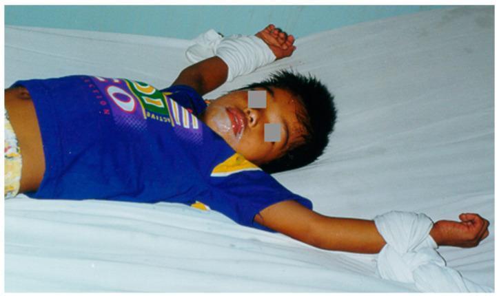
This clinical photograph displays a pediatric patient in a supine position on a white surface, illustrating the clinical manifestations of rabies encephalitis. A defining feature is the presence of thick, frothy salivary secretions around the mouth, a characteristic sign of autonomic dysfunction and pharyngeal spasms in rabies. The child's arms are abducted and secured with white cloth wrist restraints, a common clinical necessity to manage severe agitation or 'furious' symptoms associated with the disease progression. The image demonstrates the devastating presentation of encephalitic rabies and highlights the requirement for palliative management and safety protocols in advanced viral neuroinvasive diseases. Key educational concepts include rabies pathology, neurological agitation management, and infectious disease recognition.
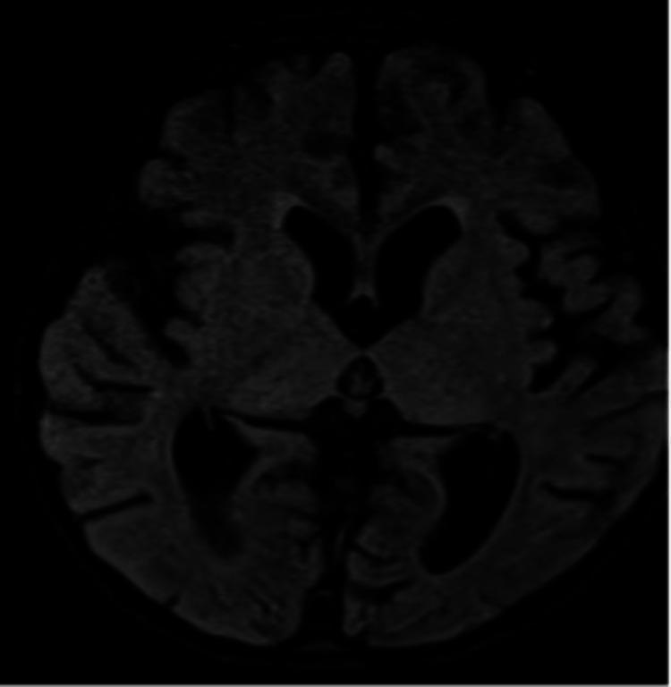
This diagnostic image is an axial MRI scan of the brain, specifically illustrating neuroanatomical changes associated with rabies encephalitis. The scan demonstrates diffuse cerebral atrophy, evidenced by prominent sulci and a generalized reduction in brain parenchyma volume. Associated with this tissue loss is ex-vacuo ventriculomegaly, characterized by the enlargement of the lateral and third ventricles. Pathological signal alterations are visible as hyperintensities in the bilateral thalami (deep gray matter nuclei) and bilateral peri-trigonal white matter, extending into the deep frontal regions. These findings represent the typical inflammatory and degenerative progression of viral encephalitis. The imaging captures the widespread nature of the pathology, involving both cortical areas and deep brain structures, which is critical for medical students and clinicians in identifying neurological manifestations of rabies infection.
Two axial T2-weighted MRI scans of the brain demonstrating neuroimaging findings in a patient with Duvenhage virus (rabies) infection. The left image shows a cross-section of the posterior fossa, featuring the medulla oblongata, pons, and cerebellum. A distinct area of hyperintensity (increased signal) is indicated by an arrow in the posterior aspect of the medulla oblongata. The right image shows a higher axial section through the cerebral hemispheres, including the lateral ventricles and deep gray matter. Multiple white arrows highlight diffuse, bilateral areas of increased signal intensity within the basal ganglia. The ventricles appear fluid-filled and dark, while the surrounding brain parenchyma shows heterogeneous signals. These findings are consistent with viral encephalitis, specifically highlighting the predilection of rabies viruses for brainstem and deep gray matter structures. These diagnostic images illustrate the progression of infectious encephalitis and provide clinical evidence of brainstem and subcortical involvement.
RABIES
1. DEFINITION
- Harrison's Principles of Internal Medicine 22E
2. EPIDEMIOLOGY
- Rabies is a zoonotic infection occurring in virtually all mammals worldwide except in Antarctica and some islands (Hawaii, UK, Australasia)
- Worldwide, endemic canine rabies causes ~59,000 human deaths annually, with most deaths in Asia and Africa; India accounts for approximately one-third of cases
- More than 3 billion people are at risk in over 100 countries
- The WHO estimates >15 million people receive post-exposure prophylaxis (PEP) annually
- Worldwide, >99% of human cases are acquired by dog bite
- Endemic canine rabies has been eliminated
- Wildlife reservoirs: bats, raccoons, skunks, foxes, coyotes
- Bat rabies virus variants are present in every state except Hawaii and are responsible for most indigenously acquired human rabies cases in the United States
- Raccoon rabies: endemic along the entire eastern coast
- Skunk rabies: midwestern states and California
- Fox rabies: New Mexico, Arizona, Alaska
- In 2021: 5 human rabies deaths; 2019, 2020, 2022: none
- 40,000-50,000 persons receive PEP annually in the US
| Vector | Region |
|---|---|
| Dogs | Asia, Latin America, Africa |
| Foxes | Europe, Arctic, North America |
| Skunks | Midwest US, Western Canada |
| Bats | North America, Latin America, Europe |
| Raccoons | Eastern US |
| Mongoose | Asia, Africa, Caribbean |
| No rabies | Hawaii, UK, Australasia, Antarctica |
- Harrison's 22E; Tintinalli's Emergency Medicine
3. ETIOLOGY
The Virus
-
Family: Rhabdoviridae
-
Genus: Lyssavirus (14 species divided into 3 phylogroups)
-
Genome: Single-stranded RNA, nonsegmented, negative-sense (antisense), 11,932 nucleotides
-
Shape: Bullet-shaped virion
-
Encodes 5 proteins:
- Nucleocapsid (N) protein
- Phosphoprotein (P)
- Matrix (M) protein
- Glycoprotein (G) - key for viral attachment and target of neutralizing antibodies
- Large polymerase (L) protein
-
Six other non-rabies lyssavirus species can cause a clinically similar picture
-
Rabies virus variants (characterized by nucleotide sequences) are associated with specific animal reservoirs - this is used epidemiologically to trace the source of infection
Transmission
-
Bite of an infected animal (main route)
-
Contamination of scratches, abrasions, or mucous membranes with saliva or neural tissue
-
Organ/corneal transplantation from patients dying of undiagnosed rabies (rare)
-
Possible aerosol exposure in bat caves and laboratories (extremely rare)
-
Person-to-person transmission by bite has NOT been documented
-
Harrison's 22E; Red Book 2021
4. PATHOPHYSIOLOGY
- Inoculation: Saliva containing infectious virus deposited in muscle and subcutaneous tissues by bite
- Initial replication: Virus remains near the wound site for much of the incubation period. Binds to nicotinic acetylcholine receptors at the neuromuscular junction (postsynaptic membrane)
- Peripheral spread: Virus spreads across the motor end plate and ascends via retrograde axoplasmic transport along peripheral nerve axons to the dorsal root ganglia and spinal cord
- CNS invasion: Rapid spread throughout the CNS via synapse-to-synapse transmission
- Centrifugal spread: Virus spreads outward from the CNS to peripheral nerves, salivary glands, cornea, skin (hair follicles), and other organs
- The salivary glands are infected, enabling transmission to the next host
- Histopathologic changes are surprisingly mild given the clinical severity
- Negri bodies - eosinophilic intracytoplasmic inclusions composed of viral proteins and RNA, found in a minority of infected neurons, most commonly in Purkinje cells of the cerebellum and hippocampal neurons - are pathognomonic but present in only ~70-80% of cases
- Perivascular cuffing and microglial nodules (Babes nodules) may be seen
5. CLINICAL PICTURE
Stage 1: Incubation Period
- Typical duration: 20-90 days (range: days to years)
- Averages 1-3 months; shorter with head/face bites (rich nerve supply, short distance to CNS)
- No symptoms
Stage 2: Prodrome (2-10 days)
- Nonspecific: Fever, malaise, headache, anorexia, nausea, vomiting, anxiety, agitation
- Specific early neurologic symptoms (pathognomonic clue): Paresthesias, pain, or pruritus near the site of the wound - occurring in 50-80% of patients; the wound has usually healed by this point
- These reflect infection/inflammation in local dorsal root or cranial sensory ganglia
Stage 3: Acute Neurologic Disease
a) Encephalitic (Furious) Rabies - 80% of cases
- Confusion, hallucinations, combativeness, bizarre behavior, seizures
- Autonomic dysfunction: Hypersalivation, lacrimation, perspiration, gooseflesh, cardiac arrhythmias, priapism
- Episodes of hyperexcitability alternating with periods of complete lucidity (becoming shorter as disease progresses)
- Hydrophobia (pathognomonic): Involuntary, painful spasm of the diaphragm and accessory respiratory, laryngeal, and pharyngeal muscles triggered by attempting to swallow liquids - due to dysfunction of brainstem neurons that normally inhibit inspiratory neurons near the nucleus ambiguus
- Aerophobia: Same spasm triggered by a draft of air
- Duration: 2-7 days
b) Paralytic (Dumb) Rabies - 20% of cases
- Flaccid paralysis beginning in the bitten limb, progressing to quadriparesis with facial paralysis
- Resembles Guillain-Barré syndrome; rabies should always be considered in the differential
- Duration: 2-10 days
Stage 4: Coma and Death
- 0-14 days after coma onset
- Death from respiratory failure, cardiac arrhythmia, or other complications
- Recovery is rare - rabies is almost universally fatal once clinical symptoms develop
| Stage | Duration | Features |
|---|---|---|
| Incubation | 20-90 days | None |
| Prodrome | 2-10 days | Fever, malaise; wound-site paresthesias/pain/pruritus |
| Encephalitic (80%) | 2-7 days | Hydrophobia, aerophobia, hyperexcitability, autonomic dysfunction |
| Paralytic (20%) | 2-10 days | Ascending flaccid paralysis, facial palsy |
| Coma/Death | 0-14 days | Respiratory failure |
- Harrison's 22E; Red Book 2021
6. DIAGNOSIS
Antemortem (Premortem) Tests
| Specimen | Test | Notes |
|---|---|---|
| Nuchal skin biopsy (nape of neck) | Direct fluorescent antibody (DFA) for viral antigen in nerve fibers around hair follicles | Most useful antemortem test |
| Saliva | RT-PCR for viral RNA; virus isolation | Virus present due to salivary gland infection |
| Serum | Rabies virus-neutralizing antibodies | Diagnostic in unvaccinated patients; may not appear until late |
| CSF | Neutralizing antibodies; RT-PCR | Antibodies in CSF are diagnostic in all patients (vaccinated or not) |
| Skin biopsy | RT-PCR |
- In a previously unimmunized patient, serum neutralizing antibodies are diagnostic
- Because rabies virus infects immunologically privileged neuronal tissues, antibodies may not develop until late in the disease
- RT-PCR plays a greater role in antemortem diagnosis in the absence of brain biopsy
Postmortem Tests
- Direct fluorescent antibody (DFA) on brain tissue - gold standard
- Immunohistochemistry or immunofluorescence on brain
- RT-PCR on brain tissue
- Negri bodies on histology (H&E stain) - pathognomonic but insensitive (~70-80%)
- Virus isolation in suckling mice or cell culture
Ancillary Tests (to exclude other diagnoses)
- CSF: Mononuclear pleocytosis, mildly elevated protein; severe pleocytosis (>1000/μL) is unusual in rabies
- CT head: Usually normal in rabies
- MRI brain: May show signal abnormalities in brainstem or gray matter (variable and nonspecific; bilateral thalami, basal ganglia, brainstem hyperintensity)
- EEG: Nonspecific abnormalities
- Harrison's 22E; Red Book 2021
7. TREATMENT
Established Disease (Symptomatic Rabies)
- There is no specific proven treatment for rabies once clinical symptoms appear
- Neither rabies vaccine nor Rabies Immune Globulin (RIG) improves prognosis once symptoms develop
- Management is supportive and palliative: sedation, pain control, intensive care
The Milwaukee Protocol
- An intensive care protocol developed at the Medical College of Wisconsin
- Uses therapeutic coma (ketamine, midazolam), amantadine, ribavirin, and other antiviral agents
- Has resulted in several survivors (see below)
- Details at www.mcw.edu/rabies
Survivors
- Approximately 19 documented survivors of rabies exist worldwide
- 11 survivors were associated with incomplete rabies vaccine schedules
- 8 survivors had received no PEP at all
- Approximately half of survivors have normal cognition; others have severe neurologic deficits
- A 15-year-old girl (Jeanna Giese, 2004) was the first documented unvaccinated rabies survivor using the Milwaukee Protocol
Isolation
-
Standard precautions with face mask, eye protection, gown, and gloves for all patient care
-
If a bite occurs from the patient or infectious material contacts a wound/mucous membrane, thorough washing with soap and water + risk assessment for PEP is required
-
Harrison's 22E; Red Book 2021
8. VACCINATION / PROPHYLAXIS
Available Vaccines (US)
- Human Diploid Cell Vaccine (HDCV) - Imovax Rabies
- Purified Chick Embryo Cell Vaccine (PCECV) - RabAvert
- Both are inactivated rabies vaccines given intramuscularly (IM) in the deltoid (adults) or anterolateral thigh (children); never in the gluteal region (poor immune response)
A. Pre-Exposure Prophylaxis (PrEP)
- Veterinarians and animal handlers
- Laboratory workers handling rabies virus
- Cave explorers
- Travelers to areas with enzootic canine rabies where access to PEP may be limited
- 3 doses of rabies vaccine: Days 0, 7, and 21 (or 28)
- Confers partial protection; if subsequently exposed, still requires PEP but no RIG needed, and only 2 booster doses required (Days 0 and 3)
- Booster doses for those at continued risk based on serology (every 2 years for high-risk; check titers every 6 months for continuous risk)
B. Post-Exposure Prophylaxis (PEP)
Step 1 - Wound Care (Most important first step)
- Immediately and thoroughly wash the wound with soap and water for at least 15 minutes
- Apply virucidal agent (povidone iodine or 70% ethanol)
- This alone can significantly reduce risk of infection
Step 2 - Assess Exposure Risk
| Animal | Action |
|---|---|
| Dogs, cats, ferrets - healthy, available for observation | Observe for 10 days; start PEP only if animal develops signs of rabies |
| Dogs, cats, ferrets - suspected rabid or escaped | Immediate immunization + RIG |
| Bats, raccoons, skunks, foxes, coyotes, mongooses | Regard as rabid; immediate immunization + RIG unless lab proves otherwise |
| Small rodents (squirrels, mice, rats, hamsters), lagomorphs | Rarely require prophylaxis; consult public health |
| Bat in room with sleeping/unaware person | Consider PEP even without confirmed bite |
Step 3 - Rabies Immune Globulin (RIG)
- 20 IU/kg of human RIG (HRIG) or equine RIG
- Infiltrate as much as possible directly into and around the wound
- Remaining volume given IM at a distant site from the vaccine
- Provides immediate passive immunity while the active vaccine response develops
- Given on Day 0 only (with first vaccine dose); NOT to be repeated
- Not indicated in previously vaccinated individuals
Step 4 - Vaccine Schedule (PEP)
- 5 doses of vaccine: Days 0, 3, 7, 14, and 28
- Given IM in the deltoid (adults) or anterolateral thigh (children)
- 2 doses only: Days 0 and 3
- RIG is NOT required
C. Animal Vaccination Programs
- Mass dog vaccination is the primary strategy for eliminating canine rabies worldwide (WHO target: zero human deaths from dog-mediated rabies by 2030)
- Oral bait vaccines have been highly effective for controlling fox rabies in Europe and raccoon rabies in North America
- Domestic animals (dogs, cats) should be vaccinated per local regulations
D. WHO Wound Categories and PEP Recommendations
| Category | Nature of Contact | Recommended Treatment |
|---|---|---|
| I | Touching or feeding animal; licks on intact skin | None (if reliable history) |
| II | Nibbling of uncovered skin; minor scratches/abrasions without bleeding | Wound treatment + vaccine |
| III | Single or multiple transdermal bites or scratches; licks on broken skin; mucous membrane contamination; bat exposures | Wound treatment + vaccine + RIG |
- Harrison's 22E; Red Book 2021; Tintinalli's Emergency Medicine
Key Points Summary
- Rabies is caused by a bullet-shaped, negative-sense ssRNA lyssavirus
- Transmitted mainly by dog bite globally; by bats in the US
- Pathognomonic finding: Negri bodies (eosinophilic intracytoplasmic inclusions in neurons, especially Purkinje cells)
- Incubation: 20-90 days (may range to years)
- Two clinical forms: Encephalitic (furious, 80%) - hydrophobia, aerophobia; Paralytic (dumb, 20%) - ascending flaccid paralysis
- Almost universally fatal once symptomatic
- Diagnosis: No single test sufficient; panel includes DFA on skin biopsy, RT-PCR on saliva/CSF, serum/CSF neutralizing antibodies
- Treatment: No proven specific therapy; Milwaukee Protocol (therapeutic coma + antivirals) used in some centers
- PEP: Immediate wound washing + RIG (20 IU/kg) + 5 doses vaccine (Days 0, 3, 7, 14, 28) for unvaccinated
- PrEP: 3 doses (Days 0, 7, 21/28) for high-risk individuals
- WHO 2030 goal: Eliminate dog-mediated human rabies deaths