
Mouse bite treatment
rodent bite wound management treatment
Mouse Bite Treatment
1. Immediate First Aid
- Wash the wound thoroughly with soap and running water for at least 5 minutes. This is the single most important step.
- Apply an antiseptic (e.g., povidone-iodine or chlorhexidine) to the wound.
- Control bleeding with gentle pressure.
- Do not close the wound with tape or sutures immediately - bite wounds are prone to infection and benefit from being left open (or loosely approximated) to allow drainage.
2. Assess Tetanus Status
- Confirm the patient's tetanus immunization history.
- Give tetanus toxoid booster if not vaccinated within the last 5 years, or the history is unknown.
- Give tetanus immunoglobulin (TIG) if the patient has never been fully immunized.
3. Antibiotic Management
| Scenario | First-Line | Alternative (Penicillin Allergy) |
|---|---|---|
| Prophylaxis (early, uninfected wound) | Amoxicillin-clavulanate 875/125 mg PO q12h | Doxycycline 100 mg PO bid |
| Established infection / Rat-bite fever treatment | Penicillin VK 500 mg PO qid OR Ceftriaxone IV | Doxycycline 100 mg PO bid |
- Antibiotic prophylaxis for rodent bites is advised sometimes (use clinical judgment based on wound severity, patient immune status, and depth of bite).
- Both Streptobacillary and Spirillary forms of rat-bite fever respond readily to penicillin or doxycycline.
4. Rat-Bite Fever - What to Watch For
- Incubation: ~10 days after bite
- Chills, fever, then morbilliform rash (spreading to palms/soles) within 2-4 days
- Arthralgia is prominent
- Complications: endocarditis, pneumonia, septic infarcts; 10% mortality if untreated
- Incubation: 1-4 weeks
- Bite site becomes inflamed or ulcerated; lymphangitis may appear
- Purplish-red indurated plaques on the abdomen
- Complications: endocarditis, meningitis, nephritis, hepatitis; ~6% mortality if untreated
5. Rabies Assessment
- Rodent (including mouse) bites rarely require rabies post-exposure prophylaxis (PEP).
- Mice, rats, squirrels, and other small rodents are very low-risk rabies vectors in most regions.
- However, assess local epidemiology and consult public health authorities if:
- The animal was behaving abnormally
- The bite was unprovoked
- You are in a high-risk geographic area
6. Wound Care Follow-Up
- Inspect daily for signs of infection: redness, swelling, warmth, purulent discharge, streaking (lymphangitis).
- Return immediately if fever develops within the first 2 weeks after the bite - this may signal rat-bite fever or bacteremia.
- Do not suture most rodent bite wounds primarily; loosely approximate if needed for cosmesis on the face only.
7. Summary Flowchart
Mouse bite
    ↓
Immediate wound wash (5 min soap + water) + antiseptic
    ↓
Tetanus assessment → boost if needed
    ↓
Wound assessment: depth, location, immune status
    ↓
Antibiotic: amoxicillin-clavulanate 875/125 mg PO q12h (prophylaxis)
    ↓
Rabies risk assessment (usually low for mice)
    ↓
Monitor 2 weeks for fever, rash, arthralgia → suspect rat-bite fever
    ↓
If rat-bite fever: Penicillin VK or doxycycline
Weils diseases
leptospirosis Weil disease treatment
leptospirosis jaundice conjunctival suffusion clinical presentation
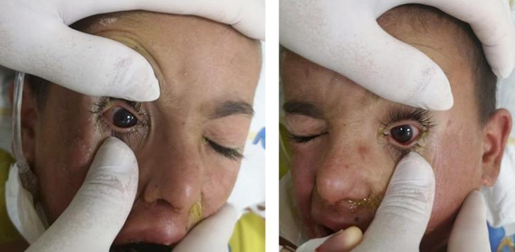
This clinical photograph displays a pediatric patient exhibiting bilateral conjunctival suffusion, a classic diagnostic sign of systemic infection such as leptospirosis. The image captures the periocular region with the eyelids being manually retracted by gloved hands to reveal marked, diffuse injection of the bulbar conjunctiva. Both eyes show prominent vascular engorgement and redness against the sclera, without evidence of purulent exudate directly on the ocular surface. Associated findings include periorbital edema and yellow-crusted discharge (serosanguinous or purulent) concentrated at the inner canthus and along the eyelid margins. The surrounding facial skin shows generalized erythema and scattered hyperpigmented macules. The clinical context suggests an acute infectious process, likely during the spirochaetemic phase of a zoonotic illness. This visual serves as a critical educational tool for identifying systemic vasculitis manifestations and differentiating conjunctival suffusion from typical bacterial conjunctivitis in a febrile, acutely ill patient.
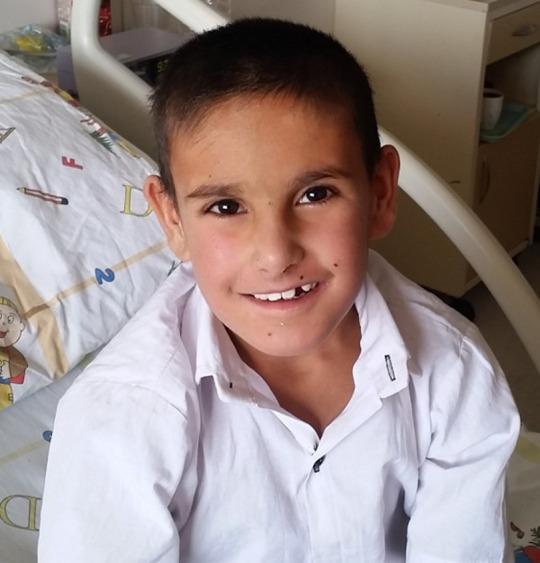
Clinical photograph of a 7-year-old male patient upon discharge from a Pediatric Intensive Care Unit following recovery from severe leptospirosis. The patient is shown in a seated position within a healthcare environment, indicated by a hospital bed with pediatric-themed linens in the background. He is alert and oriented, exhibiting a normal skin tone with mild residual malar flushing. A small benign-appearing nevus is noted adjacent to the left nasal ala. The patient's smile reveals typical mixed dentition for his age group, with a missing primary tooth in the upper right quadrant. No visible signs of the previously reported diffuse erythema or conjunctival suffusion remain. The image serves as a visual documentation of clinical improvement and physiological stability following treatment for multisystem involvement, including acute kidney injury and hemodynamic instability caused by Leptospira infection.
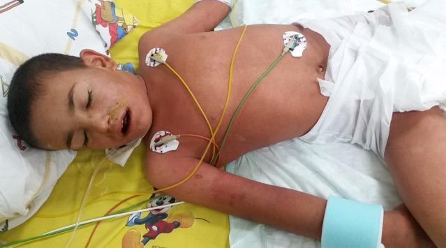
This clinical photograph displays a pediatric patient in an intensive care setting presenting with generalized erythema. The patient exhibits a diffuse, confluent macular rash characterized by intense redness across the face, torso, abdomen, and upper extremities. Notable clinical signs include bilateral conjunctival suffusion and crusting/secretions around the nasal and oral orifices. The image documents several medical interventions and monitoring devices, including a nasogastric or feeding tube, multiple ECG adhesive electrodes on the chest for continuous cardiac monitoring, and a soft blue safety restraint on the left wrist. The clinical context suggests a systemic inflammatory or infectious process, such as severe leptospirosis or toxic shock syndrome (STSS). This photograph is an educational example of multisystem disease manifestation in a pediatric patient, emphasizing the dermatological and clinical findings associated with severe systemic infections and the requirements for critical care monitoring.
Weil's Disease (Icteric Leptospirosis)
Definition
- Jaundice (hepatocellular + cholestatic)
- Acute kidney injury (AKI)
- Hemorrhagic diathesis (bleeding tendency)
Causative Organism
- Spirochete: Leptospira interrogans (17 pathogenic species described)
- Primary reservoir: rats (and other rodents, cattle, dogs)
- Transmission: contact with water/soil contaminated with infected animal urine
- High-risk exposures: flooding, sewage workers, farmers, adventure racers, swimmers in natural water bodies
Biphasic Clinical Course
Phase 1 - Leptospiraemic Phase (Days 1-7)
- High fever, chills, severe headache
- Myalgia (especially calf muscles - a hallmark)
- Conjunctival suffusion (not conjunctivitis - no exudate, just redness)
- Nausea, vomiting, abdominal pain
Phase 2 - Immune / Weil's Phase (Days 7-14+)
- Jaundice - deep, with elevated direct bilirubin
- Oliguria/anuria - non-oliguric renal insufficiency progressing to AKI
- Hemorrhage - petechiae, ecchymoses, GI bleeding, hemoptysis
- Altered mental status, arrhythmias, hypotension
- Severe pulmonary hemorrhage syndrome - increasingly recognized, mortality up to 50%
Pathophysiology
- Leptospires penetrate mucous membranes or abraded skin and enter the bloodstream
- They invade endothelial cells, disrupting barrier function and causing vasculitis
- Endothelial activation elevates soluble E-selectin and von Willebrand factor
- Thrombocytopenia from platelet consumption contributes to hemorrhage
- DIC can develop (elevated D-dimer, thrombin-antithrombin complexes, reduced antithrombin and protein C)
- Hepatic injury: disruption of bile canaliculi causes cholestasis
- Renal: tubular injury leads to potassium wasting, hypokalemia, and AKI
Investigations
| Test | Finding |
|---|---|
| WBC | Leukocytosis >80% neutrophils |
| Platelets | Thrombocytopenia (<100,000/µL in severe cases) |
| Coagulation | Elevated INR, aPTT, D-dimer |
| Bilirubin | Elevated (direct > indirect) |
| Liver enzymes | Moderately elevated ALT/AST |
| Creatinine/BUN | Elevated; pyuria, hematuria on urinalysis |
| Procalcitonin/CRP | Elevated (helps differentiate from dengue) |
| Chest CT | Ground-glass opacities, airspace nodules (diffuse alveolar hemorrhage) |
| CSF (if meningism) | Aseptic meningitis: lymphocytes ++, elevated protein, normal glucose |
Diagnostic Confirmation
- PCR on blood, urine, or CSF - most sensitive in early disease (method of choice)
- Culture from blood/urine - specialized media (EMJH), positive early before antibiotics
- Microscopic Agglutination Test (MAT) - gold standard serology; titre ≥1:400 single sample, or 4-fold rise diagnostic
- IgM ELISA/rapid test - IgM detectable from week 1; useful in resource-limited settings
- Suspect: febrile illness ≥38.5°C + epidemiologic exposure
- Probable: suspect + positive rapid IgM + 3 of: urinary abnormalities, neutrophilia >80%, platelets <100,000, bilirubin >2 mg/dL
- Confirmed: isolation, positive PCR, or MAT seroconversion
Differential Diagnosis
| Condition | Distinguishing Feature |
|---|---|
| Dengue fever | No jaundice, rash pattern differs, negative MAT |
| Malaria | Blood film positive, no conjunctival suffusion |
| Typhoid fever | Rose spots, positive Widal/blood culture |
| Viral hepatitis | No myalgia/conjunctival suffusion, exposure history |
| Hantavirus | Pulmonary-predominant, renal, no jaundice |
| Scrub typhus | Eschar, different geographic exposure |
Treatment
Mild / Outpatient
| Agent | Dose |
|---|---|
| Doxycycline (1st line) | 100 mg PO twice daily x 7 days |
| Azithromycin | 500 mg PO once daily x 3 days |
| Pregnant women | Azithromycin 500 mg OD x 3 days OR Amoxicillin 500 mg TDS x 7 days |
Severe / Inpatient (Weil's Disease)
| Agent | Dose |
|---|---|
| Penicillin G (1st line) | 1.5 million units IV every 6 hours x 7 days |
| Ampicillin | 0.5-1 g IV every 6 hours x 7 days |
| Ceftriaxone (alternative) | 1 g IV every 24 hours x 7 days |
Jarisch-Herxheimer reaction may occur after starting antibiotics (fever, rigors, tachycardia, hypotension) - manage supportively, do NOT stop antibiotics.
Supportive Care
- IV fluids to prevent AKI and correct dehydration
- Potassium supplementation for hypokalemia
- Dialysis (peritoneal or haemodialysis) for oliguric AKI - usually short-term
- Protective ventilation (low tidal volumes <6 mL/kg, high PEEP) for pulmonary hemorrhage
- ECMO has been used successfully in severe respiratory failure
- Therapeutic plasma exchange - a 2025 systematic review (PMID: 39574344) investigated its use in leptospirosis-associated severe pulmonary hemorrhage syndrome
Prognosis
| Factor | Odds Ratio of Death |
|---|---|
| Pulmonary involvement | 6.0 |
| Oliguria | 3.0 |
| Creatinine >3.0 mg/dL | 2.3 |
| Age >40 years | 2.2 |
| Platelets <70,000/µL | 2.2 |
| Age >60 vs ≤19 years | ~11x higher |
- Overall hepatorenal failure mortality: ~10%
- Overall hospitalized case fatality in one large study: 24%
- Severe pulmonary hemorrhage syndrome mortality: up to 50%
- Altered mental status is the strongest predictor of death
Prevention
- Avoid contact with contaminated water/soil; use PPE (boots, gloves, goggles)
- Rodent control
- Chemoprophylaxis for high-risk exposure (flooding, water sports, endemic workers): Doxycycline 200 mg or Azithromycin 500 mg orally once weekly, starting 1-2 days before exposure
- Animal vaccination in endemic settings
make a banner like photo in which you will describe about hpv vaccination 2026 and details regarding it add one or two photos for campaign program from community medicine department vimsar odisha
HPV vaccination India 2026 schedule guidelines girls cervical cancer
A professional wide-format medical campaign banner for HPV Vaccination 2026, landscape orientation. TOP SECTION: Bold header text "HPV VACCINATION CAMPAIGN 2026" in large white letters on a deep teal/green gradient background. Below it in smaller text: "Department of Community Medicine, VIMSAR, Burla, Odisha" LEFT SIDE: A realistic illustration of a female healthcare worker in white coat giving an injection to a teenage girl (around 14 years old), both smiling, in a clinic/community health setting with warm lighting. CENTER SECTION (white/light background panel): Key information displayed clearly: - "CERVAVAC - India's Own HPV Vaccine" (with small Indian flag icon) - "Protect Against Cervical Cancer" - Target: Girls aged 9-14 years (Priority: 14-year-olds under UIP) - 2-Dose Schedule: 0 & 6 months (ages 9-14) | 3 doses (ages 15+) - FREE under Universal Immunization Programme - Launched: 28 February 2026 by Government of India RIGHT SIDE: A second illustration showing a community health worker educating a group of women/mothers in a village setting, with awareness posters in background. BOTTOM STRIP: Deep teal banner reading "ELIMINATE CERVICAL CANCER | VACCINATE TODAY | For appointments contact: Department of Community Medicine, VIMSAR Burla - 768004, Odisha" Small ribbon symbol (teal) for cervical cancer awareness. Clean, professional, government health campaign aesthetic with orange and teal color scheme (Odisha state colors). Include the caduceus/medical cross symbol.
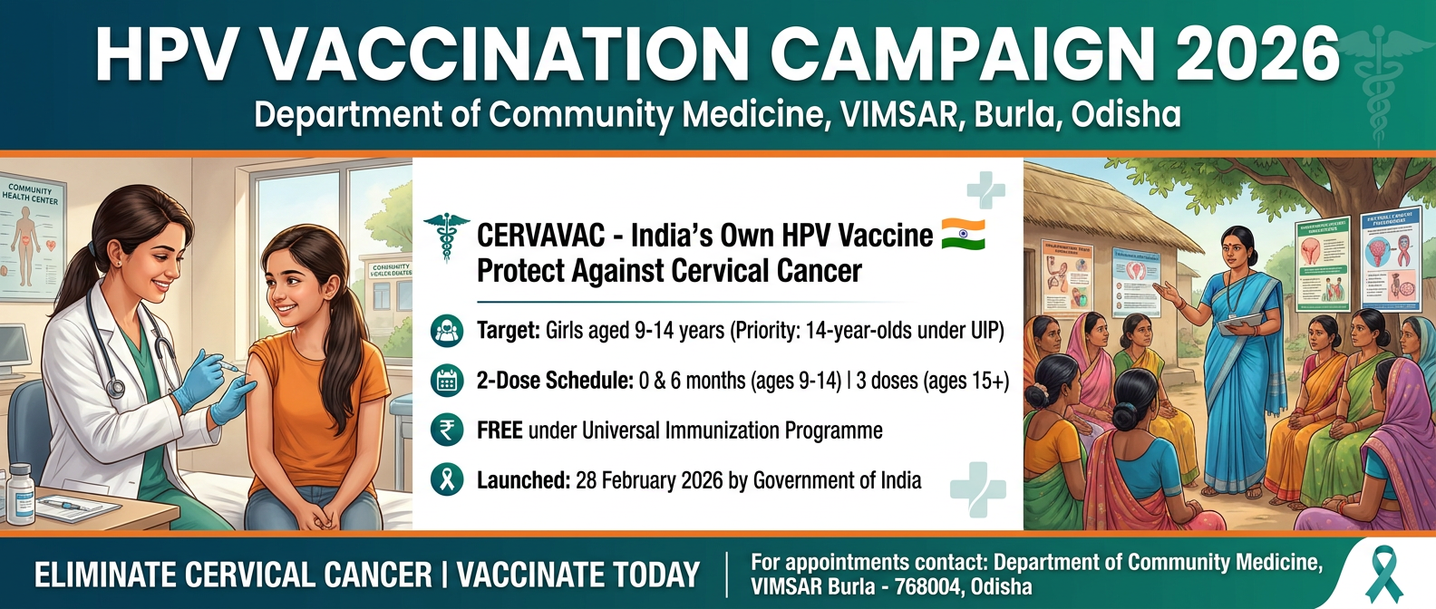
- Campaign title - HPV Vaccination Campaign 2026
- Department credit - Community Medicine, VIMSAR Burla, Odisha 768004
- Vaccine - CERVAVAC (India's indigenously developed quadrivalent HPV vaccine by Serum Institute of India)
- National launch - 28 February 2026, launched by PM Modi under the Universal Immunization Programme (UIP)
- Target group - Girls aged 9-14 years (priority: 14-year-olds); catch-up for girls/women up to 26-45 years
- Schedule:
- Ages 9-14: 2 doses at 0 and 6 months
- Ages 15 and above: 3 doses at 0, 1-2, and 6 months
- Cost - FREE under UIP (Government of India)
- Goal - Eliminate cervical cancer (India accounts for ~25% of global cervical cancer deaths; ~1 in 50 Indian girls is at risk of developing cervical cancer)
- Two campaign-style illustrations (vaccination scene + community education scene)
Include everything in the banner target group schedule etc and make a wonderful banner 4×4
A stunning 4x4 square format professional medical awareness banner for HPV Vaccination Campaign 2026. Rich, vibrant design with deep teal and saffron orange gradient background with subtle geometric patterns. Layout divided into clear sections: TOP BAND (deep teal, full width): Large bold white text: "HPV VACCINATION CAMPAIGN 2026" Subtitle: "Department of Community Medicine, VIMSAR, Burla, Odisha - 768004" Small teal ribbon (cervical cancer awareness) and medical cross icon on corners. UPPER MIDDLE ROW - Two illustrated panels side by side: LEFT PANEL: Realistic illustration of a smiling female doctor in white coat injecting HPV vaccine into the arm of a 14-year-old Indian schoolgirl in uniform, bright clinic background. RIGHT PANEL: Realistic illustration of a community health worker (ASHA worker in saree) educating a group of Indian village women and mothers about HPV vaccination, with a poster visible behind them. CENTER WHITE INFO PANEL (clean white background, teal borders): ★ VACCINE: CERVAVAC (Made in India 🇮🇳) — Quadrivalent HPV Vaccine ★ LAUNCHED: 28 February 2026 under Universal Immunization Programme (UIP) ★ FREE OF COST under Government of India TARGET GROUP (bold teal header): • PRIMARY: Girls aged 9–14 years (Priority: Class 9 girls, age 14) • SECONDARY: Girls/Women aged 15–26 years • CATCH-UP: Women up to 45 years VACCINATION SCHEDULE (orange header): • Ages 9–14: 2 DOSES → Dose 1 (Day 0) + Dose 2 (6 months later) • Ages 15 and above: 3 DOSES → Dose 1 (Day 0) + Dose 2 (1–2 months) + Dose 3 (6 months) • Immunocompromised: Minimum 2 doses, preferably 3 doses PROTECTION AGAINST (green header): ✔ Cervical Cancer (HPV types 16, 18) ✔ Genital Warts (HPV types 6, 11) ✔ Vaginal, Vulvar & Anal Cancers WHY VACCINATE? Box: India = 25% of world's cervical cancer deaths 1 in 50 Indian girls at risk of cervical cancer >95% of cervical cancers caused by HPV Vaccinating 1.15 crore girls every year under UIP BOTTOM ORANGE BAND (full width): "PROTECT HER TODAY. ELIMINATE CERVICAL CANCER TOMORROW." Contact: Dept. of Community Medicine, VIMSAR Burla | Phone: [Contact Dept.] WHO logo area + Government of India health mission logo area Overall aesthetic: Bold, colorful, professional government health campaign style. Warm Indian tones (saffron, teal, white, green). Clean typography. Inspiring and trustworthy feel. Square 4x4 format, print-quality resolution.
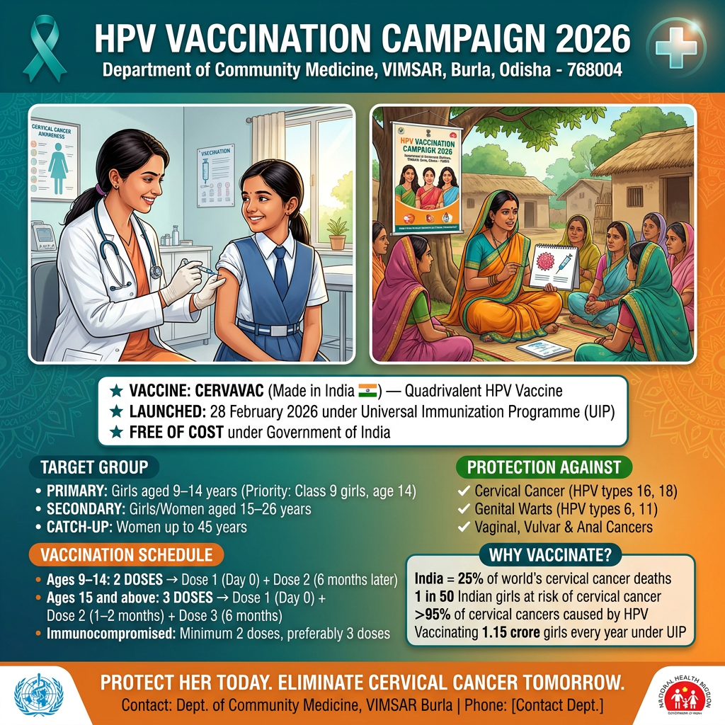
| Section | Details |
|---|---|
| Header | HPV Vaccination Campaign 2026 - VIMSAR, Burla |
| Vaccine | CERVAVAC - Made in India (Quadrivalent) |
| Launch | 28 February 2026 under UIP, Govt. of India - FREE |
| Target Group | Girls 9-14 (primary), 15-26 (secondary), up to 45 (catch-up) |
| Schedule | 2 doses (9-14 yrs) / 3 doses (15+ yrs) with timings |
| Protects Against | Cervical cancer (HPV 16,18), Genital warts (HPV 6,11) |
| Key Stats | India = 25% world cervical cancer deaths, 1.15 crore girls/yr |
| Tagline | "Protect Her Today. Eliminate Cervical Cancer Tomorrow." |
| Illustrations | Doctor vaccinating schoolgirl + ASHA worker community session |
HPV vaccination 2026 Banner in details for awareness in dept of community medicine vimsar burla
A highly detailed, visually rich, professional vertical awareness banner (portrait format, tall) for HPV Vaccination 2026. Design inspired by Indian government health campaign posters — vibrant, warm, trustworthy. Color palette: deep teal, saffron orange, white, and gold accents. ═══ TOP SECTION ═══ Full-width deep teal header band with gold border lines top and bottom. Center: Large bold white text "HPV VACCINATION AWARENESS CAMPAIGN 2026" Below in smaller gold text: "An Initiative Under Universal Immunization Programme (UIP), Govt. of India" Bottom of header: "Department of Community Medicine | VIMSAR, Burla, Odisha – 768004" Top-left corner: Small Indian flag. Top-right corner: Teal awareness ribbon + medical cross symbol. ═══ HERO IMAGE SECTION ═══ Full-width illustration: A warm, realistic scene inside a bright community health centre. A smiling female doctor in white coat with stethoscope is vaccinating a 14-year-old Indian schoolgirl in blue school uniform. The girl's mother stands beside her, looking reassured. In the background, an ASHA worker in a pink saree points to a wall chart showing the female reproductive system and cervical cancer prevention. Sunlight streams through windows. Indian rural clinic aesthetic. ═══ TEAL DIVIDER BAND ═══ Bold white text: "🛡️ CERVAVAC — INDIA'S OWN HPV VACCINE | Made by Serum Institute of India 🇮🇳" ═══ MAIN INFORMATION PANEL (white background, clean layout) ═══ SECTION 1 — WHAT IS HPV? (orange left accent bar) "Human Papillomavirus (HPV) is a sexually transmitted virus responsible for:" • 99% of Cervical Cancers • Genital Warts • Vaginal, Vulvar, Penile & Anal Cancers • Oropharyngeal Cancers Small diagram icon of virus particle next to text. SECTION 2 — WHY VACCINATE? (teal left accent bar) ★ India accounts for 25% of world's cervical cancer deaths ★ 1 in 50 Indian girls will develop cervical cancer in her lifetime ★ Over 1,25,000 new cases & 77,000 deaths annually in India ★ HPV vaccine prevents up to 90% of cervical cancers ★ 1.15 CRORE girls to be vaccinated every year under UIP ★ FREE vaccination under Government of India programme SECTION 3 — TARGET GROUP (saffron orange header box) ┌─────────────────────────────────────────┐ │ PRIMARY TARGET: Girls aged 9–14 years │ │ (Priority focus: 14-year-old girls, │ │ Class 9 students under school programme)│ │ │ │ SECONDARY TARGET: Girls/Women 15–26 yrs │ │ │ │ CATCH-UP VACCINATION: Women up to 45 yrs│ │ │ │ SPECIAL GROUP: Immunocompromised │ │ individuals (HIV+, transplant patients) │ └─────────────────────────────────────────┘ SECTION 4 — VACCINATION SCHEDULE (teal header box) Visual timeline/table format: AGE 9–14 YEARS → 2 DOSES 📅 Dose 1: Day 0 📅 Dose 2: 6 months after Dose 1 (Flexibility: Dose 2 can be given up to 12 months after Dose 1) AGE 15 YEARS & ABOVE → 3 DOSES 📅 Dose 1: Day 0 📅 Dose 2: 1–2 months after Dose 1 📅 Dose 3: 6 months after Dose 1 IMMUNOCOMPROMISED → Minimum 2 doses, preferably 3 doses SECTION 5 — PROTECTS AGAINST (green header box) Two-column icon list: ✅ Cervical Cancer (HPV types 16 & 18) ✅ Genital Warts (HPV types 6 & 11) ✅ CIN 1, 2 & 3 (pre-cancerous lesions) ✅ Vaginal & Vulvar Cancers ✅ Anal & Penile Cancers ✅ Oropharyngeal Cancers SECTION 6 — VACCINE SAFETY (gold header box) • Approved by DCGI, WHO prequalified • Safe for girls aged 9 years and above • Most common side effects: mild arm pain, slight fever (resolve in 1–2 days) • Do NOT give to pregnant women or those with severe allergy to vaccine components • Can be given with other UIP vaccines SECTION 7 — WHERE TO GET VACCINATED 🏥 Government hospitals & PHCs (FREE under UIP) 🏫 Schools – Class 9 girls (School Health Programme) 🏨 Private clinics and vaccination centres ═══ SECOND ILLUSTRATION PANEL ═══ Wide illustration: An outdoor community camp scene under a banner. ASHA workers and ANMs registering young teenage girls. A group of village mothers watching. Colourful India village background with trees. Professional health camp feel. ═══ BOTTOM FOOTER BAND (deep teal, gold border) ═══ Large bold white quote: "VACCINATE TODAY. PROTECT FOR LIFE." "HPV Vaccination — A Gift of Health for Every Girl" Second line in gold: "Organised by: Department of Community Medicine, VIMSAR, Burla, Sambalpur, Odisha – 768004" "For information & appointments: Dept. of Community Medicine, VIMSAR | UIP Helpline: 1800-11-0015" Small logos row at bottom: Government of India health mission seal placeholder, WHO logo placeholder, NHM logo placeholder. Overall: Rich, detailed, print-quality portrait banner. All text must be crisp and readable. Warm inspiring Indian health campaign aesthetic. Teal, saffron, white, gold color theme throughout.
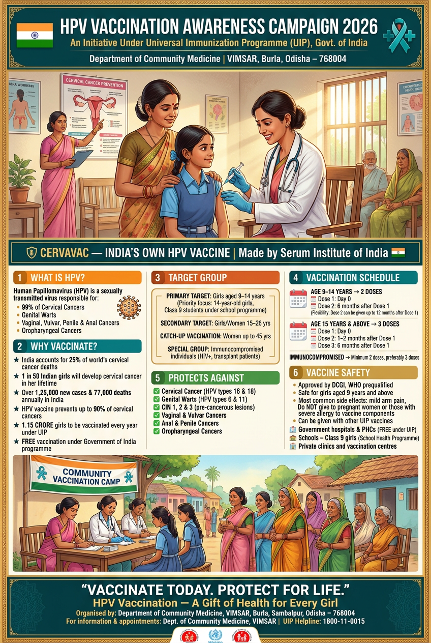
| Section | Content |
|---|---|
| 🏥 Department | Community Medicine, VIMSAR, Burla, Odisha - 768004 |
| 💉 Vaccine | CERVAVAC - Made in India, Serum Institute of India |
| 🎯 Target Group | Girls 9-14 (primary/free UIP), 15-26 (secondary), up to 45 yrs (catch-up) |
| 📅 Schedule | 2 doses (9-14 yrs) / 3 doses (15+ yrs) with exact timing |
| 🛡️ Protection | Cervical cancer, genital warts, CIN lesions, vaginal/vulvar/anal cancers |
| 📊 India Stats | 25% of world's cervical cancer deaths, 1.25 lakh new cases/yr |
| ✅ Safety | DCGI approved, WHO prequalified, mild side effects only |
| 🏕️ Two illustrations | Clinic vaccination scene + outdoor community camp scene |
| 📞 Contact/Footer | Dept. Community Medicine VIMSAR + UIP Helpline 1800-11-0015 |