
Conventional technique in microbiology
conventional techniques in microbiology overview
https://www.linkedin.com/pulse/conventional-microbiological-…
conventional microbiology techniques culture media streak plate gram stain
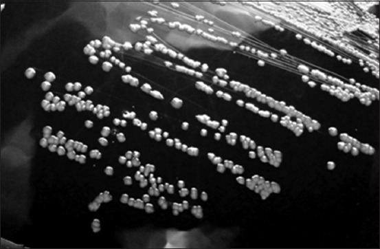
**Modality:** Microbiology culture photograph (grayscale). **Specimen:** *Corynebacterium pseudotuberculosis* (formerly *Corynebacterium ovis*) isolated on blood agar medium. **Morphological Features:** The image displays a streak plate demonstrating isolated bacterial colonies. The colonies are characterized by a distinct circular morphology with an entire margin. They exhibit a convex elevation and appear opaque with a grayish-white coloration against the dark background of the agar. The surface texture of the colonies appears slightly dry or waxy, which is characteristic of this genus. **Distribution:** Streaking pattern shows confluent growth in the primary sectors transitioning to well-defined, discrete individual colonies in the peripheral sectors, allowing for the observation of specific colony macro-morphology. **Diagnostic Context:** These visual features are consistent with the growth characteristics of *Corynebacterium* species, typically found in veterinary clinical microbiology samples. Key differentiating features include the specific convex profile and circular uniformity of the colonies on enriched media.
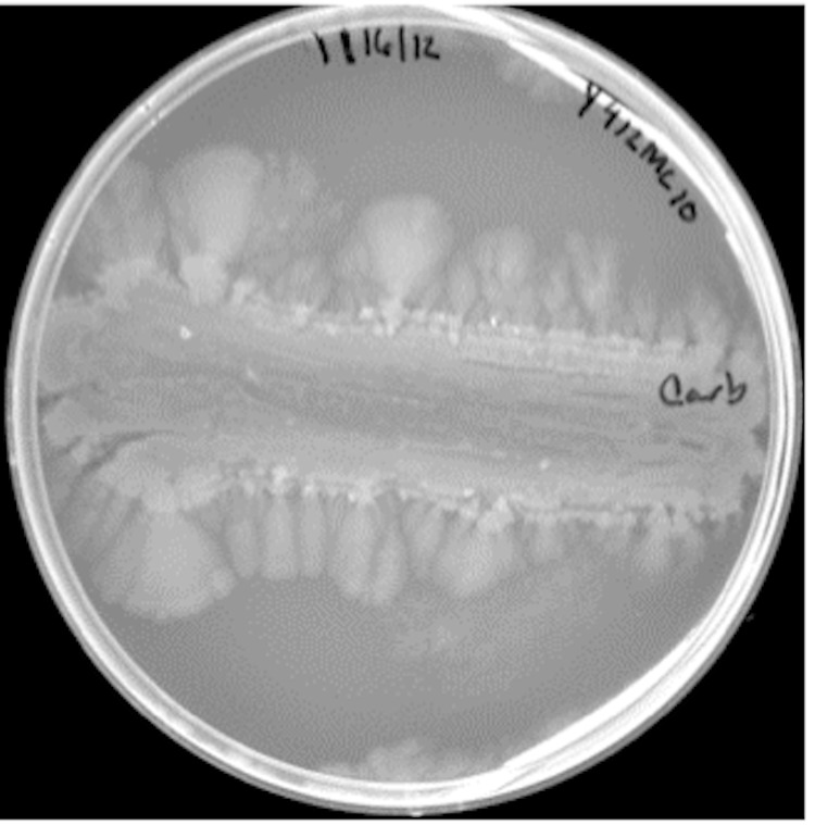
**Modality:** Microbiology culture photograph. **Specimen/Entity:** *Paenibacillus* sp. (strain Y412MC10). **Culture Conditions:** Growth on Yeast Extract-Tryptone (YT) agar medium, following 168 hours of incubation at 37°C. **Morphological Features:** The image displays a macro-view of a Petri dish containing a bacterial culture with a distinctive growth pattern. A horizontal streak serves as the primary inoculation site, showing central clearing or lysis. From the primary streak, there is significant lateral spreading of the culture toward the periphery of the plate. **Growth Pattern:** The organism exhibits characteristic colonial expansion and motility typical of *Paenibacillus* species. The leading edges of the culture demonstrate a dendritic or lobate-like spreading pattern, transitioning from a dense primary inoculum to feathered, translucent fan-like extensions. **Diagnostic Features:** Key visual indicators include high peripheral motility (swarming/spreading) and the progressive thinning or clearing of the central biomass over a prolonged incubation period. These features are critical for identifying motile, pattern-forming Gram-positive bacilli within a laboratory setting.
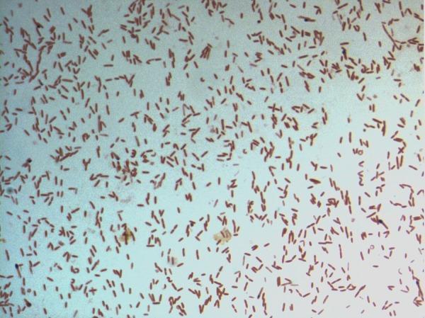
This diagnostic image shows a light microscopy view of a bacterial culture following Gram staining. The specimen displays numerous small, rod-shaped (bacilli) bacteria distributed across the field. The microorganisms exhibit a consistent pink-to-red hue, identifying them as Gram-negative. This staining characteristic indicates a cell wall structure with a thin peptidoglycan layer and an outer membrane that does not retain the crystal violet stain. The arrangement of the bacilli is predominantly individual or scattered, with occasional small clusters and pairs. Specifically identified as D. fastidiosa strain JC13T, this image serves as a primary microbiological reference for the morphological and staining properties of this novel genus within the family Erysipelotrichaceae. The visual demonstrates key diagnostic features including cell morphology, arrangement, and Gram reaction, which are essential for clinical microbiology classification and initial pathogen identification.
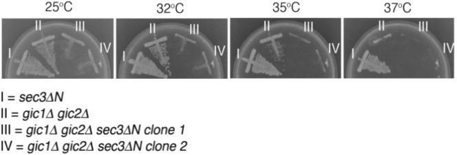
A comparison chart consisting of four clinical microbiology photographs showing yeast culture growth on YPD agar plates at different temperatures: 25°C, 32°C, 35°C, and 37°C. Each plate displays four different strains of Saccharomyces cerevisiae (labeled I-IV) via a streak-plate method to evaluate synthetic growth defects and temperature sensitivity. Strain I (sec3∆N single mutant) shows robust, consistent growth across all temperatures. Strain II (gic1∆ gic2∆ double mutant) demonstrates normal growth at 25°C but displays a progressive reduction in colony density as temperature increases, with minimal viability at 37°C. Strains III and IV (two independent clones of the sec3∆N gic1∆ gic2∆ triple mutant) exhibit a severe synthetic growth defect, characterized by significantly reduced colony density at 25°C and a complete loss of viability at temperatures of 32°C and above. This diagnostic image illustrates the essential parallel roles of Sec3 and Gic proteins in maintaining cell viability and polarized secretion, particularly under thermal stress.
Conventional Techniques in Microbiology
1. The Five "I's" of Microbiology
| Step | Description |
|---|---|
| Inoculation | Introducing a specimen (inoculum) into a suitable culture medium |
| Incubation | Maintaining cultures at optimal temperature/conditions for growth |
| Isolation | Separating individual organisms from a mixed population |
| Inspection | Observing gross and microscopic morphology |
| Identification | Determining species by biochemical, morphological, and serological tests |
2. Specimen Collection and Aseptic Technique
- Samples must be collected before antibiotic therapy when possible.
- Aseptic technique is mandatory - prevents contamination of cultures, personnel, specimens, and the environment.
- Transport media (e.g., Stuart's, Amies) preserve organisms during transit.
3. Sterilization and Disinfection
| Method | Definition | Examples |
|---|---|---|
| Sterilization | Destroys ALL microorganisms including endospores | Autoclave (121°C, 15 psi, 15 min), dry heat, filtration |
| Disinfection | Destroys vegetative microbes and viruses, NOT endospores | Chemical agents (bleach, phenolics, alcohols) |
| Sanitization | Mechanical removal of microbes to safe levels | Washing, scrubbing |
4. Culture Media
A. Physical State
- Liquid (broth) - e.g., nutrient broth, thioglycollate broth
- Semi-solid - for motility testing
- Solid (agar plates/slopes) - for isolation and colony morphology
B. Nutritional Composition
- General purpose - Nutrient agar, Blood agar, Tryptic soy agar
- Enriched - Blood agar, Chocolate agar (fastidious organisms like Neisseria, Haemophilus)
- Enrichment broth - Selectively favors target organisms (e.g., Selenite F for Salmonella)
C. Selective and Differential Media
- Selective - Inhibits unwanted flora while allowing target organism growth (e.g., MacConkey agar inhibits Gram-positives)
- Differential - Distinguishes organisms by reactions visible on the medium (e.g., blood agar for hemolysis patterns, MacConkey agar for lactose fermentation - pink vs. colorless colonies)
- Combined selective + differential - CLED (Cystine Lactose Electrolyte Deficient) agar for UTI pathogens
D. Special Media
- Löffler's serum slope - for Corynebacterium diphtheriae
- Tinsdale's / tellurite media - for diphtheria; colonial morphologies distinguish C. diphtheriae from nondiphtherial coryneforms
- Löwenstein-Jensen (LJ) agar - for Mycobacteria
- Sabouraud's dextrose agar - for fungi
- Charcoal yeast extract (BCYE) agar - for Legionella
5. Isolation Techniques
Streak Plate Method
- A loopful of specimen is streaked across an agar plate in successive sections, progressively diluting the inoculum.
- Each section is streaked at 90° to the previous.
- Organisms separate into individual colonies that represent a pure culture.
Pour Plate Method
- Inoculum is diluted and mixed with molten agar (kept at ~45°C) before pouring into a petri dish.
- As agar solidifies, organisms are trapped and form individual colonies throughout the medium.
- Useful for counting colony-forming units (CFUs).
Spread Plate Method
- A small volume of liquid inoculum is placed in the center of a solid agar plate and spread evenly with a glass spreader (Drigalski spatula) or wire spreader.
- Produces surface colonies for easy isolation.
6. Microscopy Techniques
Direct Microscopy (Wet Mount / Hanging Drop)
- Unstained preparation examined under phase contrast or dark-field.
- Used for motility, protozoa (e.g., Trichomonas), fungal elements, cells.
Staining Methods
A. Gram Staining (Most important routine stain)
- Crystal violet - Primary stain (all bacteria stain purple)
- Gram's iodine - Mordant (fixes dye)
- Decolorizer (acetone/alcohol) - Gram-negative organisms lose the purple dye
- Safranin - Counter-stain (Gram-negatives stain pink/red)
- Gram-positive = Purple (thick peptidoglycan wall)
- Gram-negative = Pink/Red (thin peptidoglycan + outer membrane)
B. Ziehl-Neelsen (ZN) / Acid-Fast Stain
- Used for Mycobacteria (M. tuberculosis, M. leprae) and Nocardia.
- Carbol fuchsin applied with heat, resists decolorization by acid-alcohol.
- Counterstained with methylene blue or malachite green.
- Acid-fast bacilli (AFB) = Red/Pink on blue background.
C. Other Important Stains
| Stain | Organism / Purpose |
|---|---|
| Albert's stain | C. diphtheriae metachromatic granules (volutin) |
| Giemsa stain | Malaria parasites, Rickettsiae, Leishmania, Chlamydia |
| Lactophenol cotton blue (LPCB) | Fungal morphology |
| India ink (negative stain) | Cryptococcus neoformans capsule |
| Periodic acid-Schiff (PAS) | Fungal cell walls |
| Modified ZN (cold method) | Cryptosporidium, Cyclospora, Cystoisospora |
| Silver stains (Gomori) | Pneumocystis jirovecii, fungi in tissue |
7. Biochemical Identification Tests
Key Tests
| Test | Principle | Example |
|---|---|---|
| Catalase test | H₂O₂ → H₂O + O₂ (bubbles) | Staph (+) vs. Strep (-) |
| Oxidase test | Cytochrome oxidase activity | Pseudomonas (+), Enterobacteria (-) |
| Coagulase test | Fibrinogen clotting | S. aureus (+) vs. CoNS (-) |
| Urease test | Urea → NH₃ + CO₂ | H. pylori, Proteus |
| Indole test (IMViC) | Tryptophan → Indole | E. coli (+), Klebsiella (-) |
| Methyl Red test | Mixed acid fermentation (acidic pH) | E. coli (+) |
| Voges-Proskauer (VP) test | Acetoin production | Klebsiella (+), E. coli (-) |
| Citrate utilization | Sole carbon source use | Klebsiella (+), E. coli (-) |
| Triple Sugar Iron (TSI) agar | Glucose/lactose/sucrose fermentation + H₂S + gas | Salmonella, Shigella differentiation |
| Optochin sensitivity | Inhibition of S. pneumoniae | S. pneumoniae sensitive; other streptococci resistant |
| CAMP test | Enhanced hemolysis with S. aureus | Group B Streptococcus positive |
8. Antimicrobial Susceptibility Testing (AST)
Kirby-Bauer Disk Diffusion Method
- Organism is inoculated onto Mueller-Hinton agar at a standardized inoculum (0.5 McFarland standard).
- Antibiotic-impregnated disks are placed on the plate.
- After 16-18 h incubation, zones of inhibition are measured.
- Results interpreted as Susceptible (S), Intermediate (I), or Resistant (R) using CLSI/EUCAST breakpoints.
Broth Dilution (MIC Determination)
- Serial two-fold dilutions of antibiotic in broth, each inoculated with the test organism.
- Minimum Inhibitory Concentration (MIC) = lowest concentration with no visible growth.
- Provides quantitative data; essential for serious infections.
E-test (Epsilometer test)
- Plastic strip with antibiotic gradient placed on inoculated agar.
- Elliptical zone of inhibition gives a direct MIC reading.
9. Animal Inoculation
- Guinea pig - M. tuberculosis, Chlamydia isolation, Rickettsia
- Mouse - Clostridium perfringens toxin, Pasteurella
- Rabbit - Virulence testing, VDRL confirmation
- Now largely replaced by molecular methods, but still used in some research settings.
10. Serological Methods (Conventional)
- Agglutination tests - Widal test (typhoid), TPHA (syphilis), slide agglutination for Salmonella/Shigella serotyping
- Precipitation tests - Ouchterlony double diffusion, immunoelectrophoresis
- Complement fixation tests - Wassermann test for syphilis
- VDRL / RPR - Non-treponemal syphilis screening
11. Limitations of Conventional Techniques
| Limitation | Impact |
|---|---|
| Time-consuming (24-72 h or longer for some organisms) | Delays treatment decisions |
| Require viable organisms | Low sensitivity for fastidious/unculturable organisms |
| Technically demanding | Inter-lab variability |
| Cannot identify all pathogens | Some viruses, intracellular bacteria missed |
| Sequential process | Each step adds time |
Despite these limitations, culture-based methods remain the gold standard because they provide viable organisms for AST, detect unexpected pathogens, and generate epidemiological data. They are now complemented by molecular (PCR, NGS) and proteomic (MALDI-TOF) methods rather than replaced by them.