
Detailed evaluation of dental implants. 10 marks. I need to write relevant for about 5 pages. Pls give me a detailed answer.
Reading File
Now let me search the library for dental implant content in parallel.
Finding Sources
Finding Sources
Searching the Web
dental implants evaluation osseointegration types components indications complications 2024
Good. Now let me read the most relevant textbook sections on dental implants in parallel.
Reading File
Reading File
Finding Sources
Finding Sources
Excellent - found a very rich section in Cummings Otolaryngology. Let me read those key sections in parallel.
Reading File
Reading File
Finding Sources
Finding Sources
I got great content from Cummings on osseointegration. Now let me read the crucial section on implant material and design more fully, plus search for types and components.
Reading File
Finding Sources
Finding Sources
I now have rich content. Let me fetch a key dental implant diagram image and do a quick PubMed search for the most recent success criteria evidence.
Loading Image
Searching PubMed
"dental implant" AND "success criteria" AND osseointegration
I now have all the content I need to write a comprehensive, detailed answer. Let me compile the full response.
Detailed Evaluation of Dental Implants
1. Introduction and Historical Background
A dental implant is an artificial device - typically a titanium screw or post - surgically placed into the jawbone to serve as a replacement root for a missing tooth. Once integrated into bone, it supports a prosthetic crown, bridge, or overdenture, restoring both function and aesthetics.
The concept of osseointegrated implants was pioneered by Per-Ingvar Brånemark of Gothenburg, Sweden, in the 1960s. During experimental studies, Brånemark discovered that titanium had a unique ability to become firmly anchored in bone without interposed soft tissue while achieving a reaction-free epithelial penetration. He termed this biological phenomenon "osseointegration" - defined as direct structural and functional connection between living bone and the surface of a load-bearing implant, without any intervening fibrous or connective tissue layer. Osseointegration is considered successful when the implant becomes rigidly anchored into adjacent bone with no reaction at the implant-bone interface (Cummings Otolaryngology, p. 3060).
Brånemark's concept was initially applied to dental rehabilitation, and by the 1980s, the application had expanded to facial prosthetics (auricular, orbital, nasal) and bone-anchored hearing devices, representing one of the most significant advances in modern reconstructive medicine.
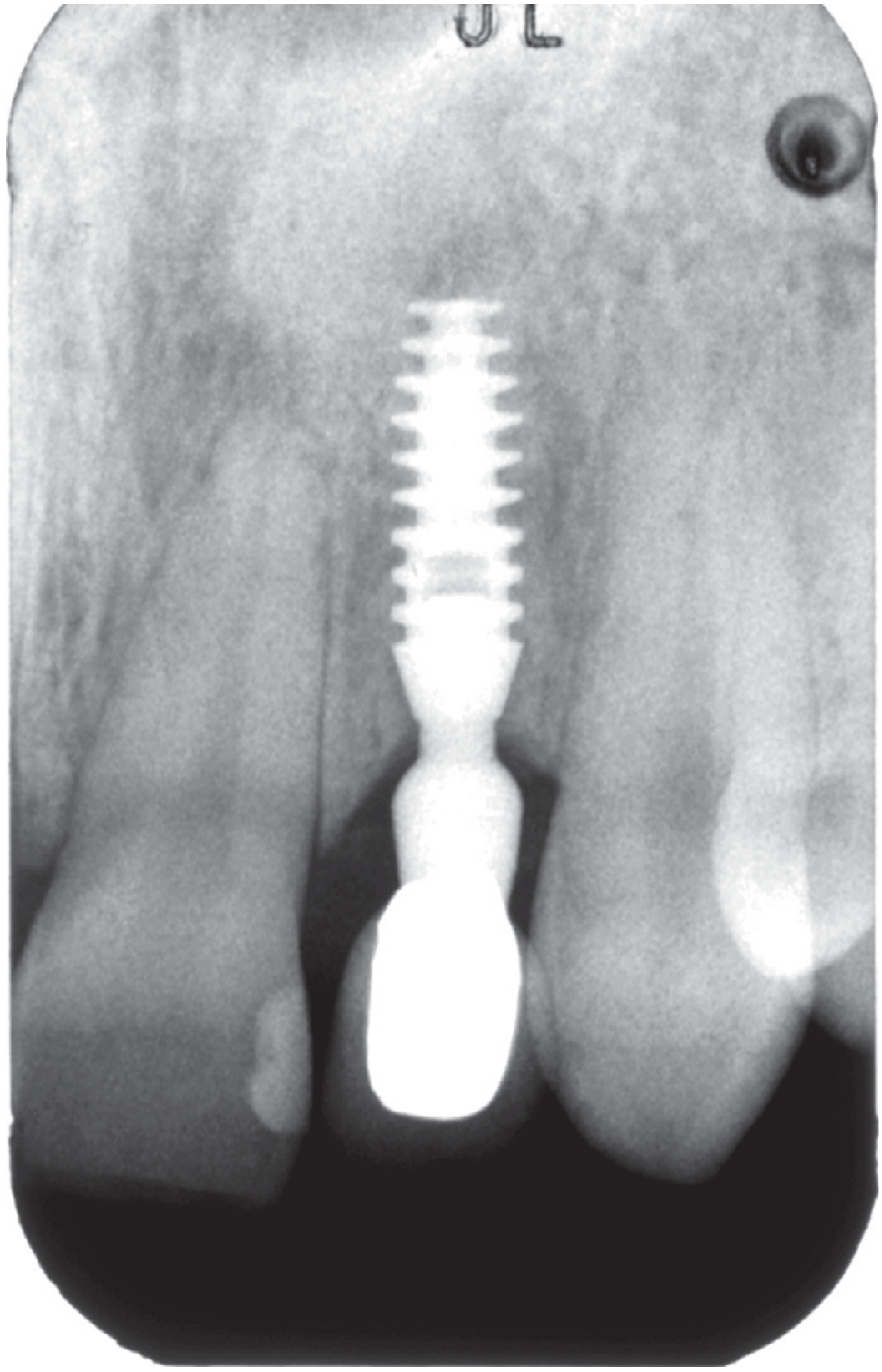
Periapical radiograph showing a threaded titanium intraosseous dental implant with crown restoration - Brogdon's Forensic Radiology
2. Components of a Dental Implant
A modern dental implant system consists of three main components:
a) The Implant Fixture (Endosseous Body)
This is the titanium post surgically embedded into the alveolar bone. It functions as the artificial tooth root. It is typically threaded/screw-shaped, with surface microarchitecture designed to promote bone ingrowth and osseointegration.
b) The Abutment
The abutment is the connecting piece that attaches to the implant fixture and protrudes through the gingiva (gum tissue) into the oral cavity. It serves as the platform onto which the prosthetic crown is mounted. Abutments may be made of titanium, zirconia, or gold alloy.
c) The Prosthetic Crown (Restoration)
The crown is the visible portion of the implant that mimics a natural tooth in form and function. It may be screwed or cemented onto the abutment. Crowns are typically fabricated from porcelain-fused-to-metal or all-ceramic materials.
3. Classification / Types of Dental Implants
By Position in Relation to Bone
| Type | Description |
|---|---|
| Endosseous (Intraosseous) | Placed directly into the jawbone; the most common type; includes screw, cylinder, and blade forms |
| Subperiosteal | Sits on top of the bone but beneath the periosteum; used when there is insufficient bone height |
| Transosseous | Passes completely through the mandible; rarely used today |
By Design of the Fixture
- Screw/threaded implants - Most widely used; the threaded design distributes stress more efficiently than T-shaped or cylindrical types. Parametric studies confirm threaded implants have a greater likelihood of osseointegration. The initial stability conferred by threads is particularly important during the early healing phase - if the implant is not absolutely stable after placement, connective tissue may form between implant and bone, preventing osseointegration (Cummings, p. 3061).
- Cylindrical/press-fit implants - Smooth cylinders that rely on surface coating for retention.
- Blade/plate-form implants - Flat, knife-like designs used in narrow ridges; largely obsolete.
By Loading Protocol
- Conventional loading - Crown placed after 3-6 months of osseointegration
- Early loading - Crown placed 1-2 months post-implant
- Immediate loading - Crown placed within 48 hours of implant placement (requires high primary stability)
- Immediate implant placement - Implant placed immediately into the extraction socket
By Staging
- Two-stage (submerged) - Implant is buried under the gingiva for 3-6 months, then uncovered in a second surgery; allows undisturbed healing
- One-stage (non-submerged) - The healing abutment is placed at the time of implant surgery; reduces total treatment time
4. Implant Materials
Titanium - The Gold Standard
The choice of implant material is the foremost determinant of successful osseointegration. Titanium has survived the test of time and represents the standard against which all other biomaterials are measured. It is available in two forms:
- Commercially pure (CP) titanium - 99.75% pure; the preferred choice for osseointegrated implants. Histologically shown to achieve direct bone-implant contact without interposed fibrous tissue (Cummings, p. 3061).
- Titanium alloy (Ti-6Al-4V) - 90% titanium, 6% aluminum, 4% vanadium; stronger but clinically inferior to CP titanium for osseointegration.
Why titanium works:
- Upon oxygen exposure, titanium forms a tightly-bonded, corrosion-resistant oxide layer on its surface. It is this titanium dioxide (TiO₂) coating - in direct contact with host tissue - that confers its remarkable biocompatibility.
- Unlike stainless steel, titanium lacks high corrosion potential and the toxicity of alloy components.
- To date, pure titanium has shown no evidence of systemic toxicity or carcinogenicity, even with decades of implantation (Cummings, p. 3060).
- Pure titanium is superior to titanium alloy in every histological criterion: no inflammatory infiltrate, no osteolysis, and no connective tissue capsule (or capsule <30 μm, which is acceptable) at the implant surface.
Other Materials
- Zirconia (ZrO₂) - Tooth-colored ceramic; used in patients with metal sensitivity; one-piece design; lower evidence base than titanium.
- PEEK (polyether ether ketone) - Experimental; used mainly in orthopedic applications.
5. Surface Design and Microarchitecture
Implant micro- and macrostructure critically impact osseointegration. Key principles:
- A rough, micropitted surface increases surface area and facilitates direct bonding between the implant's oxide surface and bone matrix. The microarchitecture should feature topography with micropits that fit the dimensions of cell membranes and large biomolecules.
- However, a surface porosity below 100 μm increases the risk of implant corrosion even with titanium.
- Surface treatments to enhance osseointegration include:
- Sandblasting and acid etching (SLA) - Creates a moderately rough surface
- Titanium plasma-spraying (TPS) - Increases surface roughness
- Hydroxyapatite (HA) coating - Mimics natural bone mineral; accelerates early osseointegration
- Anodization - Creates a thicker, more stable oxide layer
- Nanocoating - Manipulation at nanoscale improves bone-implant bonding and accelerates healing
6. Biology of Osseointegration
The biological process of osseointegration proceeds through distinct phases:
Phase 1: Blood Clot Formation (Days 0-3)
Immediately after implant placement, a blood clot forms at the bone-implant interface. Platelets release growth factors (PDGF, TGF-β) that initiate healing.
Phase 2: Inflammatory/Immune Response (Days 1-7)
Osseointegration starts as a foreign body reaction to biomaterials. Peri-implant immune cells modulate the local microenvironment - a process called osteoimmunomodulation - which is essential for favorable osseointegration. NF-κB and Wnt signaling pathways play key roles in this cellular cascade.
Phase 3: Woven Bone Formation (Weeks 1-4)
Osteoblasts migrate to the implant surface and begin depositing immature woven bone. Angiogenesis is another important component, providing vascular supply to the new bone.
Phase 4: Bone Remodeling and Maturation (Months 2-6)
Woven bone is replaced by mature lamellar bone. This phase establishes the rigid bone-implant contact required for load bearing.
Histological criteria for successful osseointegration:
- Direct contact between bone matrix and the implant without interposed fibrous or soft tissue
- No evidence of inflammation at the implant site (no infiltrate, no osteolysis)
- No connective tissue capsule on the implant surface (or capsule <30 μm) (Cummings Otolaryngology, p. 3061)
7. Indications for Dental Implants
Dental implants are indicated in the following situations:
- Single tooth replacement - Missing tooth where adjacent teeth are healthy and should not be prepared as bridge abutments
- Multiple tooth replacement - Implant-supported fixed bridges
- Complete edentulism - Implant-retained overdentures (2 or 4 implants) or full-arch fixed restorations (All-on-4, All-on-6)
- Oncological reconstruction - After jaw resection (mandibulectomy/maxillectomy) for oral cancer; implants placed primarily at the time of resection or secondarily after healing and radiotherapy
- Congenital tooth absence (agenesis)
- Craniofacial prosthesis retention - Ear (auricular), eye (orbital), nose (nasal) prostheses retained by bone-anchored implants
- Bone-anchored hearing devices (BAHA) - Osseointegrated implants in the mastoid to couple bone vibration for patients with conductive or mixed hearing loss
For oncological patients, a recent systematic review has shown that osseointegrated implants are highly effective at facilitating dental rehabilitation with high levels of implant survival, despite subsequent post-operative radiotherapy - with 75% of patients completing dental rehabilitation and showing improvements in quality of life (Scott-Brown's Otorhinolaryngology, p. 549).
8. Contraindications
Absolute Contraindications
- Active malignancy at the implant site
- Insufficient bone volume/density (relative - can be addressed with bone grafting)
- Active uncontrolled periodontal disease
Relative Contraindications
- Radiation therapy to the jaw - Reduces bone vascularity; increases risk of osteoradionecrosis. Typically, implants should be placed either before or >6 months after radiotherapy.
- Uncontrolled diabetes mellitus - Impairs osseointegration and healing
- Bisphosphonate therapy - Risk of medication-related osteonecrosis of the jaw (MRONJ)
- Smoking - Significantly increases implant failure rate; nicotine impairs bone healing
- Immunosuppression - Increases infection risk
- Bruxism (parafunctional habits) - Excessive occlusal load disrupts osseointegration; requires occlusal guards
- Poor oral hygiene - Pre-existing or uncontrolled periodontitis increases peri-implantitis risk
- Incomplete skeletal growth - Implants are typically deferred until skeletal maturity (18+ years)
9. Surgical Procedure
Pre-operative Planning
- Clinical examination: assessment of bone volume, gingival health, occlusion, opposing dentition
- Radiographic assessment: periapical radiographs, panoramic radiograph, CBCT (cone beam computed tomography) for 3D bone morphology
- Diagnostic study models (digital or conventional) to evaluate occlusion and plan prosthetic outcome
- Medical history review for systemic contraindications
Stage 1 - Implant Placement
- Local anesthesia; mucoperiosteal flap elevation to expose bone
- Osteotomy: a series of drills of increasing diameter create the implant bed. Controlled temperature during drilling is essential - without adequate irrigation, high-speed drilling can cause temperature elevations up to 89°C, which kills osteocytes and prevents subsequent osseointegration (Shambaugh Surgery of the Ear, p. 5097)
- Implant insertion to achieve primary stability (insertion torque typically 20-45 Ncm)
- In a two-stage protocol, the implant is covered with a healing screw and gingival flap closed
Healing Phase (Osseointegration)
- 3-6 months in the mandible; 4-6 months in the maxilla (less dense bone)
- Implant must be kept immobile during healing to prevent fibrous encapsulation
Stage 2 - Second-Stage Surgery (Two-Stage Protocol)
- Minor surgery to uncover the implant and attach a healing abutment
- Soft tissue allowed to mature around the abutment for 2-4 weeks
Prosthetic Phase
- Impression-making (conventional or digital)
- Custom abutment and crown fabrication
- Final crown placement
10. Evaluation of Implant Success - Success Criteria
The evaluation of dental implant success has evolved over decades. The most widely cited criteria are:
Albrektsson Criteria (1986) - Classic
- No clinical mobility (zero tolerance; any movement = failure)
- Absence of peri-implant radiolucency on radiograph
- Marginal bone loss <1.5 mm in the first year, <0.2 mm/year thereafter
- No pain, discomfort, or infection
- Implant survival rate >85% at 5 years, >80% at 10 years
Radiographic Evaluation
The marginal bone level (MBL) is the most important radiographic parameter of implant health. It is measured from the implant's crestal level during initial surgery and monitored using periapical radiographs. Studies show acceptable annual bone loss of 0-0.2 mm after the first year. Excessive marginal bone loss signals peri-implantitis or implant failure (Lee et al., 2024, KAOMI).
Clinical Mobility Assessment
Ensuring zero implant mobility is paramount. Any movement indicates failed osseointegration. The Resonance Frequency Analysis (RFA) with the Osstell device provides a non-invasive quantitative measure of implant stability, expressed as an Implant Stability Quotient (ISQ) score (scale 1-100; >70 is favorable). This is now standard in modern implant monitoring.
Peri-implant Tissue Assessment
- Probing depth around the implant (normal <3-4 mm)
- Presence/absence of bleeding on probing
- Presence/absence of suppuration
- Soft tissue color, contour, and texture
11. Complications
Early Complications (within 3 months)
- Osseointegration failure - Due to infection, excessive micromotion, overheating of bone during drilling, or poor bone quality. The implant fails to rigidly anchor into bone.
- Fibrous encapsulation - The body encapsulates the implant in fibrous tissue when it faces excessive micromotion or infection during healing; results in implant mobility and eventual failure.
- Wound dehiscence / flap necrosis
- Nerve injury - Inferior alveolar nerve damage during mandibular implant placement (paresthesia)
Late Complications (after osseointegration)
- Peri-implant mucositis - Reversible inflammatory condition of the soft tissue surrounding an implant; analogous to gingivitis. Caused by plaque accumulation.
- Peri-implantitis - Irreversible inflammatory condition involving the soft tissue AND bone around an implant, leading to progressive marginal bone loss. It is the most significant long-term complication. Clinically resembles a periodontal abscess. Pathologic changes around an implant are collectively termed "peri-implant disease." Treatment involves:
- Gentle debridement and plaque removal around the implant
- Irrigation with normal saline or 0.12% chlorhexidine solution
- Analgesia as needed
- Surgical intervention (implantoplasty, bone grafting) for advanced cases
- Referral to specialist for definitive care (Tintinalli's Emergency Medicine, p. 1625)
- Implant fracture - Fatigue fracture of the implant body, usually at the implant-abutment junction; related to excessive occlusal load
- Screw loosening - Abutment or crown screw loosening; requires re-tightening
- Esthetic failure - Soft tissue recession, visible metal collar, implant mismatch
Risk Factors for Failure
- Smoking (most significant modifiable risk factor)
- Poor oral hygiene
- Uncontrolled diabetes
- Previous radiotherapy
- Bruxism
- Thin biotype (thin gingival tissue)
- Posterior maxilla (poor bone density, proximity to sinus)
12. Implant-Supported Prosthetic Designs
| Prosthesis Type | Description | Implants Required |
|---|---|---|
| Single crown | Replaces one tooth | 1 |
| Fixed partial denture (bridge) | Replaces 2-3 teeth | 2 |
| Full-arch fixed bridge (All-on-4) | Full jaw rehabilitation | 4 (or 6) |
| Implant overdenture | Removable denture retained by implants | 2-4 (mandible), 4-6 (maxilla) |
| Bar-retained overdenture | Clips to metal bar connecting multiple implants | 3-4 |
13. Advances and Future Directions
- Zirconia implants - One-piece ceramic implants for patients with titanium allergy or esthetic demands in the anterior zone
- Nanotechnology - Nanocoated implant surfaces improve osseointegration and accelerate healing
- 3D printing / CAD-CAM - Custom-fabricated implants and prosthetics tailored to each patient's anatomy
- Smart implants - Embedded sensors for real-time monitoring of bite force, temperature, and early signs of peri-implantitis
- Growth factors (BMP, PRP) - Bone Morphogenetic Proteins and Platelet-Rich Plasma used to stimulate bone growth and enhance osseointegration in compromised sites
- Immediate load protocols - Same-day implant and crown placement reduces treatment time and patient burden
- Digital workflow - Intraoral scanners, CBCT, and guided surgical templates increase precision and reduce complications
14. Radiological Considerations
A chief practical consideration for implant patients is the effect on radiological investigations. Titanium produces relatively little degradation of CT or MRI images:
- CT: Shadowing and scatter effects cause minimal degradation of CT image quality; pure titanium has little scatter effect.
- MRI: Not contraindicated with a titanium implant in place. However, any attached metallic components (abutments, healing caps) must be removed prior to the scan (Cummings, p. 3061).
15. Outcomes and Prognosis
Dental implants are among the most predictable and well-documented interventions in modern dentistry:
- Overall 10-year survival rates: 90-96% in healthy patients
- Mandibular implants have slightly higher success rates than maxillary implants (denser bone)
- Anterior implants perform better than posterior maxillary implants
- Smokers have approximately 2-3x higher failure rates than non-smokers
- Implant-retained overdentures in edentulous patients show dramatic improvements in quality of life, masticatory function, and patient satisfaction compared to conventional dentures
The success of oncological reconstruction with dental implants has been well-established - patients receiving implants at the time of jaw resection (primary placement) or after healing (secondary placement) demonstrate high implant survival rates despite subsequent radiotherapy, with 75% completing full dental rehabilitation and showing measurable QOL improvements (Scott-Brown's, p. 549).
Summary Table: Key Evaluation Parameters
| Parameter | Method | Success Threshold |
|---|---|---|
| Implant mobility | Clinical/RFA (ISQ) | Zero mobility; ISQ >70 |
| Marginal bone loss | Periapical radiograph | <1.5 mm (Year 1), <0.2 mm/year thereafter |
| Peri-implant pocket depth | Probing | <3-4 mm |
| Bleeding on probing | Clinical | Absent |
| Radiolucency | Radiograph | Absent |
| Pain/infection | Clinical history | Absent |
| 5-year survival | Follow-up | >85% |
| 10-year survival | Follow-up | >80% |
Conclusion
Dental implants represent the current standard of care for tooth replacement, offering superior long-term outcomes compared to conventional dentures and bridges. Their success depends on a thorough understanding of osseointegration biology, careful patient selection, meticulous surgical technique, and rigorous long-term maintenance. The evaluation of implant success is multi-dimensional - incorporating clinical stability, radiographic bone levels, peri-implant tissue health, and patient-reported outcomes. Complications, especially peri-implantitis, remain a significant challenge and require proactive preventive strategies and early intervention when detected.
References:
- Cummings Otolaryngology Head and Neck Surgery, 7th Ed. - Osseointegration chapters (pp. 3060-3061)
- Scott-Brown's Otorhinolaryngology Head & Neck Surgery - Dental implant placement in reconstruction (p. 549)
- Tintinalli's Emergency Medicine - Peri-implantitis management (p. 1625)
- Brogdon's Forensic Radiology - Dental restorative materials
- Lee DJ et al. Evaluation of Criteria for Dental Implant Success: A Systematic Literature Review. J Implantol Appl Sci, 2024
This is a shared conversation. Sign in to Orris to start your own chat.