
Rheumatic fever vs Rheumatic heart disease
rheumatic fever Jones criteria carditis
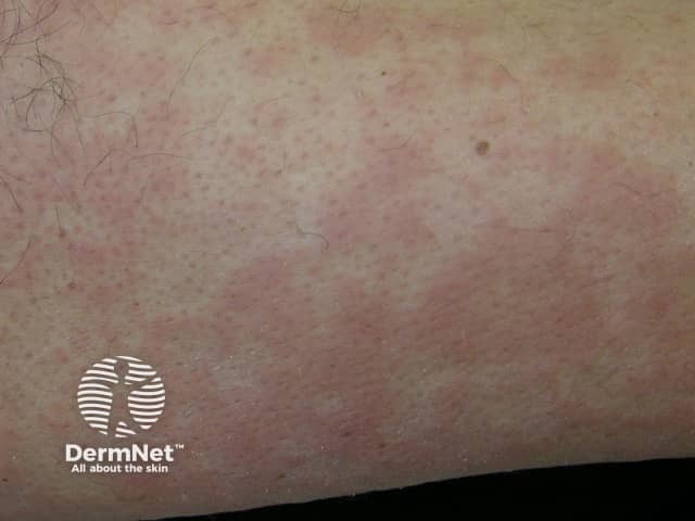
This is a clinical dermatology photograph capturing erythema marginatum on the skin of the lower limb. The modality is Clinical photography; technique: naked-eye, color-balanced image. The observed lesions are erythematous, annular plaques with central clearing and pale, sometimes hypopigmented centers. Lesions appear as rings with slightly raised margins and a uniform pink-to red hue; they range from 0.5 to 2 cm in diameter and may coalesce into larger plaques. The rash is typically non-pruritic and evanescent, with temporary eruption that may migrate or fade over hours to days; distribution commonly involves the trunk and proximal limbs, with sparing of the face. The image may reflect erythema marginatum, a cutaneous manifestation associated with acute rheumatic fever and autoimmune inflammatory processes. In longer-standing presentations, the rash signals preceding or concurrent carditis and migratory arthritis; therefore clinical correlation with recent group A Streptococcus infection, elevated ASO titers, and echocardiography is essential. Differential diagnoses include urticaria, erythema multiforme, pityriasis rosea, and drug-induced eruptions. Clinically, this finding has diagnostic significance as a major criterion in rheumatic fever per Jones criteria; management focuses on antibiotic treatment of streptococcal infection and secondary prophylaxis. This image serves educational purposes for medical students, residents, and researchers studying connective tissue disease and pediatric rheumatology.
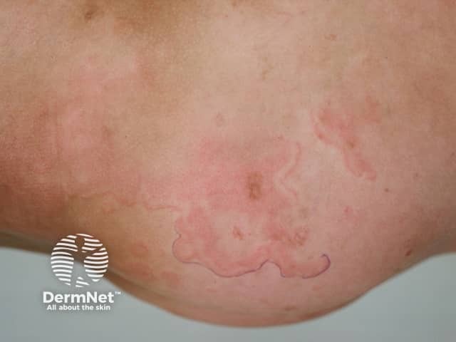
Clinical photograph (dermatology) showing erythema marginatum, an annular, non-itchy rash characteristic of acute rheumatic fever. The image depicts multiple pink to red circular to oval patches on the skin of the lower extremity with sharply demarcated, serpiginous margins and centers that appear pale or clearing. Lesions are non-vesicular, smooth-surfaced, and vary in size, with a tendency to migrate or transiently flare. The distribution favors the trunk and proximal limbs and typically spares the face. On close inspection the patches lack scaling and crusting, consistent with erythema marginatum rather than dermatitis or fungal infection. This cutaneous manifestation is part of the Jones criteria and may correlate with concurrent migratory polyarthritis, fever, and evidence of preceding group A Streptococcus infection. Histologic features (if biopsied) would show superficial perivascular lymphocytic infiltrate and dermal edema with minimal epidermal changes; clinically, the rash is a marker for systemic inflammatory involvement. Differential considerations include urticaria, erythema multiforme, tinea corporis, pityriasis rosea, and other annular dermatoses. Clinically, recognizing erythema marginatum prompts evaluation for acute rheumatic fever and guides management including antibiotic therapy and anti-inflammatory treatment. The image serves educational and diagnostic-reference purposes for dermatology, pediatrics, infectious diseases, and rheumatology training.
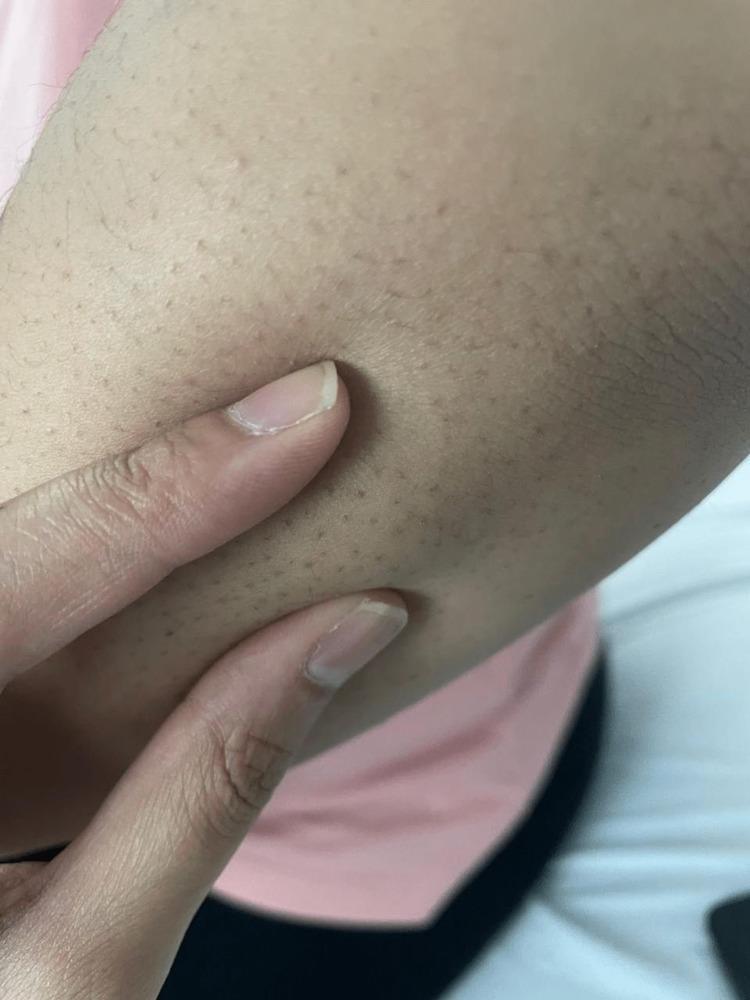
This clinical photograph displays a close-up view of the right upper extremity, specifically the extensor surface of the elbow region. The primary clinical finding is the presence of multiple, small subcutaneous nodules. These nodules appear as firm, rounded, non-tender-looking elevations beneath the skin surface, typically measuring less than 1 cm in diameter. The overlying skin is intact, displaying normal pigmentation and texture without visible erythema, warmth, or ulceration. Prominent hair follicles are visible as dark punctate spots across the skin. In a clinical context involving polyarthralgia and carditis, these nodules are a major Jones criterion for the diagnosis of acute rheumatic fever (ARF). This image serves as a teaching tool for medical students and clinicians to recognize the dermatological manifestations of systemic streptococcal-related diseases and rheumatological disorders.
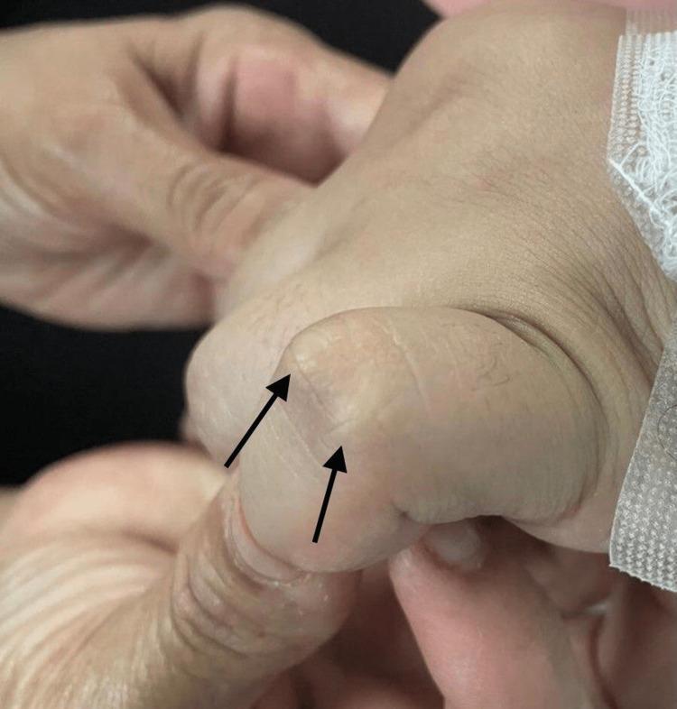
Clinical photograph showing the dorsal surface of a human hand, specifically focusing on the interphalangeal joints. Two prominent subcutaneous nodules are visible over the joint space, indicated by black arrows. These nodules appear as firm, well-circumscribed, painless elevations under the skin. They are flesh-colored to slightly pale, with smooth overlying skin and no signs of acute inflammation, erythema, or ulceration. This finding is a characteristic major Jones criterion for the diagnosis of acute rheumatic fever. The image serves as an educational tool for identifying extra-articular manifestations of rheumatological and post-streptococcal conditions in a clinical setting.
rheumatic heart disease mitral stenosis valve pathology
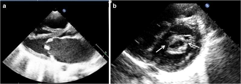
Two-panel diagnostic imaging featuring a 2D transthoracic echocardiogram (TTE) of severe mitral stenosis, typically associated with rheumatic heart disease. Panel (a) shows a parasternal long-axis view during diastole, highlighting significant thickening and increased echogenicity of the mitral valve leaflets. The anterior leaflet demonstrates characteristic 'doming' (hockey-stick deformity) with restricted excursion, indicating limited valve opening. Panel (b) presents a parasternal short-axis view at the level of the mitral valve orifice. White arrows point to commissural fusion, a hallmark of stenotic valvular pathology. The mitral valve area (MVA) is manually traced via planimetry (indicated by the dotted line), yielding a measurement of 0.73 cm², which falls into the category of severe stenosis (MVA < 1.0 cm²). These images demonstrate the classic morphology used in cardiology for assessing valvular narrowing, leaflet mobility, and subvalvular involvement to determine the clinical severity of mitral stenosis.
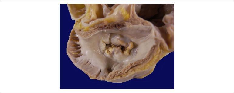
This clinical photograph shows a gross pathological specimen of a human heart, specifically an atrial view of the mitral valve. The image demonstrates hallmark signs of chronic rheumatic heart disease. Key features include significant thickening and fibrosis of the valve leaflets, resulting in a 'fish-mouth' or 'buttonhole' appearance of the valvular orifice. There is evidence of extensive commissural fusion and multiple focal areas of yellowish-white calcification along the leaflet margins and within the valve body. These structural changes characterize severe mitral stenosis, where the restricted opening of the valve impairs blood flow from the left atrium to the left ventricle. The surrounding atrial endocardium appears thickened, consistent with chronic pressure overload. This visual is highly representative for educational purposes in cardiology and pathology to illustrate the long-term sequelae of rheumatic fever on cardiac valves.
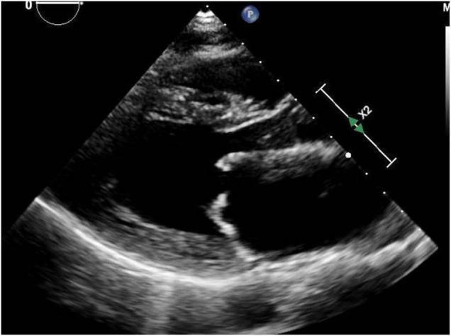
This diagnostic image is a transthoracic echocardiogram in the parasternal long-axis (PLAX) view, demonstrating classic features of mitral stenosis secondary to rheumatic heart disease. The image shows the left ventricle (LV), left atrium (LA), and the aortic root. Key pathology is localized to the mitral valve, where both the anterior and posterior leaflets exhibit significant thickening and increased echogenicity. The leaflets demonstrate restricted diastolic opening, resulting in a narrow valve orifice. The anterior mitral valve leaflet shows a characteristic 'doming' appearance (hockey-stick deformity), which is pathognomonic for rheumatic involvement. The LA appears moderately dilated, a common consequence of chronic mitral stenosis. This educational visual is intended for cardiology and radiology students to illustrate the valvular morphology and mechanical obstruction characteristic of rheumatic mitral valve disease, emphasizing the role of non-invasive ultrasound imaging in assessing valvular heart disease.
Rheumatic Fever vs. Rheumatic Heart Disease
1. Definitions at a Glance
| Feature | Acute Rheumatic Fever (ARF) | Rheumatic Heart Disease (RHD) |
|---|---|---|
| Nature | Acute inflammatory disease | Chronic structural valvular disease |
| Cause | Immune response to GAS pharyngitis | Sequela of ARF (single or recurrent) |
| Timing | Onset 2-3 weeks post-strep infection | Years to decades after ARF |
| Reversibility | Mostly reversible (except valve damage) | Irreversible structural changes |
| Peak age | 5-15 years | 3rd-4th decade (peak prevalence) |
| Sex | Equal in men and women | More common in women |
2. Pathogenesis
ARF - Molecular Mimicry
- Carditis: antibody-binding and T-cell infiltration of the heart
- Arthritis: immune complex deposition in joints
- Chorea: antibody binding to basal ganglia
- Skin manifestations: delayed hypersensitivity reactions
RHD - Progressive Valvular Destruction
3. Clinical Manifestations of ARF
Major Criteria
| Criterion | Low-Risk Populations | Moderate/High-Risk Populations |
|---|---|---|
| Carditis (clinical or subclinical echo) | Yes | Yes |
| Arthritis | Polyarticular only | Mono- or polyarticular |
| Chorea | Yes | Yes |
| Erythema marginatum | Yes | Yes |
| Subcutaneous nodules | Yes | Yes |
Minor Criteria
| Criterion | Low-Risk | Moderate/High-Risk |
|---|---|---|
| Arthralgia | Polyarthralgia | Monoarthralgia |
| Fever | ≥38.5°C | ≥38.5°C |
| ESR/CRP | ESR ≥60mm, CRP ≥3.0 mg/dL | ESR ≥30mm, CRP ≥3.0 mg/dL |
| Prolonged PR interval | Yes (if not already a major criterion) | Yes |
Individual Features of ARF in Detail
- Migratory, asymmetric polyarthritis - knees (76%), ankles (50%), elbows/wrists (12-15%)
- Exquisitely tender, often disproportionate to physical signs
- Sterile synovial fluid with lymphocyte predominance
- Rapid, dramatic response to NSAIDs/aspirin is characteristic
- Pancarditis - affects all layers (pericardium, myocardium, endocardium/valves)
- Most significant: mitral valvulitis (initially regurgitation), then aortic
- Tachycardia out of proportion to fever; rapid sleeping pulse
- ECG: prolonged PR interval
- Involuntary, non-rhythmic, purposeless movements of limbs, body, face
- Emotional lability, personality changes, muscular weakness
- Asymmetric; stops during sleep
- Latency of 4-8 weeks after GAS infection
- Firm, painless nodules 0.5-2 cm over bony prominences or extensor tendons
- Pink, non-pruritic, blanching macules/papules in a serpiginous pattern
- Distributed on trunk and proximal limbs (spares face)
- Evanescent - may appear and disappear over hours to days
4. Rheumatic Heart Disease
Pathological Sequence of Valve Damage
| Stage | Changes |
|---|---|
| Acute valvulitis | Edema, inflammatory cell infiltration, small vegetations along valve closure line |
| Healing | Progressive fibrosis, neovascularization |
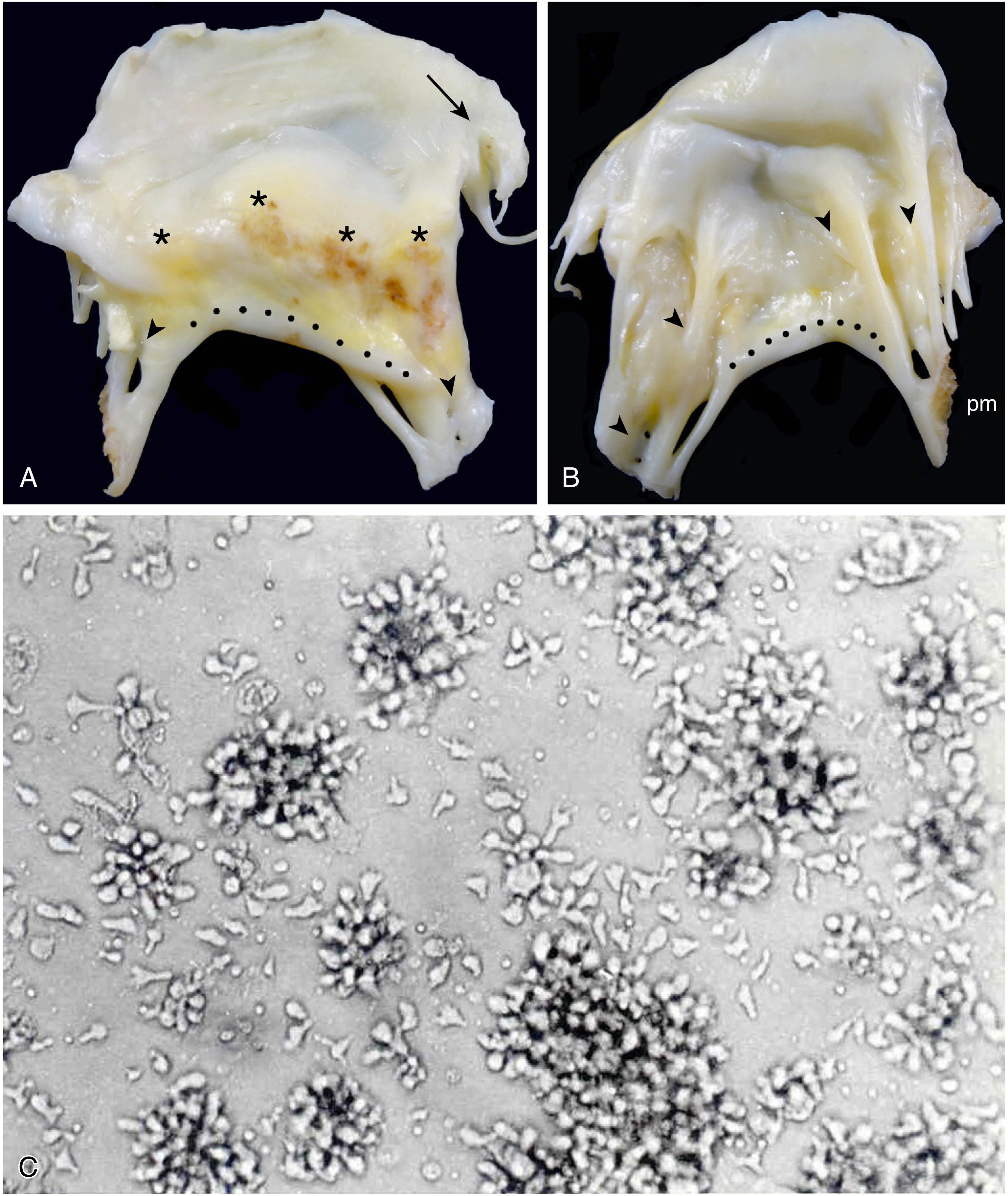
| Chronic RHD | Leaflet thickening, calcification, commissural fusion, chordal fusion/shortening |
| End-stage | "Fish-mouth" or "buttonhole" stenosis; mixed stenosis + regurgitation |
Valve Involvement Patterns
- Mitral valve: most commonly and severely affected
- Aortic valve: second most common
- Tricuspid: less frequent
- Pulmonary: rarely affected
Age-Dependent Presentation
- Young patients: pure mitral regurgitation
- Middle-age adults: mitral stenosis
- Older patients: mixed mitral valve disease
Clinical Features of RHD
- Often oligosymptomatic for many years after ARF
- Symptoms emerge when HF establishes: dyspnea, rales, edema, tachycardia, S3
- 82.5% of affected children in endemic areas are previously undiagnosed (South Africa data)
- 60% of oligosymptomatic patients have no progression in 10 years
- Once NYHA class III-IV: 0-15% survival at 10 years without surgery
- Severe pulmonary hypertension: <3 years survival without surgery
5. Treatment
ARF Treatment
| Goal | Agent |
|---|---|
| Eradicate GAS | IM benzathine penicillin G 1.2 million units stat |
| Arthritis | Aspirin 80-100 mg/kg/day (or naproxen); NSAIDs give dramatic relief |
| Carditis (mild-moderate) | NSAIDs |
| Carditis (severe) | Corticosteroids (prednisolone) |
| Chorea | Valproate, carbamazepine, or haloperidol if severe |
| Hospitalization | Required for all ARF cases |
Secondary Prophylaxis (to prevent RHD)
| Indication | Regimen | Duration |
|---|---|---|
| ARF without carditis | Benzathine penicillin G 1.2 MU IM every 4 weeks | 5 years or until age 21 |
| ARF with carditis (no residual valve disease) | Same | 10 years or until age 21 |
| ARF with carditis + residual valve disease | Same | 10 years or until age 40, sometimes lifelong |
RHD Management
- Medical: diuretics, rate control, anticoagulation (especially with mitral stenosis + AF)
- Surgical/interventional: balloon mitral valvuloplasty for suitable mitral stenosis; valve repair or replacement for severe regurgitation/stenosis
- Antibiotic prophylaxis continued
6. Key Distinguishing Points - Summary
| Point | ARF | RHD |
|---|---|---|
| Trigger | GAS pharyngitis | Recurrent ARF episodes |
| Time course | Acute (weeks) | Chronic (years-decades) |
| Arthritis | Present, migratory, reversible | Absent |
| Chorea | Present | Absent |
| Valve pathology | Acute valvulitis (regurgitation initially) | Stenosis, calcification, fusion |
| ECG | Prolonged PR interval | Signs of LA/LV enlargement, AF |
| Echo | Subclinical valvulitis detectable | Structural valve changes |
| Prevention | Treat strep pharyngitis promptly | Secondary prophylaxis post-ARF |
| Prognosis | Good if no valve damage established | Poor without surgery once symptomatic |