
Give oriGin course and termination relations and branches individual images of musculocutaneous nerve
musculocutaneous nerve origin course branches termination anatomy diagram
musculocutaneous nerve lateral cutaneous forearm termination anatomy image
musculocutaneous nerve brachial plexus origin lateral cord C5 C6 C7 anatomy image diagram
Musculocutaneous Nerve - Complete Anatomy
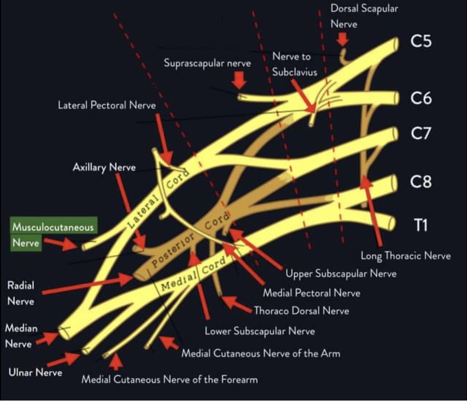
1. Origin
2. Course and Relations
| Segment | Course | Relations |
|---|---|---|
| Axilla | Arises from lateral cord at inferior border of pectoralis minor | Lateral to axillary artery; anterior to subscapularis |
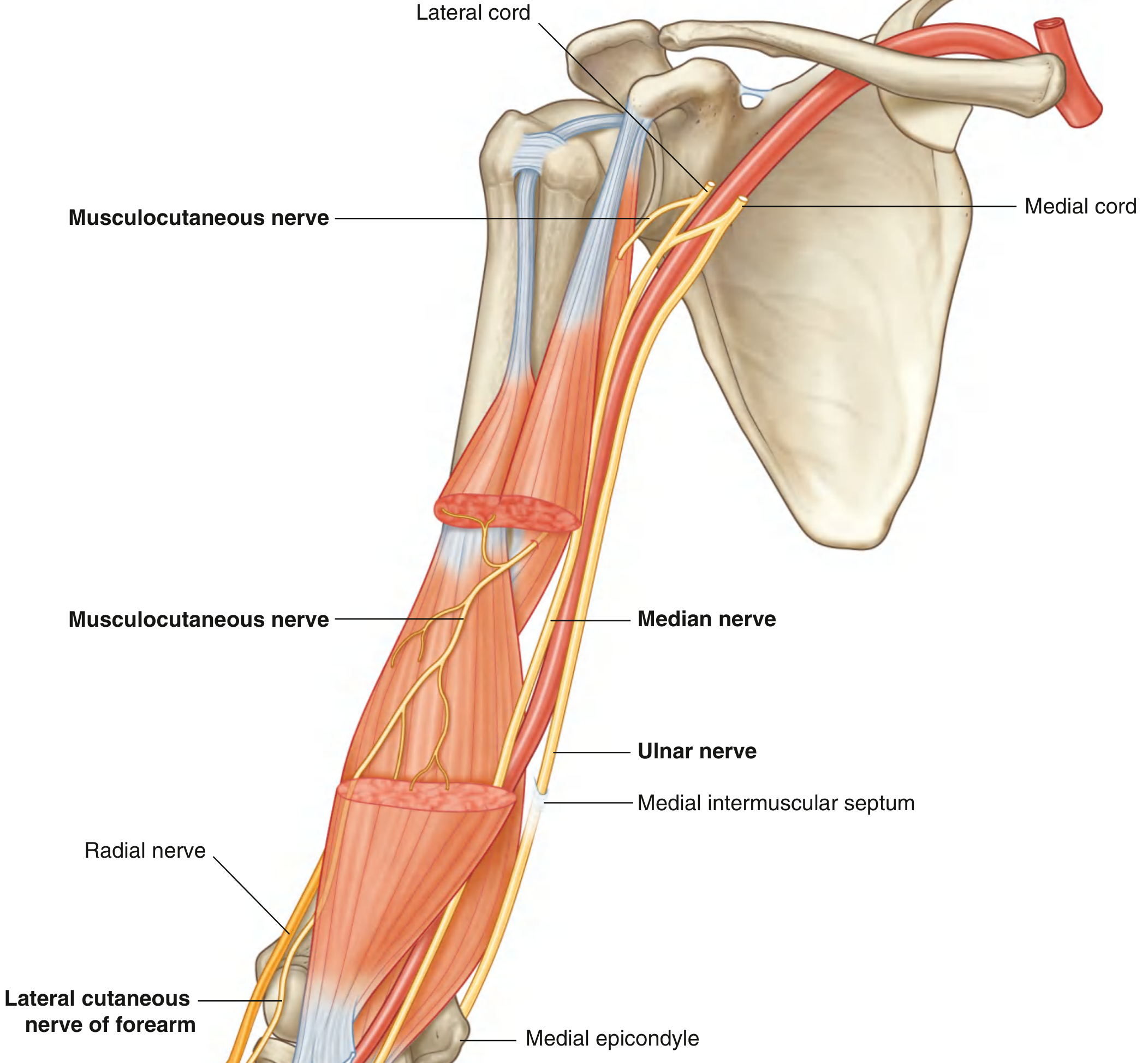
| Coracobrachialis | Pierces the coracobrachialis muscle ~3-8 cm distal to the coracoid process tip | Passes through the substance of coracobrachialis |
| Arm (upper) | Passes diagonally downward between biceps brachii and brachialis | Deep to biceps brachii, superficial to brachialis |
| Arm (lower) | Continues obliquely toward the lateral side | Still deep to biceps, superficial to brachialis |
| Elbow | Pierces the deep fascia lateral to the biceps brachii tendon, ~2 cm above the elbow | Becomes subcutaneous; lateral to biceps tendon |
| Forearm | Continues as the lateral cutaneous nerve of the forearm | Subcutaneous; anterior and posterior divisions |

3. Branches
Motor Branches (Muscular Branches)
| Branch | Muscle Supplied | Level Given Off |
|---|---|---|
| Branch to coracobrachialis | Coracobrachialis | Given off within/before piercing coracobrachialis; often the most proximal branch |
| Branch to biceps brachii | Both heads of biceps brachii | Given off as nerve runs between biceps and brachialis |
| Branch to brachialis | Brachialis (medial portion) | Given off as nerve descends; lateral aspect also receives radial nerve |
Note: The brachialis has dual innervation - medial part by musculocutaneous nerve (C5, C6), lateral part by radial nerve (C7).

Sensory Branch (Terminal)
- Anterior division - runs in subcutaneous tissue along anterolateral forearm from elbow to wrist
- Posterior division - runs along posterolateral forearm
4. Termination
5. Summary Table
| Feature | Detail |
|---|---|
| Origin | Lateral cord of brachial plexus |
| Roots | C5, C6, C7 |
| Emerges | Inferior border of pectoralis minor |
| Pierces | Coracobrachialis (3-8 cm distal to coracoid) |
| Course in arm | Between biceps brachii (superficial) and brachialis (deep) |
| Pierces deep fascia | Lateral to biceps tendon, ~2 cm above elbow |
| Termination | Lateral cutaneous nerve of forearm |
| Motor supply | Coracobrachialis, biceps brachii, brachialis |
| Sensory supply | Anterolateral + posterolateral forearm (elbow to wrist) |
| Function | Elbow flexion, forearm supination (via biceps), shoulder flexion (via coracobrachialis) |
I want only easy images
Simple clean anatomy diagram showing the origin of the musculocutaneous nerve. Show the brachial plexus with roots C5, C6, C7 labeled on the right side, forming the lateral cord, and the musculocutaneous nerve branching off from the lateral cord on the left. Use simple yellow lines for nerves on a white background. Bold text labels. Easy to understand for medical students.
Simple clean anatomy diagram of the musculocutaneous nerve course in the arm. Show a front view of the arm with: the nerve entering the axilla from the lateral cord, piercing through the coracobrachialis muscle, then traveling between biceps brachii (front) and brachialis (back) muscles down the arm. Use simple colors - yellow for nerve, red for muscles, white background. Large bold labels for each structure. Easy medical student diagram.
Simple easy anatomy diagram showing the 3 muscular branches of the musculocutaneous nerve. Draw a front view of the arm. Show a yellow nerve line going down the arm giving off 3 labeled branches: Branch 1 going to Coracobrachialis muscle (top), Branch 2 going to Biceps Brachii muscle (middle), Branch 3 going to Brachialis muscle (lower). Each branch clearly labeled with arrows. White background, simple bold text, color-coded muscles. Easy to understand medical diagram.
Simple clean anatomy diagram showing the termination of the musculocutaneous nerve. Show a front view of the arm and forearm. At the elbow level, the musculocutaneous nerve (yellow line) pierces the deep fascia lateral to the biceps tendon and becomes the Lateral Cutaneous Nerve of the Forearm. Then show it splitting into 2 divisions: Anterior division (covering the front-outer forearm) and Posterior division (covering the back-outer forearm). Show the sensory skin area shaded in light blue on the lateral forearm from elbow to wrist. White background, large bold labels, simple drawing.
429 Too Many Requests. {'message': '{\n "error": {\n "code": 429,\n "message": "Resource exhausted. Please try again later. Please refer to https://cloud.google.com/vertex-ai/generative-ai/docs/error-code-429 for more details.",\n "status": "RESOURCE_EXHAUSTED"\n }\n}\n', 'status': 'Too Many Requests'}
We encountered an error while processing your request. Please try again or reach out to us for help.