

IMG_4571.jpg
Check right nostrils 2 week blood clots comes lesion is there inside what could be possible diagnosis
analyse_media
Reading File
Now let me search the medical library and web for the relevant differential diagnoses.
Finding Sources
Searching the Web
right nostril recurrent blood clot 2 weeks nasal lesion differential diagnosis ENT
Finding Sources
Finding Sources
Finding Sources
Searching PubMed
unilateral nasal lesion blood clot recurrent epistaxis differential diagnosis
Finding Sources
Finding Sources
Reading File
Finding Sources
Reading File
Reading File
Now I have a solid picture. Let me compile the comprehensive answer.
Based on the image analysis and my medical library research, here is a thorough differential diagnosis for your situation:
What the Image Shows
The photo shows the underside of the nose. In the anatomical right nostril (left side of the image), there is a reddish-pinkish area of tissue visible deeper in the nasal passage, behind the nasal hairs. This is consistent with an abnormal or vascular lesion at the anterior nasal cavity/septum area. The shadows from nasal hairs limit how much detail can be seen, but the reddened tissue is notable.
Differential Diagnosis: Unilateral Right Nostril Lesion with Recurrent Blood Clots (2 Weeks)
1. Anterior Septal Ulcer / Erosion (Most Common)
The nasal septum at the front of the nose has a rich blood vessel network called the Kiesselbach plexus. Repeated nose-picking, dry air, or minor trauma can cause a crusting ulcer there that repeatedly bleeds and forms clots. This is the single most common cause of recurrent unilateral nosebleeds with visible lesion.
2. Nasal Vestibulitis / Folliculitis
Bacterial infection (usually Staph. aureus) of the nasal hair follicles causes painful redness, swelling, and crust formation that can bleed. It appears as a red, tender inflamed area just inside the nostril opening.
3. Nasal Hemangioma / Pyogenic Granuloma
A fleshy, bright-red vascular growth (pyogenic granuloma or lobular capillary hemangioma) that bleeds very easily with minimal touch. These are common in adults, can appear on the nasal septum or vestibule, and recurrently form blood clots over the surface. This is a strong possibility given the 2-week history of blood clots with a visible lesion.
4. Nasal Polyp (Less Likely)
Nasal polyps are pale/grey grape-like growths arising from inflamed mucosa. They usually cause obstruction and can bleed if traumatized. Typically pale, not dark red.
5. Nasal Septal Perforation
Chronic ulceration can lead to a hole in the septum, with crusting and recurrent bleeding on the edges. Associated with nose-picking, cocaine use, nasal sprays, or Wegener's granulomatosis.
6. Juvenile Nasopharyngeal Angiofibroma (if adolescent male)
As noted in the Textbook of Family Medicine 9e, recurrent epistaxis plus nasal obstruction in adolescent boys should specifically raise concern for this benign but locally aggressive vascular tumor. It causes significant, recurrent bleeding.
7. Nasal Malignancy (Rare but Must Exclude)
Squamous cell carcinoma or other nasal tumors can present as a bleeding lesion inside one nostril. The 2-week duration is short, but if the lesion is firm, ulcerated, irregular, or non-healing, this must be ruled out - especially in smokers or those over 40.
8. Hereditary Hemorrhagic Telangiectasia (HHT / Osler-Weber-Rendu)
A genetic condition causing multiple tiny vascular malformations on the nasal mucosa. Presents with recurrent spontaneous nosebleeds, often unilateral at first. Look for similar spots on lips or fingertips.
9. Coagulopathy / Blood Thinners
Any bleeding disorder (low platelets, hemophilia, von Willebrand disease) or use of anticoagulants (aspirin, warfarin, clopidogrel) can cause recurrent nasal bleeding from a previously minor vessel.
Red Flags - See a Doctor Urgently If:
- Bleeding does not stop after 20-30 minutes of firm pressure
- The lesion is growing, firm, irregular, or painless
- You have foul-smelling discharge from one side only
- You have facial numbness, vision changes, or swelling
- You are an adolescent male with nasal obstruction (possible angiofibroma)
- The bleeding is heavy or you feel dizzy
What You Need
This requires a direct examination by an ENT (Ear, Nose & Throat) specialist. They will perform:
- Anterior rhinoscopy (nasal speculum + light) to clearly view the lesion
- Possibly a nasal endoscopy for deeper views
- Biopsy if the lesion looks suspicious
- Blood tests if a coagulopathy is suspected
A photo cannot replace this - especially because shadows from nasal hair may be hiding the true extent of the lesion.
Disclaimer: This is educational information only, not a medical diagnosis. Please see an ENT or your doctor for proper evaluation. - Cummings Otolaryngology Head and Neck Surgery; Textbook of Family Medicine 9e
What is treatment for ant septal ulcer
Treatment for anterior septal ulcers involves moisturizing the mucosa, treating infection, and stopping the bleeding cycle. The main approaches are saline irrigation, antibiotic ointments, and avoiding trauma (nose-picking). Cautery is used for persistent bleeding.
Searching sources...
Finding Sources
Searching the Web
anterior nasal septal ulcer treatment management ENT guidelines
Finding Sources
Finding Sources
Reading File
Finding Sources
Reading File
Reading File
Loading Image
Here is a complete, structured guide to treating anterior nasal septal ulcers:
Treatment of Anterior Nasal Septal Ulcer
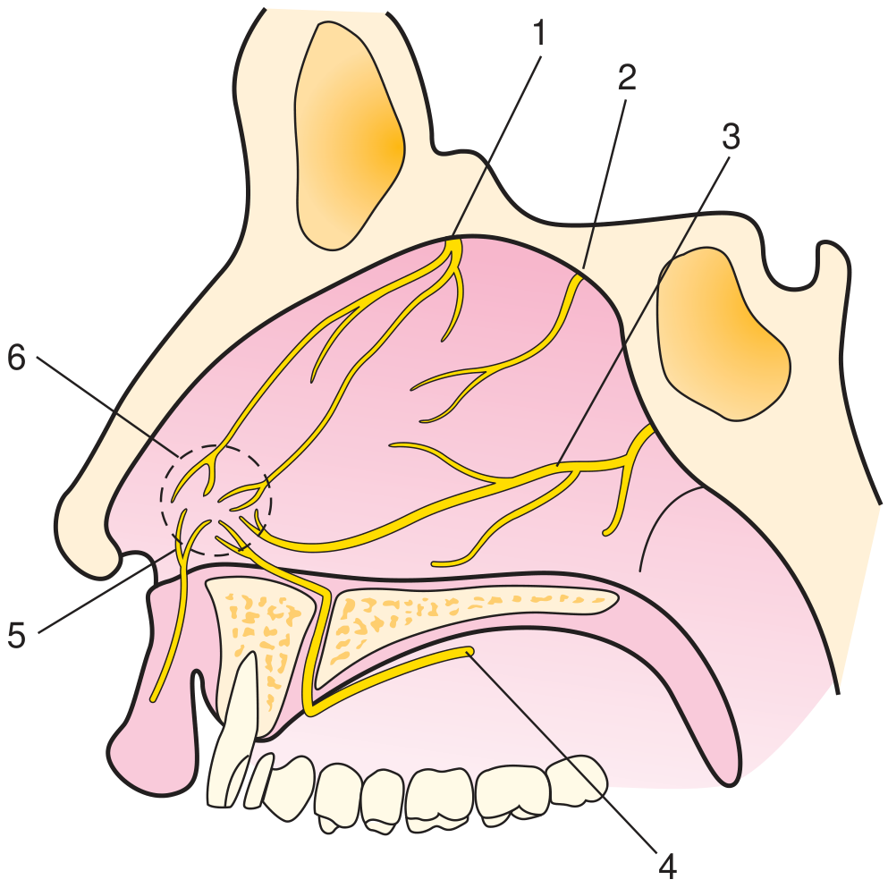
The Kiesselbach plexus at the front of the nasal septum (labeled 6 in the diagram below) is where most anterior septal ulcers form. Treatment follows a step-up approach from conservative care to procedural interventions.
Step 1 - Conservative / First-Line Treatment
These are the foundation of management and address the root cause:
| Measure | Details |
|---|---|
| Stop trauma | No nose-picking, no dry nose-blowing; this is the single most important step |
| Saline nasal irrigation | Twice daily with isotonic saline (e.g. NeilMed, saline spray) - softens crusts, promotes healing |
| Nasal moisturizing | Apply a water-based emollient (nasal gel, saline gel) or petroleum jelly on a cotton swab to the ulcer 2-3x daily to keep the mucosa moist |
| Antibiotic ointment | Mupirocin (Bactroban) nasal ointment applied topically - treats secondary Staph. aureus infection, promotes healing; used twice daily for 2 weeks |
| Home humidifier | Particularly helpful in dry climates or air-conditioned rooms; reduces mucosal desiccation |
| Avoid irritants | Stop nasal decongestant sprays (e.g. xylometazoline if overused), nasal steroid sprays on the ulcer site, cocaine if applicable |
Step 2 - If Bleeding Persists (Cautery)
When the ulcer continues to bleed despite conservative measures, chemical or electrical cautery is the standard next step:
- Silver nitrate cauterization - the most common office procedure. A silver nitrate stick is applied directly to the bleeding vessels in Kiesselbach's area under local anaesthesia. It destroys the prominent superficial vessels causing the bleeding. Simple, quick, and effective.
- Electrocautery - used if chemical cautery fails; done by an ENT under local anaesthetic.
- Important rule: never cauterize both sides of the septum at the same time - this risks septal perforation by cutting off blood supply from both sides simultaneously.
- Gray's Anatomy for Students; Textbook of Family Medicine 9e
Step 3 - Nasal Packing (for Active Bleeding Episodes)
If an acute bleed from the ulcer won't stop with pressure:
- Suction any clots first with a Frazier-tip suction
- Decongest and anesthetize with 4% lidocaine + phenylephrine
- Place a MeroCel or Rapid Rhino anterior nasal pack coated in antibiotic ointment
- Pack stays in for 2-5 days, then remove
- After removal: continue saline and nasal ointment for 2 more weeks
- Give prophylactic antibiotics (to prevent toxic shock syndrome and sinusitis while the pack is in)
- Textbook of Family Medicine 9e
Step 4 - Underlying Cause Treatment
Critically, if an ulcer is not healing, the underlying cause must be treated:
| Cause | Specific Treatment |
|---|---|
| Hypertension | Blood pressure control |
| Blood thinners (aspirin, warfarin) | Review with prescribing doctor - dose adjustment if possible |
| Staph infection / vestibulitis | Oral flucloxacillin or trimethoprim-sulfamethoxazole + topical mupirocin |
| Autoimmune (Wegener's/GPA, lupus) | Systemic immunosuppression (rheumatology referral) |
| Nasal steroid spray causing ulcer | Switch spray technique, use with saline, or stop temporarily |
Step 5 - Persistent/Non-Healing Ulcer (Advanced)
For ulcers that don't heal after weeks of conservative treatment:
- Biopsy - mandatory if the ulcer is firm, irregular, or growing (to exclude malignancy or granulomatous disease)
- Extracellular matrix scaffold (e.g. Biodesign/Surgisis) - a newer technique where a scaffold is placed over the ulcer to promote tissue regeneration; effective in cases resistant to all other treatment
- Surgical septoplasty with excision of ulcerated tissue - a last resort
- Arterial embolization (sphenopalatine artery) - for severe recurrent bleeding uncontrollable by all other means
When to See an ENT Urgently
- Bleeding that does not stop after 20-30 minutes of firm pinching
- Ulcer present for more than 3-4 weeks with no improvement
- Ulcer growing, firm, or painless (possible malignancy)
- Foul-smelling discharge from one nostril
- You are on blood thinners and bleeding is heavy
Disclaimer: This is educational information only. Please have an ENT specialist examine and treat the ulcer directly - especially if it has not healed after 2 weeks of conservative care.

IMG_4573.jpg
Should i use this cream
analyse_media
Finding Sources
Searching the Web
Celestoderm-B betamethasone gentamicin ointment use on nasal septal ulcer safe or not
About This Cream: Celestoderm-B with Garamycin
This is Betamethasone + Gentamicin ointment - a combination of:
- Betamethasone - a potent topical corticosteroid (anti-inflammatory)
- Gentamicin - an aminoglycoside antibiotic (kills bacteria)
Short Answer: No - do NOT use this on your nasal septal ulcer without a doctor's advice
Here is why:
Why the gentamicin part is potentially helpful...
Gentamicin is an antibiotic that could theoretically help treat secondary bacterial infection (like Staph. aureus) in a nasal ulcer. However, mupirocin (Bactroban) is the preferred and specifically recommended antibiotic ointment for nasal use - it is safer, better absorbed by nasal mucosa, and directly targets the most common bacteria involved.
Why the betamethasone part is the problem
Betamethasone is a potent corticosteroid - and steroids are contraindicated on ulcers. Here is what it would do to your septal ulcer:
| Effect | Why It's Harmful |
|---|---|
| Suppresses healing | Steroids slow tissue repair - your ulcer will take longer to heal or may never heal |
| Thins the mucosa | Steroid atrophy of already fragile nasal lining increases the risk of septal perforation |
| Suppresses immunity locally | Makes a bacterial or fungal infection worse, not better |
| Masks worsening | Reduces pain/redness without fixing the cause - you won't notice the ulcer is getting worse |
The NHS betamethasone guidance specifically states: do not use betamethasone on broken skin or ulcers.
What You Should Use Instead
For your anterior septal ulcer, the correct treatment is:
- Mupirocin 2% ointment (Bactroban) - apply a tiny amount with a clean cotton swab twice daily. This is the right antibiotic for nasal use.
- Saline nasal spray - 2x daily to keep the area moist and clean
- Plain petroleum jelly / nasal gel - keeps the mucosa from drying out
- No picking or rubbing
Bottom Line
Put the Celestoderm away for this problem. The steroid in it will actively harm a nasal ulcer. See an ENT so they can examine the lesion directly and prescribe the correct treatment. If there is a Staph infection, they may give you mupirocin or oral antibiotics.
Disclaimer: This is educational information, not a prescription. Please consult your doctor before applying any medication inside your nose.
This is a shared conversation. Sign in to Orris to start your own chat.