
Lymphagiosarcoma
Reading File
Lymphangiosarcoma is a rare malignant tumor of lymphatic endothelial cells, classically associated with chronic lymphedema (Stewart-Treves syndrome). Let me pull up the authoritative textbook content.
Searching sources...
Finding Sources
Finding Sources
Searching PubMed
lymphangiosarcoma Stewart-Treves syndrome
Good results. Now let me pull the detailed content from the most informative sources.
Reading File
Reading File
Reading File
Reading File
Now let me fetch the clinical image and also get the Dermatology textbook content for more detail.
Loading Image
Finding Sources
Now let me pull the immunohistochemistry/pathology detail and the Sabiston content on radiation-associated Stewart-Treves.
Reading File
Reading File
I now have comprehensive content from multiple authoritative textbooks. Here is the full, detailed answer:
Lymphangiosarcoma (Stewart-Treves Syndrome)
Definition
Lymphangiosarcoma is a rare, highly malignant tumor arising from lymphatic endothelial cells. It is a variant of angiosarcoma, classified under the broader "malignant angioendothelioma" umbrella along with hemangiosarcoma. The eponym Stewart-Treves syndrome specifically refers to lymphangiosarcoma arising in postmastectomy lymphedema, first described by Stewart and Treves in 1948. - Schwartz's Principles of Surgery, 11e, p. 1596
Pathogenesis & Etiology
The key predisposing factor is chronic, long-standing lymphedema (typically >10 years). The underlying mechanism involves:
- Prolonged lymphatic stasis creating an immunologically impaired microenvironment
- Local tissue hypoxia and chronic inflammation promoting malignant endothelial transformation
- The tumor arises from lymphatic endothelium and proliferates rapidly with early systemic spread
Causes of the underlying lymphedema include:
| Cause | Notes |
|---|---|
| Post-mastectomy / axillary node dissection | Classic; occurs in ~0.07% of patients after axillary dissection |
| Postmastectomy + radiation (Stewart-Treves syndrome) | Latency ~10 years; tumor occurs outside radiation field but within the zone of lymphedema |
| Filarial infections | Chronic filariasis causing lower extremity lymphedema |
| Congenital lymphedema | Lower extremity involvement |
| Primary or secondary lymphedema (any cause) | Can affect both upper and lower extremities |
The development of angiosarcoma from radiation therapy alone (with minimal/no lymphedema) is called "cutaneous postradiation angiosarcoma of the breast" - a distinct but related entity. - Sabiston Textbook of Surgery, p. 1294; Andrews' Diseases of the Skin
Clinical Features
- Acute worsening of pre-existing chronic lymphedema
- Appearance of bluish or reddish-purple subcutaneous nodules on the affected extremity (typically the arm in postmastectomy cases)
- Nodules have a propensity toward hemorrhage and ulceration
- Multiple lesions are common
- Rapid proliferation with early systemic metastasis
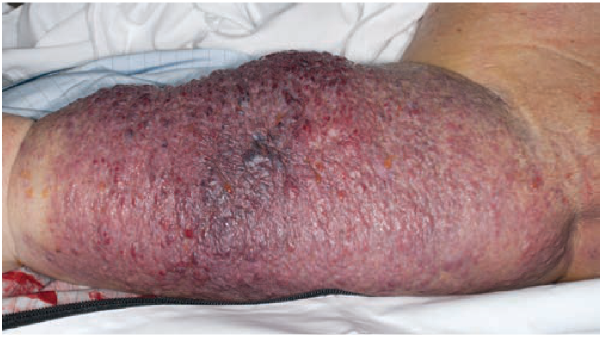
Figure: Lymphangiosarcoma developing 10 years after breast cancer treatment (Schwartz's Principles of Surgery, 11e)
Immunohistochemistry (Pathology)
Diagnosis is confirmed by tissue biopsy with immunohistochemical staining:
- Positive markers: CD34, CD31, Ulex europaeus lectin (endothelial markers)
- Negative: Antikeratin antibodies (useful to exclude metastatic breast carcinoma, which stains keratin-positive)
This panel differentiates lymphangiosarcoma from metastatic carcinoma in the skin of a patient with breast cancer history. - Andrews' Diseases of the Skin
Prognosis
Extremely poor:
- Average survival after diagnosis: ~19 months
- Early and aggressive metastatic spread is the rule
- Schwartz's Principles of Surgery reports the tumor is "generally aggressive"
Treatment
- Radical surgery - often requires radical amputation of the affected extremity; this may be lifesaving if performed early
- Preoperative chemotherapy - used in the multimodal approach, as for other high-grade sarcomas
- Radiation therapy - combined with surgery in a multimodal approach
- The standard sarcoma approach: neoadjuvant chemoradiation followed by surgical excision is the framework - Sabiston Textbook of Surgery
Related/Differential Entities
| Entity | Key Differentiator |
|---|---|
| Cutaneous postradiation angiosarcoma of breast | From radiation alone, minimal lymphedema, arises in breast |
| Kaposi's sarcoma | HIV/HHV-8 associated, blood vessel endothelium, different distribution |
| Hemangiosarcoma | Arises from blood vascular endothelium (not lymphatic) |
| Metastatic breast carcinoma to skin | Antikeratin positive, CD31/CD34 negative |
Key Facts Summary
- First described: Stewart & Treves, 1948 (6 cases of postmastectomy lymphangiosarcoma)
- Incidence: ~0.07% after axillary node dissection
- Latency: Usually >10 years of chronic lymphedema before malignant transformation
- Site: Most common in the upper extremity (postmastectomy), but can occur in any chronically lymphedematous region
- Predisposing conditions listed by Washington Manual: Age, prior radiation, chemical/chemotherapy exposure, genetic syndromes, Paget's disease of bone, HIV/HHV-8 (Kaposi), and chronic lymphedema (lymphangiosarcoma/Stewart-Treves)
Recent evidence note: A 2025 systematic review on MRI features of Stewart-Treves syndrome (PMID 39907978, Jpn J Radiol, Jun 2025) provides updated imaging characterization - worth consulting if radiological diagnosis is relevant to your context.
Multinodular goitre
Multinodular goitre (MNG) is a thyroid gland enlarged by multiple nodules, resulting from follicular hyperplasia and colloid accumulation, often due to iodine deficiency or TSH-driven stimulation. Let me pull comprehensive textbook content.
Searching sources...
Finding Sources
Finding Sources
Searching PubMed
multinodular goiter management guidelines
Good. Now let me read the detailed sections from the most comprehensive sources.
Reading File
Reading File
Reading File
Reading File
Good content so far. Let me now read the continuation from Bailey & Love (investigations/treatment) and S Das (clinical features/treatment), plus the non-toxic MNG section from Scott-Brown's.
Reading File
Reading File
Reading File
Excellent content. Let me now fetch the clinical images and get the rest of the Bailey & Love surgical treatment section.
Reading File
Loading Image
Reading File
I now have rich, comprehensive content from multiple authoritative surgical and ENT textbooks. Here is the complete structured answer:
Multinodular Goitre (MNG)
Definition
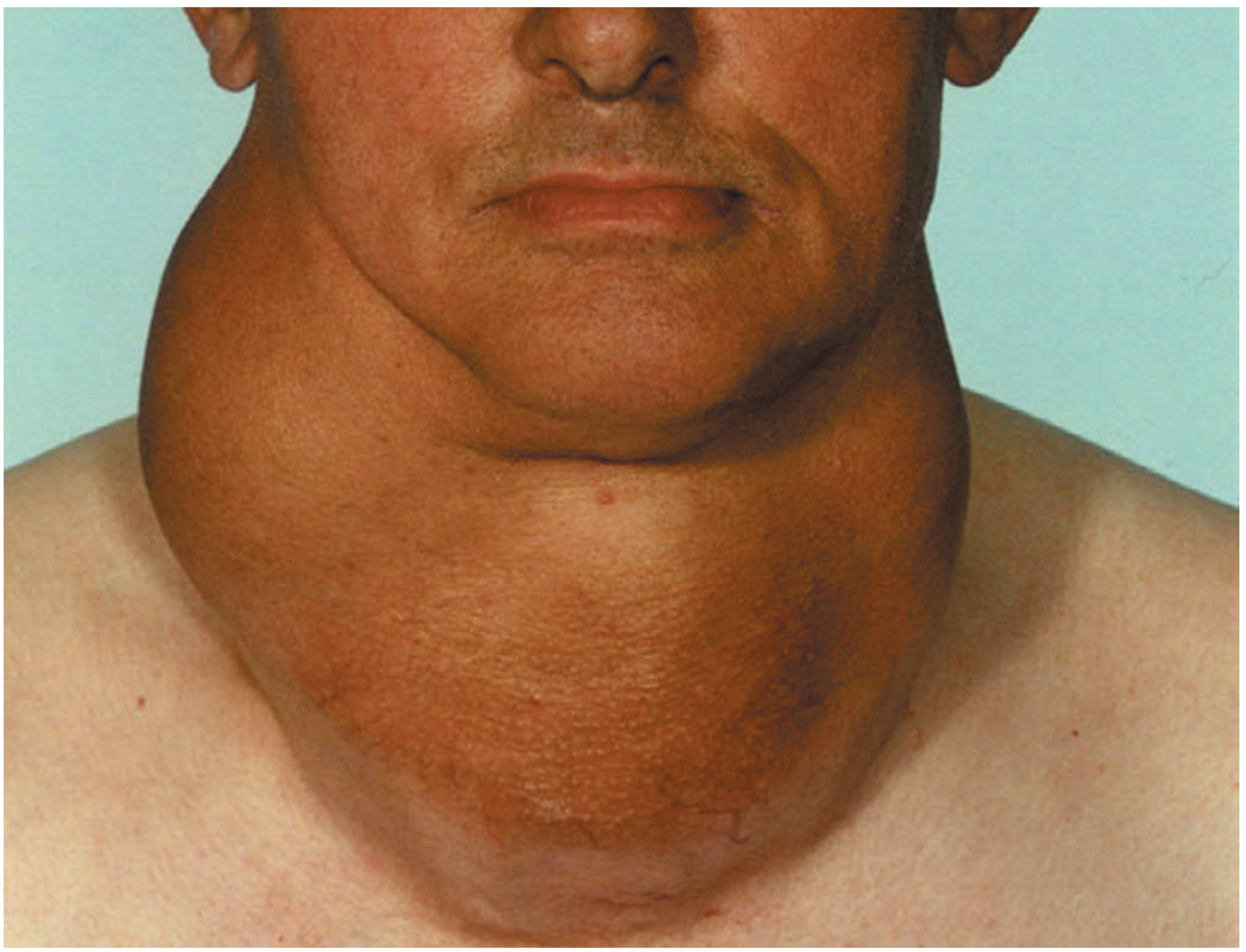
A multinodular goitre is an enlargement of the thyroid gland characterised by the presence of multiple nodules within its substance, resulting from repeated cycles of follicular hyperplasia, colloid accumulation, haemorrhage, necrosis, and fibrosis. It is also known as adenoparenchymatous goitre. - S Das, Manual on Clinical Surgery, 13e
Classification
Goitres as a whole are classified as:
| Type | Subtypes |
|---|---|
| Non-toxic (Simple) | Diffuse parenchymatous, Colloid, Multinodular, Solitary nodular |
| Toxic | Diffuse (Graves' disease), Multinodular (toxic MNG), Toxic adenoma (Plummer's) |
| Neoplastic | Benign (adenoma), Malignant (carcinoma) |
| Thyroiditis | Acute bacterial, Granulomatous, Autoimmune (Hashimoto's), Riedel's |
S Das, Manual on Clinical Surgery, 13e
Pathogenesis
The sequence of events:
- Iodine deficiency (or goitrogenic substances, genetic enzyme defects) → low circulating thyroid hormones → raised TSH
- TSH drives diffuse follicular hyperplasia (parenchymatous/hyperplastic goitre)
- With fluctuating TSH levels, some lobules remain active, others become inactive - creating areas of active and inactive tissue
- Inactive follicles fill with colloid → colloid goitre
- Repeated cycles of hyperplasia, necrosis, haemorrhage, fibrosis → multinodular stage (irreversible)
The cut surface shows nodules with haemorrhagic and necrotic areas separated by normal tissue containing active follicles. Nodules may be colloid or cellular; cystic degeneration, haemorrhage, and calcification are common. - S Das; Bailey & Love, 28e
Other contributing factors:
- Goitrogenic substances: cabbage, turnips, brassica vegetables, soya, antithyroid drugs, para-aminosalicylates
- Physiological demands: puberty, pregnancy (increased metabolic demand)
- Genetic mutations within specific nodules can drive clonal expansion and autonomous function (in toxic MNG) - Scott-Brown's ENT HNS, Vol 1
Epidemiology
- 6 times more common in females than males (oestrogen receptors in thyroid tissue)
- In endemic areas: nodulation appears early, ages 20-30 years
- In sporadic areas: nodulation appears later, ages 30-40 years (patient may be unaware until their 40s or 50s)
- Approximately 50% of patients who present with a "solitary nodule" actually have MNG on further evaluation
Bailey & Love, 28e; S Das, 13e
Clinical Features
Symptoms
- Slowly enlarging, painless lump in the neck
- Sudden pain and enlargement if haemorrhage occurs into a nodule
- Pressure symptoms when the goitre becomes large:
- Dyspnoea (tracheal compression/deviation)
- Dysphagia to solids
- Stridor
- Engorged neck veins (superior mediastinal compression)
- Symptoms worse at night or in the recumbent position
Signs
- Gland has an asymmetrical shape with a smooth, nodular surface
- Nodule consistency varies: soft to hard (hard nodules = calcification or tense haemorrhage)
- Moves upwards on swallowing (confirming thyroid origin)
- Hardness and irregularity due to calcification can simulate carcinoma
Functional Status
- Most patients are euthyroid (non-toxic MNG)
- Secondary thyrotoxicosis occurs in ~25% of long-standing cases (autonomous nodule function)
- In very long-standing disease, most nodules become inactive → myxoedema may develop by age 60-70
S Das, 13e; Bailey & Love, 28e
Toxic Multinodular Goitre
- Occurs when two or more autonomously functioning nodules secrete excess thyroid hormone
- The most common cause of hyperthyroidism in the elderly in iodine-replete areas; in iodine-deficient areas it is the most common cause of hyperthyroidism overall
- Thyroid hyperfunction is less marked than in Graves' disease
- Cardiovascular effects predominate: palpitations, atrial fibrillation, other tachyarrhythmias
- Classical eye signs (exophthalmos), tremor may be absent
- Jod-Basedow effect: pharmacological iodine doses (e.g., IV contrast media) can precipitate acute thyrotoxicosis in MNG, especially in iodine-deficient areas
- Definitive treatment with radioiodine is required
Scott-Brown's ENT HNS, Vol 1
Complications of MNG
| Complication | Notes |
|---|---|
| Haemorrhage into a nodule | Sudden pain, rapid enlargement; if impacted at thoracic inlet - acute respiratory obstruction requiring emergency tracheostomy |
| Tracheal obstruction | Gross lateral displacement or retrosternal extension compressing AP diameter |
| Secondary thyrotoxicosis | Transient mild episodes in up to 30%; overt in ~25% |
| Malignant transformation | Increased incidence (usually follicular carcinoma) reported in endemic areas; dominant/rapidly growing nodules warrant aspiration cytology |
| Calcification | Common; can simulate carcinoma clinically |
| Myxoedema | Late complication when nodules become inactive |
Investigations
- Thyroid function tests (TFTs) - to exclude hypo- or hyperthyroidism
- Thyroid antibodies - to differentiate from autoimmune (Hashimoto's) thyroiditis (the two can coexist)
- Ultrasound - gold standard for nodule assessment; multiple isoechoic nodules with varying cystic change throughout both lobes = almost certainly benign; US outperforms CT/MRI for nodule characterisation
- FNAC (Fine Needle Aspiration Cytology) - only required for nodules with suspicious ultrasonographic features (not necessarily the largest/dominant nodule); must be US-guided
- CT scan of chest and neck - if swallowing or breathing symptoms are present; best to assess tracheal/oesophageal compression or retrosternal extension
- MRI - can identify cystic/solid components and haemorrhage in retrosternal goitre
- Radionuclide scan (¹³¹I) - helpful for retrosternal extension; hot vs cold nodule determination
Bailey & Love, 28e; Scott-Brown's, Vol 1; Grainger & Allison's Diagnostic Radiology
Treatment
Non-Toxic MNG
Most patients with MNG are asymptomatic and do NOT require surgery.
Surgery is indicated for:
- Features suggesting underlying malignancy
- Pressure symptoms (after excluding other causes)
- Tracheal compression
- Cosmetic reasons (patient preference)
- FNAC suspicious of malignancy
Medical (non-surgical) options:
- Iodised salt - primary prevention in endemic areas; dramatically reduces incidence
- Thyroxine suppression therapy - now considered outdated; leads to subclinical hyperthyroidism with risks of reduced bone mineral density and cardiovascular disease; nodules often regrow on discontinuation
- Radioactive iodine (¹³¹I) - used when surgery is refused or contraindicated by age/frailty/comorbidities; can reduce goitre volume by up to 60%, with most shrinkage occurring soon after treatment; better outcomes in younger patients with smaller goitres receiving higher doses
Surgical options:
| Procedure | Indication |
|---|---|
| Total thyroidectomy | Bilateral symmetric disease; younger patients; prevents recurrence; treats incidental carcinoma |
| Total lobectomy (dominant side) | Asymmetric disease (more common); particularly in older patients |
| Dunhill procedure | Total lobectomy one side + subtotal resection contralateral - largely replaced by total thyroidectomy |
| Subtotal thyroidectomy | Historically used; significant problem is propensity for regrowth |
Key surgical principles:
- More than half of benign nodules regress over 10 years without surgery
- Reoperation for recurrent goitre is more difficult and hazardous - hence preference for total thyroidectomy in younger patients at first operation
- Total thyroidectomy requires lifelong thyroxine replacement
Bailey & Love, 28e; Scott-Brown's, Vol 1
Toxic MNG
- Antithyroid drugs (carbimazole/propylthiouracil) - initial medical control
- Definitive treatment: radioiodine (¹³¹I) - treatment of choice; older patients respond well
- Surgery if radioiodine contraindicated or goitre is large
Key Points Summary
| Feature | Non-toxic MNG | Toxic MNG |
|---|---|---|
| Age | Any; endemic 20-30y, sporadic 30-40y | Elderly predominance |
| Thyroid function | Euthyroid | Hyperthyroid |
| Dominant symptoms | Neck lump, pressure | Cardiovascular (AF, palpitations) |
| Exophthalmos/tremor | Absent | Often absent |
| Definitive treatment | Surgery / ¹³¹I | ¹³¹I (preferred) / surgery |
| Risk of malignancy | Present (follicular > papillary) | Present |
This is a shared conversation. Sign in to Orris to start your own chat.