
Reproductive physiology notes for professional exam mbbs 1st yr
Reproductive Physiology - MBBS 1st Year Notes
1. OVERVIEW OF REPRODUCTIVE PHYSIOLOGY
- GnRH (from hypothalamus) - pulsatile release
- FSH and LH (from anterior pituitary)
- Testosterone (testes), Estrogen + Progesterone (ovaries)
2. MALE REPRODUCTIVE PHYSIOLOGY
A. Structure of the Testes
| Component | Proportion | Function |
|---|---|---|
| Seminiferous tubules | 80% of testis | Spermatogenesis |
| Leydig cells | In connective tissue | Testosterone synthesis |
| Sertoli cells | Line seminiferous tubules | Support spermatogenesis |
B. Sertoli Cell Functions (4 key functions)
- Nutritive - provide nutrients to developing sperm (isolated from bloodstream)
- Blood-testis barrier - tight junctions between Sertoli cells; protects developing sperm from noxious substances while allowing testosterone to cross
- Fluid secretion - aqueous fluid into lumen of seminiferous tubules; transports sperm toward epididymis
- Androgen-binding protein (ABP) - secreted into lumen; keeps local testosterone levels high near developing sperm
Exam tip: Sertoli cells are regulated by FSH. Leydig cells are regulated by LH.
C. Spermatogenesis
Duration: ~64 days for one full cycle
Output: ~128 million sperm/day (each spermatogonium produces 64 spermatozoa)
- Mitotic phase - Spermatogonia (stem cells) divide mitotically to produce spermatocytes
- Meiotic phase - Primary spermatocytes (2n) undergo meiosis I → secondary spermatocytes (n) → meiosis II → spermatids (haploid)
- Spermiogenesis - Spermatids transform into mature spermatozoa (loss of cytoplasm, development of flagella, acrosome formation)
D. Sperm Transport and Accessory Glands
- Epididymis: Primary site for sperm maturation and storage; sperm remain viable here for several months
- Seminal vesicles: Contribute ~60% of ejaculate volume (fructose - energy source for sperm, prostaglandins)
- Prostate gland: Alkaline secretions (neutralize vaginal acidity, zinc, citric acid)
- Bulbourethral (Cowper's) glands: Pre-ejaculatory fluid (lubrication, neutralizes urethral acidity)
E. Testosterone
Stimulus: LH from anterior pituitary
| Mediated by Testosterone | Mediated by DHT (5α-reduced) |
|---|---|
| Differentiation of epididymis, vas deferens, seminal vesicles | Differentiation of penis, scrotum, prostate |
| Increased muscle mass | Male hair pattern |
| Pubertal growth spurt | Male pattern baldness |
| Epiphyseal closure | Sebaceous gland activity |
| Deepening of voice | Growth of prostate |
| Spermatogenesis | |
| Negative feedback on anterior pituitary | |
| Libido |
- Testosterone → negative feedback on both hypothalamus (↓GnRH) and anterior pituitary (↓LH)
- Inhibin (from Sertoli cells) → negative feedback on anterior pituitary (↓FSH)
3. FEMALE REPRODUCTIVE PHYSIOLOGY
A. Anatomy of the Ovary
- Cortex (outer/largest) - lined by germinal epithelium; contains all oocytes in follicles
- Medulla (middle) - mixture of cell types
- Hilum (inner) - blood vessels and lymphatics
B. Oogenesis
| Stage | Event |
|---|---|
| Gestational weeks 8-9 | Oogonia enter meiosis → become primary oocytes |
| Weeks 20-24 gestation | Peak ~7 million oogonia |
| Birth | ~2 million oocytes remain |
| Puberty | ~400,000 oocytes remain |
| Menopause | Few/none remain |
- Primary oocytes are arrested in prophase of Meiosis I from fetal life until ovulation
- First meiotic division completed just before ovulation
- Second meiotic division completed only upon fertilization
- Females do NOT produce new oocytes after birth (unlike males who continuously produce sperm)
4. THE MENSTRUAL CYCLE
Day 1 = First day of menstrual bleeding
A. Phases of the Menstrual Cycle
Uterine (Endometrial) Cycle: Menstrual phase → Proliferative phase → Secretory phase
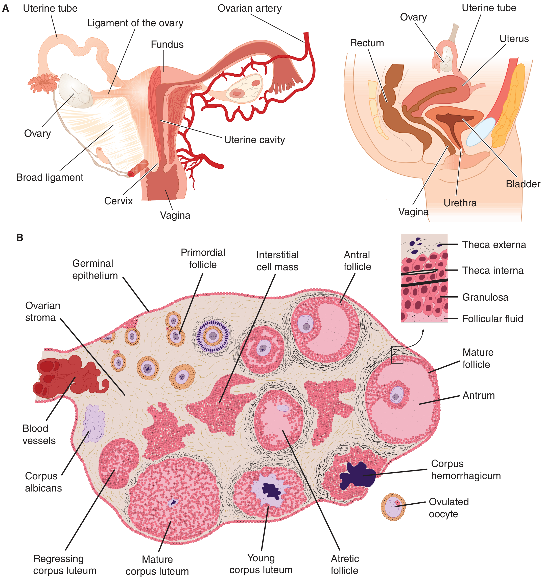
B. Follicular (Proliferative) Phase - Days 1-14
- FSH (slightly elevated) stimulates 6-12 primary follicles to grow
- One dominant (Graafian) follicle selected by day ~6 (related to ability to secrete estrogen); others undergo atresia (apoptosis)
- Dominant follicle produces increasing estradiol (E2)
- Primordial follicle (oocyte + single granulosa cell layer)
- Primary follicle (multiple granulosa cell layers)
- Secondary follicle (theca interna + theca externa form)
- Antral (Graafian) follicle (antrum fills with estrogen-rich follicular fluid)
- Theca interna cells (stimulated by LH) → produce androgens (androstenedione)
- Granulosa cells (stimulated by FSH) → aromatize androgens → estrogens
- Estradiol → endometrial growth, gland elongation, spiral artery growth
- Cervical mucus: copious, watery, elastic, shows "ferning" pattern on glass slide
- Ferning creates channels in the cervix for sperm penetration
C. Ovulation - Day 14 (of 28-day cycle)
Ovulation occurs ~36 hours after onset of the LH surge
- High estrogen (sustained >200 pg/mL for >50 hours) → switches from negative to positive feedback on anterior pituitary
- Massive LH surge → proteolytic enzyme activation, prostaglandin secretion
- Follicle ruptures → oocyte extruded into peritoneal cavity
- Fimbriated ends of fallopian tube pick up the oocyte
D. Luteal (Secretory) Phase - Days 14-28
- Ruptured follicle → granulosa + theca cells proliferate → filled with lipid-rich luteal cells → Corpus luteum
- Corpus luteum secretes estrogen + progesterone (VEGF needed for its blood supply)
- If no pregnancy: Corpus luteum degenerates → corpus albicans → ↓ estrogen + progesterone → menstruation (Day 28)
- If pregnancy: hCG from trophoblast (has same α-subunit as LH) maintains corpus luteum until placenta takes over (~10 weeks)
- Progesterone: glands become tortuous, accumulate glycogen, ↑mucus secretions; stroma edematous; spiral arteries elongate + coil
- Cervical mucus: thick, non-elastic, does not fern (blocks sperm - fertilization window closed)
- Progesterone has mild thermogenic effect → basal body temperature rises ~0.5°C in luteal phase (basis of rhythm contraception method)
E. Hormone Levels Summary During the Cycle
| Phase | Dominant Hormone | FSH | LH | Estrogen | Progesterone |
|---|---|---|---|---|---|
| Menstruation (D1-5) | Low all | ↑ starts | Low | Low | Low |
| Follicular (D5-14) | Estrogen | ↑ then ↓ | Low then surge | Rising → peak | Low |
| Ovulation (D14) | LH surge | Mid-cycle surge | Surge | Peak just before | Low |
| Luteal (D14-28) | Progesterone | Low | Low | Moderate | High |
5. HORMONAL CONTROL - HPG AXIS
Hypothalamus
     |
  GnRH (pulsatile)
     |
Anterior Pituitary
   /        \
 FSH         LH
  |            |
Granulosa    Theca/Leydig
cells        cells
  |            |
Inhibin      Estrogen/
             Testosterone
     |
  Negative feedback (usually)
  Positive feedback at mid-cycle surge (estrogen only)
- GnRH must be pulsatile to stimulate FSH/LH (continuous GnRH → downregulates receptors → no effect → basis of GnRH agonist therapy for endometriosis, prostate cancer)
- Inhibin (from granulosa/Sertoli cells) selectively suppresses FSH
- Activin (also from granulosa cells) stimulates FSH release
6. PUBERTY
- Thelarche (breast development) - first sign
- Hair (pubic and axillary)
- Adrenarchy (adrenal androgens)
- Rapid growth spurt
- Birth (menarche) - average age ~12-13 years
- Testicular enlargement - first sign
- Pubic hair
- Growth spurt
- Voice change
- Spermarche
7. FERTILIZATION AND IMPLANTATION
- Chemoattraction of sperm to ovum
- Sperm bind to zona pellucida (ZP3 receptor)
- Acrosomal reaction - breakdown of acrosome, release of acrosin (trypsin-like protease) → facilitates zona penetration
- Sperm head fuses with oocyte membrane via fertilin protein → sperm nucleus released into oocyte cytoplasm
- Cortical reaction → zona pellucida hardens → blocks polyspermy
- Fast block: Immediate change in oocyte membrane potential
- Slow block: Structural change in zona pellucida (cortical reaction, zona hardening)
- Fertilized ovum (zygote) → travels down fallopian tube over ~3 days → reaches 8-16 cell stage → becomes blastocyst
- Implantation occurs in uterine endometrium ~6-7 days after fertilization (decidualized endometrium in secretory phase)
- Trophoblast cells of blastocyst secrete hCG → maintains corpus luteum → continued progesterone → protects pregnancy
8. ESTROGEN AND PROGESTERONE - ACTIONS SUMMARY
Estrogen actions:
- Uterus: endometrial proliferation, growth of glands and spiral arteries
- Cervix: watery, elastic mucus with ferning (sperm-friendly)
- Breast: ductal growth, areolar pigmentation, fat deposition
- Bone: promotes pubertal growth spurt; closes epiphyses at end of puberty
- Feedback: negative (usually), positive at mid-cycle surge
- Stimulates prolactin secretion from pituitary
- Enlarges external genitalia
- Female fat distribution (subcutaneous)
Progesterone actions:
- Uterus: converts proliferative → secretory endometrium; maintains pregnancy; raises threshold to myometrial contractions
- Cervix: thick, non-elastic mucus (no ferning - sperm-hostile)
- Breast: stimulates secretory activity in mammary ducts (with estrogen)
- Temperature: mild thermogenic effect (↑BBT in luteal phase by ~0.5°C)
- Feedback: negative on pituitary; responsible for FSH mid-cycle surge
9. QUICK REVISION TABLES
Cells and Hormones at a Glance
| Cell | Hormone Produced | Regulated by |
|---|---|---|
| Leydig cells | Testosterone | LH |
| Sertoli cells | Inhibin, ABP | FSH |
| Theca interna | Androgens | LH |
| Granulosa cells | Estrogen, Inhibin | FSH |
| Corpus luteum | Progesterone + Estrogen | LH (then hCG) |
| Trophoblast | hCG | - |
Key Numbers to Remember
| Fact | Number |
|---|---|
| Duration of spermatogenesis | 64 days |
| Sperm produced per day | 128 million |
| Oogonia at peak (gestation weeks 20-24) | 7 million |
| Oocytes at birth | 2 million |
| Oocytes at puberty | 400,000 |
| Average menstrual cycle length | 28 days |
| Ovulation occurs after LH surge | ~36 hours |
| Fertilization to implantation | ~6-7 days |
| Time for blastocyst to reach uterus | ~3 days |
| Temperature rise at ovulation (BBT) | ~0.5°C |
| Testicular temperature | 35-36°C (1-2°C below core) |
10. HIGH-YIELD EXAM POINTS
- Blood-testis barrier is formed by tight junctions between Sertoli cells (not Leydig cells)
- FSH regulates Sertoli cells (spermatogenesis); LH regulates Leydig cells (testosterone)
- Female meiosis is arrested in prophase of Meiosis I until ovulation; completed at fertilization
- Ferning of cervical mucus = follicular/estrogenic phase; no ferning = luteal/progestogenic phase
- Mittelschmerz = mid-cycle pain at ovulation
- Positive feedback of estrogen → LH surge → ovulation
- hCG = "pregnancy hormone"; same α-subunit as LH/FSH/TSH; rescues corpus luteum
- Inhibin suppresses FSH specifically (not LH)
- Testosterone → muscle, voice, libido; DHT → prostate, hair, genitalia
- GnRH must be pulsatile - continuous = down-regulation (used therapeutically)
- Corpus luteum → progesterone dominance → if no pregnancy → luteolysis → withdrawal bleeding
- Spermatogonia (2n) → Primary spermatocytes (2n) → Secondary spermatocytes (n) → Spermatids (n) → Spermatozoa
1
What did you mean by '1'?
Create notes as flowchart and diagram create it into doc format
mkdir -p /home/daytona/workspace/repro-physiology && ls /home/daytona/workspace/repro-physiology && node -e "require('docx'); console.log('docx ok')"
~/repro-physiology/build.js
const {
  Document, Packer, Paragraph, TextRun, Table, TableRow, TableCell,
  HeadingLevel, AlignmentType, BorderStyle, WidthType, ShadingType,
  VerticalAlign, PageNumber, Header, Footer, PageBreak,
  convertInchesToTwip, UnderlineType
} = require('docx');
const fs = require('fs');

// ─── colour palette ──────────────────────────────────────────────────
const BLUE      = "1F4E79";
const LBLUE     = "2E75B6";
const TEAL      = "17375E";
const LTBLUE    = "BDD7EE";
const LLBLUE    = "DEEAF1";
const PINK      = "C00000";
const GREEN     = "375623";
const LGREEN    = "E2EFDA";
const LYELLOW   = "FFF2CC";
const LORANGE   = "FCE4D6";
const LGREY     = "F2F2F2";
const WHITE     = "FFFFFF";
const DARKGREY  = "404040";
const BLACK     = "000000";

// ─── helper builders ─────────────────────────────────────────────────

function heading1(text) {
  return new Paragraph({
    text,
    heading: HeadingLevel.HEADING_1,
    spacing: { before: 360, after: 120 },
    shading: { type: ShadingType.SOLID, color: BLUE },
    run: { color: WHITE, bold: true },
  });
}

function heading2(text) {
  return new Paragraph({
    text,
    heading: HeadingLevel.HEADING_2,
    spacing: { before: 240, after: 80 },
  });
}

function heading3(text) {
  return new Paragraph({
    text,
    heading: HeadingLevel.HEADING_3,
    spacing: { before: 160, after: 60 },
  });
}

function para(text, opts = {}) {
  return new Paragraph({
    children: [new TextRun({ text, color: opts.color || BLACK, bold: opts.bold, italics: opts.italic, size: opts.size || 22 })],
    spacing: { before: 60, after: 60 },
    alignment: opts.align || AlignmentType.LEFT,
    indent: opts.indent ? { left: convertInchesToTwip(0.3) } : undefined,
  });
}

function bullet(text, level = 0, color = BLACK) {
  return new Paragraph({
    children: [new TextRun({ text, color, size: 22 })],
    bullet: { level },
    spacing: { before: 40, after: 40 },
  });
}

function keyPoint(label, value) {
  return new Paragraph({
    children: [
      new TextRun({ text: label + ": ", bold: true, color: LBLUE, size: 22 }),
      new TextRun({ text: value, color: BLACK, size: 22 }),
    ],
    spacing: { before: 50, after: 50 },
    indent: { left: convertInchesToTwip(0.2) },
  });
}

function divider() {
  return new Paragraph({
    text: "",
    border: { bottom: { color: LBLUE, size: 6, style: BorderStyle.SINGLE } },
    spacing: { before: 120, after: 120 },
  });
}

function pageBreak() {
  return new Paragraph({ children: [new PageBreak()] });
}

function centeredBold(text, size = 28, color = WHITE, shade = LBLUE) {
  return new Paragraph({
    children: [new TextRun({ text, bold: true, color, size })],
    alignment: AlignmentType.CENTER,
    spacing: { before: 80, after: 80 },
    shading: { type: ShadingType.SOLID, color: shade },
  });
}

// ─── TABLE helpers ────────────────────────────────────────────────────

function makeCell(text, opts = {}) {
  return new TableCell({
    children: [new Paragraph({
      children: [new TextRun({
        text,
        bold: opts.bold || false,
        color: opts.textColor || BLACK,
        size: opts.size || 20,
      })],
      alignment: opts.align || AlignmentType.LEFT,
      spacing: { before: 40, after: 40 },
    })],
    shading: opts.shade ? { type: ShadingType.SOLID, color: opts.shade } : undefined,
    verticalAlign: VerticalAlign.CENTER,
    width: opts.width ? { size: opts.width, type: WidthType.PERCENTAGE } : undefined,
    columnSpan: opts.span || 1,
  });
}

function headerRow(cells, shade = LBLUE) {
  return new TableRow({
    children: cells.map(c =>
      typeof c === 'string'
        ? makeCell(c, { bold: true, textColor: WHITE, shade, align: AlignmentType.CENTER })
        : makeCell(c.text, { bold: true, textColor: WHITE, shade, align: AlignmentType.CENTER, width: c.w })
    ),
    tableHeader: true,
  });
}

function dataRow(cells, altShade) {
  return new TableRow({
    children: cells.map(c =>
      typeof c === 'string'
        ? makeCell(c, { shade: altShade })
        : makeCell(c.text, { shade: altShade || c.shade, bold: c.bold, textColor: c.color, align: c.align, width: c.w })
    ),
  });
}

function simpleTable(headers, rows, colWidths) {
  const tRows = [headerRow(headers.map((h, i) => ({ text: h, w: colWidths ? colWidths[i] : undefined })))];
  rows.forEach((r, idx) => {
    tRows.push(dataRow(r.map((c, i) => ({
      text: c,
      w: colWidths ? colWidths[i] : undefined,
      shade: idx % 2 === 0 ? LGREY : WHITE,
    })));
  });
  return new Table({
    rows: tRows,
    width: { size: 100, type: WidthType.PERCENTAGE },
    margins: { top: 80, bottom: 80, left: 100, right: 100 },
  });
}

// ─── FLOWCHART via ASCII-art table ───────────────────────────────────

function flowBox(text, shade = LTBLUE, textColor = TEAL, bold = true) {
  return new Table({
    rows: [new TableRow({ children: [makeCell(text, { shade, textColor: textColor, bold, align: AlignmentType.CENTER, size: 22 })] })],
    width: { size: 60, type: WidthType.PERCENTAGE },
    float: { horizontalAnchor: "margin", absoluteHorizontalPosition: convertInchesToTwip(1.5) },
  });
}

function arrow() {
  return new Paragraph({
    children: [new TextRun({ text: "↓", color: LBLUE, bold: true, size: 32 })],
    alignment: AlignmentType.CENTER,
    spacing: { before: 60, after: 60 },
  });
}

function flowStep(label, detail, shade = LLBLUE) {
  return [
    new Table({
      rows: [new TableRow({
        children: [
          makeCell(label, { shade, bold: true, textColor: TEAL, align: AlignmentType.CENTER, size: 20, w: 30 }),
          makeCell(detail, { shade: WHITE, textColor: BLACK, align: AlignmentType.LEFT, size: 20, w: 70 }),
        ],
      })],
      width: { size: 100, type: WidthType.PERCENTAGE },
    }),
    arrow(),
  ];
}

// ─── CONTENT BUILDER ─────────────────────────────────────────────────
const children = [];

// ══════════════════════════════════════════════════════════════════════
// TITLE PAGE
// ══════════════════════════════════════════════════════════════════════
children.push(
  new Paragraph({ spacing: { before: 600 } }),
  new Paragraph({
    children: [new TextRun({ text: "REPRODUCTIVE PHYSIOLOGY", bold: true, color: WHITE, size: 52 })],
    alignment: AlignmentType.CENTER,
    shading: { type: ShadingType.SOLID, color: BLUE },
    spacing: { before: 200, after: 60 },
  }),
  new Paragraph({
    children: [new TextRun({ text: "MBBS 1st Year — Professional Exam Notes", bold: false, color: WHITE, size: 28 })],
    alignment: AlignmentType.CENTER,
    shading: { type: ShadingType.SOLID, color: LBLUE },
    spacing: { before: 0, after: 200 },
  }),
  new Paragraph({
    children: [new TextRun({ text: "Flowcharts • Diagrams • Tables • High-Yield Points", italics: true, color: LBLUE, size: 24 })],
    alignment: AlignmentType.CENTER,
    spacing: { before: 200, after: 60 },
  }),
  new Paragraph({
    children: [new TextRun({ text: "Sources: Costanzo Physiology 7e | Ganong's 26e | Guyton & Hall | Berek & Novak", italics: true, color: DARKGREY, size: 20 })],
    alignment: AlignmentType.CENTER,
    spacing: { before: 60, after: 400 },
  }),
  pageBreak()
);

// ══════════════════════════════════════════════════════════════════════
// SECTION 1 — HPG AXIS FLOWCHART
// ══════════════════════════════════════════════════════════════════════
children.push(
  heading1("1. HYPOTHALAMO-PITUITARY-GONADAL (HPG) AXIS"),
  para("The entire reproductive system is governed by a 3-tier hormonal hierarchy."),
  divider(),
);

// Flowchart built as a vertical table
const hpgRows = [
  ["HYPOTHALAMUS", "Arcuate nucleus\nPulse generator → GnRH (pulsatile, q 60-90 min)\nContinuous GnRH → downregulation (used therapeutically)", BLUE, WHITE],
  ["↓  GnRH  ↓", "Gonadotropin-Releasing Hormone (decapeptide)", LBLUE, WHITE],
  ["ANTERIOR PITUITARY", "Gonadotroph cells\nFSH + LH secreted in response to GnRH", TEAL, WHITE],
  ["↓  FSH + LH  ↓", "Gonadotropins travel via bloodstream", LTBLUE, TEAL],
  ["GONADS (Testes / Ovaries)", "Testes: Sertoli cells (FSH) + Leydig cells (LH)\nOvaries: Granulosa cells (FSH) + Theca cells (LH)", LGREEN, GREEN],
  ["↑  Negative Feedback  ↑", "Testosterone / Estrogen / Inhibin → suppress hypothalamus & pituitary", LORANGE, PINK],
  ["⚡ EXCEPTION: Positive Feedback", "High sustained estrogen (>200 pg/mL, >50 hrs) → triggers LH SURGE → Ovulation", LYELLOW, PINK],
];

const hpgTable = new Table({
  rows: hpgRows.map(([label, detail, shade, tc]) =>
    new TableRow({
      children: [
        makeCell(label, { shade, bold: true, textColor: tc, align: AlignmentType.CENTER, size: 22, w: 30 }),
        makeCell(detail, { shade: label.includes("↓") || label.includes("↑") ? shade : WHITE, textColor: BLACK, align: AlignmentType.LEFT, size: 20, w: 70 }),
      ],
    })
  ),
  width: { size: 100, type: WidthType.PERCENTAGE },
});
children.push(hpgTable, para(""), divider());

// Key regulators table
children.push(heading2("Key Regulatory Hormones"));
children.push(simpleTable(
  ["Hormone", "Source", "Target", "Key Action"],
  [
    ["GnRH", "Hypothalamus", "Ant. Pituitary", "Stimulates FSH + LH (must be pulsatile)"],
    ["FSH", "Ant. Pituitary", "Sertoli / Granulosa cells", "Spermatogenesis / Follicle development"],
    ["LH", "Ant. Pituitary", "Leydig / Theca cells", "Testosterone / Androgen synthesis"],
    ["Inhibin", "Sertoli / Granulosa", "Ant. Pituitary", "Selectively suppresses FSH"],
    ["Testosterone", "Leydig cells", "Multiple", "Secondary sex characteristics, spermatogenesis"],
    ["Estradiol (E2)", "Granulosa cells", "Multiple", "Follicular phase, positive feedback → LH surge"],
    ["Progesterone", "Corpus luteum", "Uterus / Pituitary", "Secretory phase, maintain pregnancy"],
    ["hCG", "Trophoblast", "Corpus luteum", "Maintains CL in early pregnancy"],
  ],
));
children.push(para(""), pageBreak());

// ══════════════════════════════════════════════════════════════════════
// SECTION 2 — MALE REPRODUCTIVE PHYSIOLOGY
// ══════════════════════════════════════════════════════════════════════
children.push(
  heading1("2. MALE REPRODUCTIVE PHYSIOLOGY"),
  heading2("A. Testicular Structure"),
);

children.push(simpleTable(
  ["Component", "% of Testis", "Cell Type", "Function"],
  [
    ["Seminiferous tubules", "80%", "Sertoli cells + Spermatogonia", "Spermatogenesis"],
    ["Connective tissue + Leydig", "20%", "Leydig cells", "Testosterone synthesis"],
    ["Epididymis", "—", "Stereocilia epithelium", "Sperm maturation & storage"],
  ],
));

children.push(
  heading2("B. Sertoli Cell Functions"),
  para("Regulated by FSH. Located in seminiferous tubule epithelium."),
);
const sertoliFn = [
  ["1. NUTRITIVE", "Provide nutrients to developing sperm (isolated from blood)", LLBLUE],
  ["2. BLOOD-TESTIS BARRIER", "Tight junctions between Sertoli cells; selective permeability; protects developing sperm", LLBLUE],
  ["3. FLUID SECRETION", "Aqueous fluid → transports sperm toward epididymis", LGREEN],
  ["4. ANDROGEN-BINDING PROTEIN (ABP)", "Secreted into tubule lumen; maintains high local testosterone near sperm", LYELLOW],
];
children.push(new Table({
  rows: sertoliFn.map(([fn, detail, shade]) =>
    new TableRow({ children: [
      makeCell(fn, { shade: LBLUE, bold: true, textColor: WHITE, align: AlignmentType.CENTER, w: 35 }),
      makeCell(detail, { shade, textColor: BLACK, w: 65 }),
    ]})
  ),
  width: { size: 100, type: WidthType.PERCENTAGE },
}));

// Spermatogenesis Flowchart
children.push(
  heading2("C. Spermatogenesis Flowchart"),
  para("Duration: ~64 days | Output: 128 million sperm/day", { bold: true }),
  para(""),
);

const spermFlow = new Table({
  rows: [
    new TableRow({ children: [
      makeCell("SPERMATOGONIUM (2n)", { shade: BLUE, bold: true, textColor: WHITE, align: AlignmentType.CENTER }),
      makeCell("Stem cell | Mitotic divisions in basal compartment of seminiferous tubule", { shade: LLBLUE }),
    ]}),
    new TableRow({ children: [
      makeCell("↓ Mitosis ↓", { shade: LTBLUE, bold: true, textColor: TEAL, align: AlignmentType.CENTER }),
      makeCell("", { shade: WHITE }),
    ]}),
    new TableRow({ children: [
      makeCell("PRIMARY SPERMATOCYTE (2n)", { shade: LBLUE, bold: true, textColor: WHITE, align: AlignmentType.CENTER }),
      makeCell("Largest cell; enters Meiosis I; arrested in prophase I for days", { shade: LLBLUE }),
    ]}),
    new TableRow({ children: [
      makeCell("↓ Meiosis I ↓", { shade: LTBLUE, bold: true, textColor: TEAL, align: AlignmentType.CENTER }),
      makeCell("Reductional division: chromosome number halved", { shade: WHITE }),
    ]}),
    new TableRow({ children: [
      makeCell("SECONDARY SPERMATOCYTE (n)", { shade: TEAL, bold: true, textColor: WHITE, align: AlignmentType.CENTER }),
      makeCell("Haploid; short-lived; quickly enters Meiosis II", { shade: LLBLUE }),
    ]}),
    new TableRow({ children: [
      makeCell("↓ Meiosis II ↓", { shade: LTBLUE, bold: true, textColor: TEAL, align: AlignmentType.CENTER }),
      makeCell("Equational division: sister chromatids separated", { shade: WHITE }),
    ]}),
    new TableRow({ children: [
      makeCell("SPERMATID (n)", { shade: GREEN, bold: true, textColor: WHITE, align: AlignmentType.CENTER }),
      makeCell("Round cell; haploid; no further division", { shade: LGREEN }),
    ]}),
    new TableRow({ children: [
      makeCell("↓ Spermiogenesis ↓", { shade: LGREEN, bold: true, textColor: GREEN, align: AlignmentType.CENTER }),
      makeCell("Morphological transformation: acrosome, flagellum, loss of cytoplasm", { shade: WHITE }),
    ]}),
    new TableRow({ children: [
      makeCell("MATURE SPERMATOZOON (n)", { shade: PINK, bold: true, textColor: WHITE, align: AlignmentType.CENTER }),
      makeCell("Head + midpiece + tail; 64 sperm from each spermatogonium", { shade: LORANGE }),
    ]}),
  ],
  width: { size: 100, type: WidthType.PERCENTAGE },
});
children.push(spermFlow);

// Testosterone table
children.push(heading2("D. Testosterone — Actions"), para(""));
children.push(simpleTable(
  ["Action mediated by TESTOSTERONE", "Action mediated by DHT (5α-reductase)"],
  [
    ["Differentiation: epididymis, vas deferens, seminal vesicles", "Differentiation: penis, scrotum, prostate"],
    ["Increased muscle mass", "Male hair pattern (beard, chest)"],
    ["Pubertal growth spurt", "Male pattern baldness"],
    ["Epiphyseal closure", "Sebaceous gland activity"],
    ["Deepening of voice", "Growth of prostate"],
    ["Spermatogenesis", ""],
    ["Negative feedback on pituitary", ""],
    ["Libido", ""],
  ],
  [50, 50]
));

// Sperm pathway diagram
children.push(heading2("E. Sperm Pathway (ASCII Flowchart)"), para(""));
const spermPath = new Table({
  rows: [
    ["TESTES (Seminiferous tubules)"],
    ["↓"],
    ["RETE TESTIS"],
    ["↓"],
    ["EFFERENT DUCTULES"],
    ["↓"],
    ["EPIDIDYMIS → Maturation + Storage (viable months)"],
    ["↓"],
    ["VAS DEFERENS → smooth muscle contractions at ejaculation"],
    ["↓"],
    ["EJACULATORY DUCT (joins seminal vesicle duct)"],
    ["↓"],
    ["URETHRA (+ prostate secretions added)"],
    ["↓"],
    ["EJACULATE: Seminal vesicles 60% + Prostate 30% + Sperm 10%"],
  ].map(([t], i) => new TableRow({ children: [
    makeCell(t, {
      shade: i % 2 === 0 ? LLBLUE : WHITE,
      bold: !t.includes("↓"),
      textColor: t.includes("↓") ? LBLUE : TEAL,
      align: AlignmentType.CENTER,
    }),
  ]})),
  width: { size: 80, type: WidthType.PERCENTAGE },
});
children.push(spermPath, para(""), pageBreak());

// ══════════════════════════════════════════════════════════════════════
// SECTION 3 — FEMALE REPRODUCTIVE PHYSIOLOGY
// ══════════════════════════════════════════════════════════════════════
children.push(heading1("3. FEMALE REPRODUCTIVE PHYSIOLOGY"));

// Oogenesis flowchart
children.push(heading2("A. Oogenesis Flowchart"), para(""));
const oogenesisFlow = new Table({
  rows: [
    ["Gestational weeks 4-6:\nPrimordial germ cells migrate to gonadal ridge → OOGONIA (2n) — multiply by MITOSIS"],
    ["Gestational weeks 8-9:\nOogonia → PRIMARY OOCYTES — enter Meiosis I — ARRESTED in Prophase I ← key exam point"],
    ["Gestational weeks 20-24:\nPeak 7 MILLION oogonia/oocytes"],
    ["AT BIRTH:\n~2 million oocytes remain (atresia begins)"],
    ["AT PUBERTY:\n~400,000 oocytes remain"],
    ["EACH CYCLE (after puberty):\nFSH recruits 6-12 follicles → dominant follicle selected → meiosis I RESUMES"],
    ["JUST BEFORE OVULATION:\nMeiosis I COMPLETED → Secondary oocyte (n) + First polar body\nSecondary oocyte ARRESTED at Metaphase II"],
    ["AT FERTILIZATION (only if sperm penetrates):\nMeiosis II COMPLETED → Mature ovum (n) + Second polar body"],
    ["AT MENOPAUSE:\nFew/no oocytes remain"],
  ].map(([t], i) => new TableRow({ children: [
    makeCell(t, {
      shade: [BLUE, LBLUE, LTBLUE, LLBLUE, LGREEN, LYELLOW, LORANGE, LGREEN, LGREY][i] || WHITE,
      bold: false,
      textColor: i < 2 ? WHITE : BLACK,
      align: AlignmentType.LEFT,
    }),
  ]})),
  width: { size: 100, type: WidthType.PERCENTAGE },
});
children.push(oogenesisFlow);

// Key numbers
children.push(heading3("Key Numbers — Oogenesis"), simpleTable(
  ["Timepoint", "Number of Oocytes"],
  [
    ["Peak (Gestational weeks 20-24)", "~7 million"],
    ["Birth", "~2 million"],
    ["Puberty", "~400,000"],
    ["Each month ovulated", "1 (usually)"],
    ["Menopause", "~0"],
  ],
  [60, 40]
));

// Follicle Development
children.push(heading2("B. Follicular Development Stages"), para(""));
children.push(new Table({
  rows: [
    new TableRow({ children: [
      makeCell("Stage", { shade: BLUE, bold: true, textColor: WHITE, align: AlignmentType.CENTER, w: 25 }),
      makeCell("Structure", { shade: BLUE, bold: true, textColor: WHITE, align: AlignmentType.CENTER, w: 40 }),
      makeCell("Stimulus", { shade: BLUE, bold: true, textColor: WHITE, align: AlignmentType.CENTER, w: 35 }),
    ]}),
    new TableRow({ children: [
      makeCell("Primordial follicle", { shade: LLBLUE, bold: true, w: 25 }),
      makeCell("Oocyte + single flat granulosa cell layer", { w: 40 }),
      makeCell("None (quiescent)", { w: 35 }),
    ]}),
    new TableRow({ children: [
      makeCell("Primary follicle", { shade: LGREEN, bold: true, w: 25 }),
      makeCell("Oocyte + cuboidal granulosa (multi-layer)", { w: 40 }),
      makeCell("FSH (early)", { w: 35 }),
    ]}),
    new TableRow({ children: [
      makeCell("Secondary follicle", { shade: LYELLOW, bold: true, w: 25 }),
      makeCell("+ Theca interna + Theca externa form", { w: 40 }),
      makeCell("FSH + local estrogen", { w: 35 }),
    ]}),
    new TableRow({ children: [
      makeCell("Antral (Graafian) follicle", { shade: LORANGE, bold: true, w: 25 }),
      makeCell("Antrum filled with estrogen-rich fluid\nZona pellucida visible", { w: 40 }),
      makeCell("FSH + LH (both needed)", { w: 35 }),
    ]}),
    new TableRow({ children: [
      makeCell("Dominant follicle", { shade: PINK, bold: true, textColor: WHITE, w: 25 }),
      makeCell("One selected ~Day 6; others → atresia", { w: 40 }),
      makeCell("Estrogen positive feedback on self (more FSH receptors)", { w: 35 }),
    ]}),
  ],
  width: { size: 100, type: WidthType.PERCENTAGE },
}));

// Two-cell two-gonadotropin
children.push(
  heading2("C. Two-Cell, Two-Gonadotropin Theory"),
  para(""),
  new Table({
    rows: [
      new TableRow({ children: [
        makeCell("THECA INTERNA CELL\n(stimulated by LH)", { shade: LBLUE, bold: true, textColor: WHITE, align: AlignmentType.CENTER }),
        makeCell("", { shade: WHITE, align: AlignmentType.CENTER }),
        makeCell("GRANULOSA CELL\n(stimulated by FSH)", { shade: GREEN, bold: true, textColor: WHITE, align: AlignmentType.CENTER }),
      ]}),
      new TableRow({ children: [
        makeCell("Produces ANDROGENS\n(androstenedione, testosterone)", { shade: LLBLUE, align: AlignmentType.CENTER }),
        makeCell("→ Diffuse across basement membrane →", { shade: LGREY, align: AlignmentType.CENTER, bold: true, textColor: LBLUE }),
        makeCell("AROMATASE converts\nAndrogens → ESTROGENS", { shade: LGREEN, align: AlignmentType.CENTER }),
      ]}),
      new TableRow({ children: [
        makeCell("", { shade: WHITE }),
        makeCell("→ ESTRADIOL (E2) released into circulation", { shade: LYELLOW, align: AlignmentType.CENTER, bold: true, textColor: PINK }),
        makeCell("", { shade: WHITE }),
      ]}),
    ],
    width: { size: 100, type: WidthType.PERCENTAGE },
  }),
  para(""),
  pageBreak()
);

// ══════════════════════════════════════════════════════════════════════
// SECTION 4 — MENSTRUAL CYCLE
// ══════════════════════════════════════════════════════════════════════
children.push(heading1("4. THE MENSTRUAL CYCLE — FLOWCHART"));
children.push(para("Average: 28 days | Day 1 = First day of menstrual bleeding", { bold: true }));
children.push(para(""));

// Menstrual cycle as a large flowchart-table
const mcRows = [
  // Headers
  null,
  // Menstrual phase
  ["DAYS 1–5\nMENSTRUAL PHASE", "Uterine endometrium sheds\n→ Menstrual bleeding\nSpiral arteries vasospasm → ischemia → necrosis", "FSH ↑ slightly (rising)\nEstrogen LOW\nProgesterone LOW", PINK, WHITE],
  ["↓", "", "", LGREY, DARKGREY],
  ["DAYS 1–14\nFOLLICULAR PHASE\n(PROLIFERATIVE)", "Dominant follicle grows\nGranulosa cells produce estradiol\nEndometrium proliferates\nCervical mucus: watery, elastic, FERNING (+)", "FSH → ↓ as E2 rises\nLH low\nE2 → rising peak", LBLUE, WHITE],
  ["↓", "", "", LGREY, DARKGREY],
  ["DAY ~12-13\nPRE-OVULATORY", "E2 > 200 pg/mL sustained >50 hrs\n→ POSITIVE FEEDBACK\n→ Anterior pituitary: massive LH SURGE", "LH SURGE ← critical\nFSH mid-cycle surge (progesterone effect)\nE2 peaks just before ovulation", TEAL, WHITE],
  ["↓", "", "", LGREY, DARKGREY],
  ["DAY 14\nOVULATION", "Dominant follicle ruptures\nOocyte released into peritoneal cavity\nPicked up by fimbriae of fallopian tube\nMittelschmerz (fleeting pelvic pain)", "LH surge → ~36 hrs → OVULATION\nFollicle wall enzymes (collagenase)\n+ Prostaglandins → rupture", BLUE, WHITE],
  ["↓", "", "", LGREY, DARKGREY],
  ["DAYS 14–28\nLUTEAL PHASE\n(SECRETORY)", "Corpus luteum forms from ruptured follicle\nSecrets progesterone + estrogen\nEndometrium: tortuous glands, glycogen, edema\nCervical mucus: thick, non-elastic, NO FERNING\nBBT rises ~0.5°C", "Progesterone HIGH\nEstrogen moderate\nFSH + LH LOW (neg. feedback)", LGREEN, BLACK],
  ["↓", "", "", LGREY, DARKGREY],
  ["DAY 28 — NO PREGNANCY", "Corpus luteum degenerates → Corpus albicans\n↓ Estrogen + Progesterone → Withdrawal bleeding\n→ NEW CYCLE BEGINS", "All hormones fall\nFSH starts rising → next cycle", LORANGE, BLACK],
  ["↓ PREGNANCY", "hCG from trophoblast (LH-like)\n→ Rescues corpus luteum\n→ Continues progesterone until placenta takes over (~10 wks)", "hCG detectable from day ~8-10 post-fertilization\nBasis of pregnancy tests", LYELLOW, BLACK],
];

const mcTable = new Table({
  rows: mcRows.slice(1).map(([phase, endo, hormones, shade, tc]) => {
    if (phase === "↓") {
      return new TableRow({ children: [
        makeCell("↓", { shade: LGREY, bold: true, textColor: LBLUE, align: AlignmentType.CENTER }),
        makeCell("", { shade: WHITE }),
        makeCell("", { shade: WHITE }),
      ]});
    }
    return new TableRow({ children: [
      makeCell(phase, { shade, bold: true, textColor: tc, align: AlignmentType.CENTER, w: 20 }),
      makeCell(endo, { shade: WHITE, textColor: BLACK, w: 50 }),
      makeCell(hormones, { shade: LGREY, textColor: DARKGREY, w: 30 }),
    ]});
  }),
  width: { size: 100, type: WidthType.PERCENTAGE },
});
children.push(mcTable);

// Hormone level summary
children.push(heading2("Hormone Levels at a Glance"), para(""), simpleTable(
  ["Phase", "FSH", "LH", "Estrogen", "Progesterone"],
  [
    ["Menstrual (D1-5)", "↑ Rising", "Low", "Low", "Low"],
    ["Follicular (D5-14)", "↑ then ↓", "Low", "Rising", "Low"],
    ["Ovulation (D14)", "Mid-surge", "⚡ SURGE", "Peak (just before)", "Low"],
    ["Luteal (D14-28)", "Low", "Low", "Moderate", "HIGH ↑"],
    ["If pregnant", "Suppressed", "Suppressed", "Rising", "HIGH (CL + placenta)"],
  ],
  [22, 15, 15, 25, 23]
));
children.push(para(""), pageBreak());

// ══════════════════════════════════════════════════════════════════════
// SECTION 5 — CERVICAL MUCUS & ENDOMETRIUM
// ══════════════════════════════════════════════════════════════════════
children.push(heading1("5. CERVICAL MUCUS & ENDOMETRIAL CHANGES"));
children.push(simpleTable(
  ["Feature", "FOLLICULAR (Estrogen dominant)", "LUTEAL (Progesterone dominant)"],
  [
    ["Cervical mucus", "Copious, watery, elastic", "Scant, thick, non-elastic"],
    ["Ferning pattern", "PRESENT (spinn-barkheit, sperm-friendly)", "ABSENT (sperm-hostile)"],
    ["Endometrium", "Proliferates (glands + stroma grow)", "Secretory (glands tortuous, glycogen, edema)"],
    ["Spiral arteries", "Elongate", "Coil + elongate further"],
    ["BBT", "Lower (pre-ovulatory baseline)", "Rises ~0.5°C (thermogenic effect of P4)"],
    ["Sperm penetration", "Easy (channels in mucus)", "Blocked"],
  ],
  [25, 37, 38]
));

children.push(heading2("Endometrial Phases in Detail"), para(""));
children.push(new Table({
  rows: [
    new TableRow({ children: [
      makeCell("Phase", { shade: BLUE, bold: true, textColor: WHITE, align: AlignmentType.CENTER, w: 20 }),
      makeCell("Days", { shade: BLUE, bold: true, textColor: WHITE, align: AlignmentType.CENTER, w: 10 }),
      makeCell("Hormone", { shade: BLUE, bold: true, textColor: WHITE, align: AlignmentType.CENTER, w: 15 }),
      makeCell("Histological Changes", { shade: BLUE, bold: true, textColor: WHITE, align: AlignmentType.CENTER, w: 55 }),
    ]}),
    new TableRow({ children: [
      makeCell("Menstrual", { shade: LORANGE, w: 20 }), makeCell("1–5", { shade: LORANGE, w: 10 }), makeCell("Low E + P", { shade: LORANGE, w: 15 }),
      makeCell("Functional layer shed; spiral arteries vasospasm; prostaglandin-induced ischemia", { w: 55 }),
    ]}),
    new TableRow({ children: [
      makeCell("Proliferative", { shade: LGREEN, w: 20 }), makeCell("5–14", { shade: LGREEN, w: 10 }), makeCell("Estrogen ↑", { shade: LGREEN, w: 15 }),
      makeCell("Straight tubular glands; mitoses in stroma; compact; stromal edema absent", { w: 55 }),
    ]}),
    new TableRow({ children: [
      makeCell("Early Secretory", { shade: LYELLOW, w: 20 }), makeCell("14–21", { shade: LYELLOW, w: 10 }), makeCell("Progesterone ↑", { shade: LYELLOW, w: 15 }),
      makeCell("Subnuclear vacuoles in glands (glycogen below nucleus) ← EARLIEST secretory change", { w: 55 }),
    ]}),
    new TableRow({ children: [
      makeCell("Late Secretory", { shade: LLBLUE, w: 20 }), makeCell("21–28", { shade: LLBLUE, w: 10 }), makeCell("P4 + E2", { shade: LLBLUE, w: 15 }),
      makeCell("Glands tortuous + serrated ('saw-tooth'); stromal edema; decidualization; coiled arteries", { w: 55 }),
    ]}),
  ],
  width: { size: 100, type: WidthType.PERCENTAGE },
}));
children.push(para(""), pageBreak());

// ══════════════════════════════════════════════════════════════════════
// SECTION 6 — FERTILIZATION & IMPLANTATION
// ══════════════════════════════════════════════════════════════════════
children.push(heading1("6. FERTILIZATION & IMPLANTATION FLOWCHART"), para(""));

const fertFlow = new Table({
  rows: [
    ["OVULATION → Oocyte released into peritoneal cavity\n(Still in Metaphase II — meiosis not yet complete)", BLUE, WHITE],
    ["↓ Fimbriae pick up oocyte → Transport in fallopian tube", LGREY, DARKGREY],
    ["CAPACITATION OF SPERM in female tract\n(Removal of cholesterol from sperm membrane → hypermotility, hyperactivation)", LBLUE, WHITE],
    ["↓ 50-100 sperm reach the ampulla", LGREY, DARKGREY],
    ["SPERM CONTACT: Zona Pellucida binding via ZP3 receptor", LTBLUE, TEAL],
    ["↓", LGREY, DARKGREY],
    ["ACROSOMAL REACTION: Acrosome releases enzymes (acrosin - trypsin-like)\n→ Zona penetration", LYELLOW, BLACK],
    ["↓", LGREY, DARKGREY],
    ["SPERM-EGG FUSION via FERTILIN protein on sperm head\n→ Sperm nucleus enters oocyte cytoplasm", LGREEN, BLACK],
    ["↓", LGREY, DARKGREY],
    ["PREVENTION OF POLYSPERMY:\n1. Fast block: Rapid change in oocyte membrane potential\n2. Slow block: Cortical reaction → Zona hardening", LORANGE, BLACK],
    ["↓", LGREY, DARKGREY],
    ["MEIOSIS II COMPLETED: Secondary oocyte → Mature ovum + 2nd polar body\nZygote (2n) formed", LGREEN, BLACK],
    ["↓ Cleavage divisions begin", LGREY, DARKGREY],
    ["Day 1-3: Zygote → 2-cell → 4-cell → 8-cell → Morula", LLBLUE, TEAL],
    ["↓ Travels down fallopian tube (~3 days)", LGREY, DARKGREY],
    ["Day 4-5: BLASTOCYST forms (inner cell mass + trophoblast)", LTBLUE, TEAL],
    ["↓ Enters uterine cavity", LGREY, DARKGREY],
    ["Day 6-7: IMPLANTATION in posterior wall of uterine body\n(Trophoblast cells invade decidualized endometrium)", BLUE, WHITE],
    ["↓", LGREY, DARKGREY],
    ["TROPHOBLAST secretes hCG → Rescues corpus luteum → Continued progesterone\n→ PREGNANCY MAINTAINED", PINK, WHITE],
  ].map(([t, shade, tc]) => new TableRow({ children: [
    makeCell(t, { shade, textColor: tc, align: t === "↓" ? AlignmentType.CENTER : AlignmentType.LEFT, bold: !t.startsWith("↓") && !t.startsWith("Day") }),
  ]})),
  width: { size: 100, type: WidthType.PERCENTAGE },
});
children.push(fertFlow);
children.push(para(""), pageBreak());

// ══════════════════════════════════════════════════════════════════════
// SECTION 7 — ESTROGEN & PROGESTERONE ACTIONS
// ══════════════════════════════════════════════════════════════════════
children.push(heading1("7. ESTROGEN & PROGESTERONE — ACTIONS DIAGRAM"));
children.push(new Table({
  rows: [
    new TableRow({ children: [
      makeCell("TARGET ORGAN", { shade: BLUE, bold: true, textColor: WHITE, align: AlignmentType.CENTER, w: 20 }),
      makeCell("ESTROGEN (E2) ACTION", { shade: LBLUE, bold: true, textColor: WHITE, align: AlignmentType.CENTER, w: 40 }),
      makeCell("PROGESTERONE (P4) ACTION", { shade: GREEN, bold: true, textColor: WHITE, align: AlignmentType.CENTER, w: 40 }),
    ]}),
    ...([
      ["Uterus", "Endometrial proliferation\nGrowth of glands + stroma\nSpiral artery elongation", "Secretory transformation\nGlycogens in glands\nRaised threshold for contractions (maintain pregnancy)"],
      ["Cervix", "Watery, elastic, copious mucus\nFerning (+) — sperm-friendly", "Thick, scant, non-elastic mucus\nFerning (−) — sperm-hostile"],
      ["Breast", "Ductal growth\nAreolar pigmentation\nFat deposition (female shape)", "Alveolar development\nSecretory activity in ducts"],
      ["Bone", "Pubertal growth spurt\nEpiphyseal closure at end of puberty", "Minimal direct effect"],
      ["Pituitary", "Negative feedback (usually)\nPositive feedback at mid-cycle surge\nStimulates prolactin secretion", "Negative feedback\nResponsible for FSH mid-cycle surge"],
      ["Fat distribution", "Subcutaneous fat (hips, thighs)", "Mild thermogenic effect (↑BBT 0.5°C)"],
      ["External genitalia", "Enlargement", "—"],
      ["Vagina", "Cornification of epithelium", "Decreased cornification"],
    ].map(([org, e, p], i) => new TableRow({ children: [
      makeCell(org, { shade: i % 2 === 0 ? LLBLUE : WHITE, bold: true, w: 20 }),
      makeCell(e, { shade: i % 2 === 0 ? LGREY : WHITE, w: 40 }),
      makeCell(p, { shade: i % 2 === 0 ? LGREEN : WHITE, w: 40 }),
    ]}))),
  ],
  width: { size: 100, type: WidthType.PERCENTAGE },
}));
children.push(para(""), pageBreak());

// ══════════════════════════════════════════════════════════════════════
// SECTION 8 — PUBERTY
// ══════════════════════════════════════════════════════════════════════
children.push(heading1("8. PUBERTY — SEQUENCE FLOWCHART"));

children.push(heading2("Female Puberty (mnemonic: THELARCHE first)"), para(""));
const femalePuberty = new Table({
  rows: [
    ["1. THELARCHE (Breast bud)", "First sign of puberty in girls\nAverage age: 8-13 years\nEstrogen-driven", LLBLUE],
    ["2. PUBARCHE", "Pubic and axillary hair growth\nAdrenal androgens (adrenarche)", LGREEN],
    ["3. PEAK GROWTH SPURT", "Estrogen + GH → rapid height gain", LYELLOW],
    ["4. MENARCHE", "First menstrual period\nAverage age: 12-13 years (range 10-16)\nInitially anovulatory cycles common", LORANGE],
    ["5. ADULT BODY COMPOSITION", "Final fat distribution, bone density\nEpiphyseal closure", LGREY],
  ].map(([step, detail, shade]) => new TableRow({ children: [
    makeCell(step, { shade: LBLUE, bold: true, textColor: WHITE, align: AlignmentType.CENTER, w: 30 }),
    makeCell(detail, { shade, w: 70 }),
  ]})),
  width: { size: 100, type: WidthType.PERCENTAGE },
});
children.push(femalePuberty);

children.push(heading2("Male Puberty"), para(""));
const malePuberty = new Table({
  rows: [
    ["1. TESTICULAR ENLARGEMENT", "First sign in boys\nAverage age: 9-14 years\nLH → Leydig cells → testosterone", LLBLUE],
    ["2. PUBARCHE + SCROTAL PIGMENTATION", "Androgens → pubic, axillary, facial hair", LGREEN],
    ["3. PENILE GROWTH + VOICE CHANGE", "Testosterone → laryngeal enlargement → deepening", LYELLOW],
    ["4. PEAK GROWTH SPURT", "Later than females; taller final height", LORANGE],
    ["5. SPERMARCHE", "First ejaculation with sperm\nMay have nocturnal emissions", LGREY],
  ].map(([step, detail, shade]) => new TableRow({ children: [
    makeCell(step, { shade: TEAL, bold: true, textColor: WHITE, align: AlignmentType.CENTER, w: 30 }),
    makeCell(detail, { shade, w: 70 }),
  ]})),
  width: { size: 100, type: WidthType.PERCENTAGE },
});
children.push(malePuberty, para(""), pageBreak());

// ══════════════════════════════════════════════════════════════════════
// SECTION 9 — HIGH-YIELD QUICK REVISION
// ══════════════════════════════════════════════════════════════════════
children.push(heading1("9. HIGH-YIELD EXAM SUMMARY"));

children.push(heading2("Key Numbers Table"), simpleTable(
  ["Fact", "Value"],
  [
    ["Duration of spermatogenesis", "64 days"],
    ["Sperm produced daily", "~128 million"],
    ["Peak oogonia (gestational wks 20-24)", "~7 million"],
    ["Oocytes at birth", "~2 million"],
    ["Oocytes at puberty", "~400,000"],
    ["Average menstrual cycle", "28 days"],
    ["Ovulation after LH surge", "~36 hours"],
    ["Fertilization → implantation", "~6-7 days"],
    ["Fallopian tube transit time", "~3 days"],
    ["BBT rise at ovulation", "~0.5°C"],
    ["Testicular temperature", "35-36°C (1-2°C below body)"],
    ["hCG detectable (post-fertilization)", "~Day 8-10"],
    ["Placenta takes over from CL", "~10 weeks gestation"],
  ],
  [70, 30]
));

children.push(heading2("Cells and Hormones — Quick Reference"), simpleTable(
  ["Cell", "Hormone Produced", "Regulated By"],
  [
    ["Leydig cells (testis)", "Testosterone", "LH"],
    ["Sertoli cells (testis)", "Inhibin, ABP, fluid", "FSH"],
    ["Theca interna (ovary)", "Androgens → aromatized to estrogen", "LH"],
    ["Granulosa cells (ovary)", "Estrogen (via aromatase), Inhibin", "FSH"],
    ["Corpus luteum", "Progesterone + Estrogen", "LH (then hCG)"],
    ["Trophoblast (early pregnancy)", "hCG", "—"],
    ["Placenta (mid-late pregnancy)", "Progesterone + Estrogen", "—"],
  ],
));

children.push(heading2("Top 12 Exam Points"), para(""));
const examPoints = [
  "Blood-testis barrier = tight junctions between SERTOLI cells (not Leydig cells)",
  "FSH → Sertoli cells (spermatogenesis) | LH → Leydig cells (testosterone) — memorize this",
  "Female meiosis arrested in PROPHASE I from fetal life until ovulation — completed at fertilization",
  "FERNING of cervical mucus = follicular/estrogenic phase; NO ferning = luteal/progestogenic phase",
  "MITTELSCHMERZ = mid-cycle pain at ovulation (minor blood irritating peritoneum)",
  "POSITIVE feedback: High sustained estrogen → LH surge → Ovulation (unique exception to negative feedback)",
  "hCG = 'pregnancy hormone'; same α-subunit as LH/FSH/TSH; rescues corpus luteum in early pregnancy",
  "INHIBIN selectively suppresses FSH (not LH) — from Sertoli cells and granulosa cells",
  "Testosterone → muscle/voice/libido; DHT (5α-reduced) → prostate/hair/genitalia",
  "GnRH must be PULSATILE — continuous GnRH = receptor downregulation = no gonadotropins (used for endometriosis, prostate CA)",
  "Corpus luteum → progesterone dominance; if no pregnancy → luteolysis → withdrawal bleeding",
  "Subnuclear vacuoles in endometrial glands = EARLIEST histological sign of secretory (post-ovulatory) phase",
];
examPoints.forEach((pt, i) =>
  children.push(new Paragraph({
    children: [
      new TextRun({ text: `${i+1}. `, bold: true, color: PINK, size: 22 }),
      new TextRun({ text: pt, size: 22, color: BLACK }),
    ],
    spacing: { before: 80, after: 60 },
    shading: { type: ShadingType.SOLID, color: i % 2 === 0 ? LGREY : WHITE },
  }))
);

// Footer note
children.push(para(""), divider(), new Paragraph({
  children: [new TextRun({ text: "Notes compiled from: Costanzo Physiology 7e | Ganong's Review of Medical Physiology 26e | Guyton & Hall Textbook of Medical Physiology | Berek & Novak's Gynecology", italics: true, size: 18, color: DARKGREY })],
  alignment: AlignmentType.CENTER,
  spacing: { before: 100, after: 40 },
}));

// ─── BUILD DOC ────────────────────────────────────────────────────────

const doc = new Document({
  styles: {
    default: {
      document: {
        run: { font: "Calibri", size: 22 },
      },
    },
    paragraphStyles: [
      {
        id: "Heading1",
        name: "Heading 1",
        basedOn: "Normal",
        run: { bold: true, size: 32, color: WHITE, font: "Calibri" },
        paragraph: { spacing: { before: 360, after: 120 }, shading: { type: ShadingType.SOLID, color: BLUE } },
      },
      {
        id: "Heading2",
        name: "Heading 2",
        basedOn: "Normal",
        run: { bold: true, size: 26, color: TEAL, font: "Calibri" },
        paragraph: { spacing: { before: 240, after: 80 }, border: { bottom: { style: BorderStyle.SINGLE, color: LTBLUE, size: 4 } } },
      },
      {
        id: "Heading3",
        name: "Heading 3",
        basedOn: "Normal",
        run: { bold: true, size: 22, color: LBLUE, font: "Calibri" },
        paragraph: { spacing: { before: 160, after: 60 } },
      },
    ],
  },
  sections: [{
    properties: {
      page: {
        margin: {
          top: convertInchesToTwip(0.8),
          right: convertInchesToTwip(0.8),
          bottom: convertInchesToTwip(0.8),
          left: convertInchesToTwip(0.8),
        },
      },
    },
    headers: {
      default: new Header({
        children: [new Paragraph({
          children: [
            new TextRun({ text: "REPRODUCTIVE PHYSIOLOGY — MBBS 1st Year Notes", bold: true, color: WHITE, size: 18 }),
            new TextRun({ text: "        Page ", size: 18, color: WHITE }),
            new PageNumber({ type: "current", color: WHITE }),
          ],
          shading: { type: ShadingType.SOLID, color: BLUE },
          spacing: { before: 80, after: 80 },
        })],
      }),
    },
    children,
  }],
});

Packer.toBuffer(doc).then(buf => {
  fs.writeFileSync("/home/daytona/workspace/repro-physiology/Reproductive_Physiology_Notes.docx", buf);
  console.log("Done: Reproductive_Physiology_Notes.docx");
}).catch(e => { console.error(e); process.exit(1); });
cd /home/daytona/workspace/repro-physiology && node build.js 2>&1
~/repro-physiology/build.js
Successfully replaced 1 instance(s) of the string in '/home/daytona/workspace/repro-physiology/build.js'
cd /home/daytona/workspace/repro-physiology && node build.js 2>&1
~/repro-physiology/build.js
Successfully replaced 1 instance(s) of the string in '/home/daytona/workspace/repro-physiology/build.js'
~/repro-physiology/build.js
Successfully replaced 1 instance(s) of the string in '/home/daytona/workspace/repro-physiology/build.js'
cd /home/daytona/workspace/repro-physiology && node build.js 2>&1
python /home/daytona/skills/docx/scripts/office/validate.py /home/daytona/workspace/repro-physiology/Reproductive_Physiology_Notes.docx 2>&1
cd /home/daytona/workspace/repro-physiology && python -c "import zipfile; z=zipfile.ZipFile('Reproductive_Physiology_Notes.docx'); print('Files:', z.namelist()[:8]); print('Size:', __import__('os').path.getsize('Reproductive_Physiology_Notes.docx'), 'bytes')"
Reproductive Physiology Notes
Word Document · DOCX
| Section | Content |
|---|---|
| Title Page | Styled cover with source references |
| 1. HPG Axis | Full flowchart table (Hypothalamus → Pituitary → Gonads → Feedback), key hormones table, positive vs. negative feedback |
| 2. Male Physiology | Testicular structure, Sertoli cell functions (4-row diagram), Spermatogenesis flowchart (Spermatogonium → Spermatozoon step-by-step), Testosterone actions table (T vs. DHT), Sperm pathway diagram |
| 3. Female Physiology | Oogenesis flowchart (fetal to menopause), follicle development stages diagram, Two-cell Two-gonadotropin theory diagram |
| 4. Menstrual Cycle | Full 28-day cycle flowchart (all 5 phases with hormones + endometrial changes), hormone levels table |
| 5. Cervical Mucus & Endometrium | Ferning vs. no ferning comparison, endometrial phases histology table |
| 6. Fertilization & Implantation | Step-by-step flowchart (capacitation → acrosomal reaction → polyspermy block → implantation → hCG) |
| 7. Estrogen & Progesterone | Side-by-side actions table (8 organ systems) |
| 8. Puberty | Separate flowcharts for female and male puberty sequences |
| 9. High-Yield Summary | Key numbers table, cells & hormones quick-ref, Top 12 exam points highlighted |