
Knee aspiration Orthopaedic from maheshwari
Reading File
Now let me search the Maheshwari orthopaedics textbook for knee aspiration content.
Finding Sources
Finding Sources
The library has Miller's and Campbell's for orthopaedics but not specifically "Maheshwari." Let me search more broadly for knee aspiration content across orthopaedic sources, and also search paragraphs for detailed content.
Finding Sources
Reading File
The library doesn't have Maheshwari specifically but has Miller's and other orthopaedic sources. Let me get the detailed knee aspiration content from Tintinalli (comprehensive procedural detail) and also search specifically for indications, technique, and synovial fluid analysis.
Reading File
Finding Sources
Now let me get the detailed synovial fluid analysis and indications from the rheumatology and orthopaedic textbooks, and also fetch the knee aspiration diagram images.
Reading File
Finding Sources
Let me get the synovial fluid analysis table and the detailed knee aspiration technique from Miller's orthopaedic review.
Reading File
Finding Sources
Let me get the comprehensive synovial fluid table from Harriet Lane and the knee aspiration technique details from Miller's orthopaedics, plus fetch the anatomical diagram images.
Reading File
Finding Sources
Now let me fetch the knee aspiration diagram images and also search for contraindications.
Loading Image
Finding Sources
Now let me get the contraindications and complications details, and also check for the Rheumatology image of knee aspiration.
Reading File
Loading Image
I now have enough comprehensive information. Note: The library does not contain Maheshwari specifically, but does have Miller's Review of Orthopaedics, Campbell's Operative Orthopaedics, Roberts and Hedges, Tintinalli, and Rheumatology textbooks - all highly authoritative orthopaedic/surgical sources covering knee aspiration thoroughly. Let me now compile a complete answer.
Knee Aspiration (Arthrocentesis of the Knee Joint)
Note: "Maheshwari's Textbook of Orthopaedics" is not in the available library. The content below is drawn from Miller's Review of Orthopaedics 9th Ed, Campbell's Operative Orthopaedics 15th Ed (2026), Roberts & Hedges' Clinical Procedures in Emergency Medicine, Tintinalli's Emergency Medicine, and Rheumatology (Elsevier, 2022) - all standard authoritative orthopaedic references that align with what Maheshwari covers on this topic.
Definition
Knee aspiration (arthrocentesis) is the percutaneous needle puncture of the knee joint to withdraw synovial fluid for diagnostic or therapeutic purposes.
Indications
Diagnostic:
- Suspected septic arthritis (most important - gold standard test)
- Crystal-induced arthropathy (gout, pseudogout)
- Unexplained acute monoarthritis
- Traumatic hemarthrosis - to diagnose ligamentous/bony injury
- Determining communication between a laceration and the joint space
Therapeutic:
- Relief of pain from tense effusion / hemarthrosis
- Evacuation of pus in septic arthritis (prevents cartilage damage)
- Intra-articular drug instillation (steroids, hyaluronic acid)
- Post-TKA suspected prosthetic joint infection - mandatory when serum biomarkers (CRP/ESR) are abnormal
"Joint aspiration has a number of major indications. In patients in whom infection, crystal, or blood is the suspected cause of monoarthritis, aspiration and synovial fluid analysis are essential and required for diagnosis." - Rheumatology, Elsevier 2022
Contraindications
| Condition | |
|---|---|
| Absolute | Overlying cellulitis / skin infection at puncture site |
| Relative | Bleeding diathesis / coagulopathy (INR up to 4.5 has been shown safe) |
| Relative | Known bacteremia (theoretical - infection may seed joint) |
| Relative | Prosthetic joint (high infection risk - avoid unless prosthetic infection suspected) |
Equipment
- Syringe: 20-50 mL (large syringes for tense effusions)
- Needle: 18G (for large effusions, thick/viscous fluid); 21G acceptable for smaller aspirations
- Antiseptic solution (povidone-iodine / chlorhexidine)
- Local anaesthetic: 1-2% lidocaine (optional - experienced operators may skip)
- Sterile gloves, drape, specimen containers (plain, EDTA, culture bottles)
Technique
Patient position: Supine, knee fully extended, quadriceps muscle relaxed.
Approaches
1. Lateral Parapatellar Approach (most common)
- Identify the midpoint of the patella
- Insert needle ~1 cm inferior to the lateral patellar edge
- Direct needle posteriorly (toward joint space behind patella), horizontally
- Angle needle posterior to patella into the suprapatellar pouch
2. Medial Parapatellar Approach
- Same level (midpoint of patella), but medial to patella
- Same posterior direction into joint
3. Suprapatellar Approach
- With knee slightly flexed (pillow under popliteal fossa)
- Enter at the superomedial or superolateral corner of patella
- Useful when large effusion is present
Key steps:
- Clean skin with antiseptic; apply local anaesthetic if needed
- Identify landmark (patellar midpoint)
- Insert needle 1 cm below and lateral (or medial) to patella midpoint
- Direct posterior to patella, horizontally toward joint space
- A soft "pop" may be felt as needle enters capsule
- Aspirate fluid freely - rotate needle if flow stops (synovial membrane may occlude tip)
- An assistant can "milk" fluid from proximal/distal sides to facilitate aspiration of small amounts
- For obese/large knees, a needle >1.5 inches may be required
- Ultrasound guidance is recommended for difficult cases or small effusions
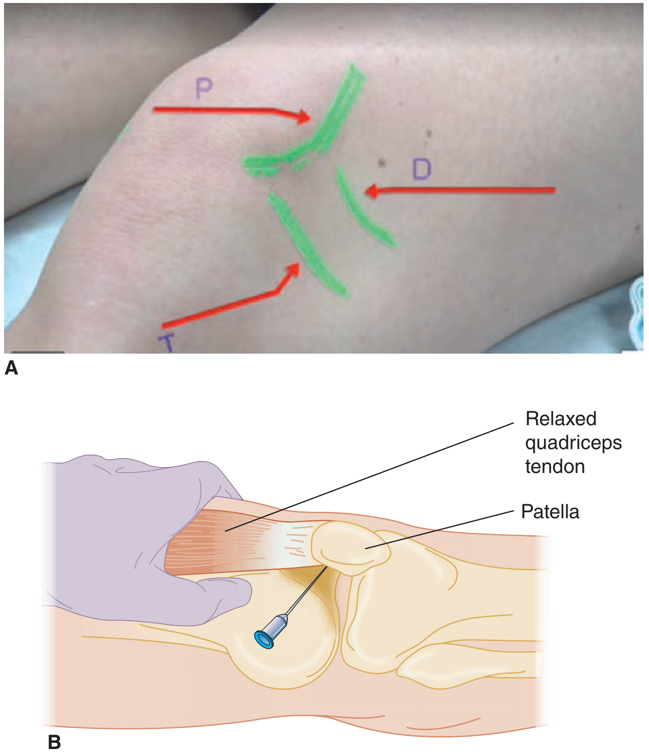
Landmarks for knee arthrocentesis. P = patella; D = distal femur; T = tibia. Needle passes posterior to patella into the joint space with quadriceps relaxed.
Synovial Fluid Analysis
Send fluid for: Cell count + differential, Gram stain, Culture & sensitivity, Crystal examination (polarised microscopy)
Classification of Synovial Fluid
| Group | Clarity | Colour | WBC (cells/mL) | PMN (%) | Glucose difference | Example |
|---|---|---|---|---|---|---|
| Normal | Transparent | Yellow | <150 | <25 | 0-10 mg/dL | - |
| Non-inflammatory | Transparent | Xanthochromic | <3,000 | <30 | 0-10 | OA, trauma |
| Inflammatory | Transparent-opaque | Xanthochromic/white | 3,000-75,000 | >50 | 0-40 | RA, gout, pseudogout, reactive arthritis |
| Infectious (Septic) | Opaque | White/yellow | 50,000-200,000 | >90 | 20-100 | Septic arthritis |
| Hemorrhagic | Opaque | Red-brown | 50-10,000 | <50 | 0-20 | Trauma, hemophilia, pigmented villonodular synovitis |
(Harriet Lane Handbook 23rd ed, Table 28.2)
Additional interpretation (Textbook of Family Medicine 9e):
| WBC/mm³ | Interpretation |
|---|---|
| <200 | Normal |
| <2,000 | Non-inflammatory |
| 2,000-20,000 | Mild inflammation (e.g., SLE) |
| 20,000-50,000 | Moderate inflammation (RA, reactive arthritis) |
| 50,000 | Severe inflammation (sepsis, gout) |
| >100,000 | Septic arthritis until proved otherwise |
Crystal Analysis
| Crystal | Shape | Birefringence | Condition |
|---|---|---|---|
| Monosodium urate | Needle-shaped | Negatively birefringent (yellow parallel) | Gout |
| Calcium pyrophosphate (CPPD) | Rhomboidal/short rods | Weakly positively birefringent (blue parallel) | Pseudogout |
Bedside test: Ability to read newsprint through the fluid = non-inflammatory (clear fluid).
"Inflammatory synovial fluid must be considered secondary to infection until proved otherwise by culture. The presence of crystals does not exclude the possibility of joint infection." - Textbook of Family Medicine 9e
Complications
- Iatrogenic septic arthritis - most feared; risk ~1/10,000; strict aseptic technique mandatory
- Hemarthrosis - especially in anticoagulated patients (very rare; INR up to 4.5 shown safe)
- Local pain / vasovagal reaction
- Cartilage damage - if repeated corticosteroid injections given (not aspiration per se)
- Post-aspiration flare (after steroid injection)
Intra-articular Lidocaine Challenge Test (Miller's, 9th ed)
After aspirating joint fluid, inject the joint with 15 mL of local anaesthetic (lidocaine or 50:50 lidocaine/bupivacaine):
- Relief of ≥90% of pain = positive test
- Indicates pain originates primarily from within the knee joint (useful in diagnosing intra-articular source vs. extra-articular pain, e.g., in post-TKA evaluation)
Sources: Miller's Review of Orthopaedics 9th Ed | Campbell's Operative Orthopaedics 15th Ed (2026) | Roberts & Hedges' Clinical Procedures | Tintinalli's Emergency Medicine | Rheumatology (Elsevier 2022) | Textbook of Family Medicine 9e | Harriet Lane Handbook 23rd ed
This is a shared conversation. Sign in to Orris to start your own chat.