
Defination of all Krishanagata rogas ( opthal bams )
Krishanagata rogas Ayurveda ophthalmology BAMS definition list
https://www.easyayurveda.com/netra-krishnagata-rogas
Krishnagata rogas 5 types Savrana Shukra Avrana Shukra Sira Shukla definition Ayurveda
https://www.easyayurveda.com/netra-krishnagata-rogas
Krishnagata Rogas - Definitions (BAMS Ophthalmology / Shalakya Tantra)
What is Krishnagata?
Total Number of Krishnagata Rogas
| Authority | Number of Diseases |
|---|---|
| Sushruta (Sushruta Samhita) | 4 diseases |
| Vagbhata (Ashtanga Hridayam) | 5 diseases (adds Siraja Shukra) |
The 4 Krishnagata Rogas (as per Sushruta)
1. Savrana Shukra (Kshata Shukra / Savrana Shukla)
- Small, pinpoint circular ulcer on cornea
- Hot (burning) tears (Ushnasrava)
- Severe eye pain
- Inability to see properly
- Nimagna Rupa (sunken appearance of the lesion)
2. Avrana Shukra (Avrana Shukla)
- Thin white opacity/covering on the cornea
- No pain (Nirveda)
- No tearing (Ashrunayana)
- No ulceration - smooth surface
- Diminished vision due to opacity
Avrana Shukra is considered a sequel or an early/less severe stage compared to Savrana Shukra - if the ulcer heals but leaves a scar, it manifests as Avrana Shukra.
3. Akshipakatyaya (Pakaatyaya)
- Severe, intense pain in the eye
- Pus formation in the anterior chamber (Hypopyon)
- Redness and swelling of the entire eye
- Complete loss of vision
- The entire eye is involved (not just cornea)
- Fever and constitutional symptoms may accompany
4. Ajakajata (Ajakaajaata)
- Dark-colored (bluish-black) protrusion of the cornea
- Cornea becomes thin and bulges outward
- Resembles the eye of a goat
- Usually painless in established stage
- Severely impaired or absent vision
- Results from weakened corneal tissue after ulceration
5. Siraja Shukra (Sira Shukla) - Additional disease per Vagbhata
- White opacity on the cornea
- Visible vascular tracks (blood vessels) running into the cornea
- Reduced transparency of the cornea
- Impaired vision
Summary Table
| # | Roga | Modern Equivalent | Dosha | Prognosis |
|---|---|---|---|---|
| 1 | Savrana Shukra | Corneal Ulcer / Ulcerative Keratitis | Raktaja | Asadhya (Incurable) |
| 2 | Avrana Shukra | Corneal Opacity / Leucoma | Raktaja | Sadhya (Curable) |
| 3 | Akshipakatyaya | Hypopyon / Panophthalmitis | Tridoshaja | Asadhya (Incurable) |
| 4 | Ajakajata | Anterior Staphyloma | Raktaja | Asadhya (Incurable) |
| 5 | Siraja Shukra | Corneal Vascularization/Pannus | - | Knowledge-level (Vagbhata only) |
Key Points for BAMS Exam
- Total Krishnagata Rogas: 4 (Sushruta) or 5 (Vagbhata)
- Only curable (Sadhya) disease: Avrana Shukra - treated by medicines only (Ashastra Krutya)
- Only Tridoshaja disease: Akshipakatyaya
- The other 3 (Savrana Shukra, Akshipakatyaya, Ajakajata) are Asadhya (incurable)
- Raktaja diseases: Savrana Shukra, Avrana Shukra, Ajakajata (all three caused by vitiated blood)
- Progression sequence: Savrana Shukra → Avrana Shukra → Akshipakatyaya → Ajakajata (stages of worsening corneal disease)
- Krishna Mandala = Cornea + Iris; formed from Vata Mahabhuta
- BAMS Detailed study required: Savrana/Kshata Shukla and Avrana Shukla
- BAMS Brief knowledge required: Sira Shukla, Akshipakatyaya, and Ajakajata
Classify Corneal Ulcers.Describe etiology, clinical features, management and complications of Bacterial, Viral and Fungal Corneal Ulcers. ( Don't make it too difficult for me haha )
Corneal Ulcers - Classification, Etiology, Features, Management & Complications
What is a Corneal Ulcer?
CLASSIFICATION
CORNEAL ULCERS
│
├── A. By Etiology (Most Important!)
│   ├── 1. INFECTIVE
│   │   ├── Bacterial (most common overall)
│   │   ├── Viral (Herpes Simplex - most common in developed countries)
│   │   ├── Fungal (most common in tropical/agricultural regions)
│   │   └── Protozoal (Acanthamoeba - contact lens wearers)
│   │
│   └── 2. NON-INFECTIVE
│       ├── Neurotrophic (loss of corneal sensation)
│       ├── Exposure keratopathy (lagophthalmos, ectropion)
│       ├── Autoimmune (Mooren's ulcer, rheumatoid arthritis)
│       ├── Nutritional (Vitamin A deficiency → xerophthalmia)
│       └── Toxic / chemical
│
├── B. By Location
│   ├── Central
│   └── Peripheral / Marginal
│
├── C. By Depth
│   ├── Superficial (epithelial)
│   ├── Stromal (anterior / middle / deep)
│   └── Descemetocele (Descemet's membrane exposed)
│
└── D. By Prognosis
    ├── Healing (responding to treatment)
    └── Non-healing / Progressive
1. BACTERIAL CORNEAL ULCER
Most common cause of infectious corneal ulcer worldwide.
Etiology (Common Organisms)
| Organism | Key Feature |
|---|---|
| Pseudomonas aeruginosa | #1 in contact lens wearers; >60% of lens-related ulcers; very aggressive |
| Staphylococcus aureus | Common skin/nasal commensal; focal well-defined yellow-white infiltrate |
| Streptococcus pneumoniae | Upper respiratory commensal; often aggressive |
| S. pyogenes | Throat/vaginal commensal; aggressive infections |
| Neisseria gonorrhoeae | Can penetrate HEALTHY epithelium (important exception!) |
| N. meningitidis, H. influenzae, C. diphtheriae | Also can penetrate intact epithelium |
Most bacteria need compromised corneal defences to cause infection. The exceptions above can breach a healthy cornea.
Risk Factors (Predisposing Conditions)
- Contact lens wear - single most important risk factor (especially soft lenses worn overnight, poor hygiene)
- Corneal trauma (including post-LASIK surgery)
- Ocular surface disease (dry eye, blepharitis)
- Systemic immunosuppression, diabetes
- Topical steroid use
- Bullous keratopathy, keratoconus
Clinical Features
- Sudden onset severe eye pain
- Redness (circumcorneal injection)
- Photophobia
- Watering / discharge (mucopurulent)
- Blurred vision
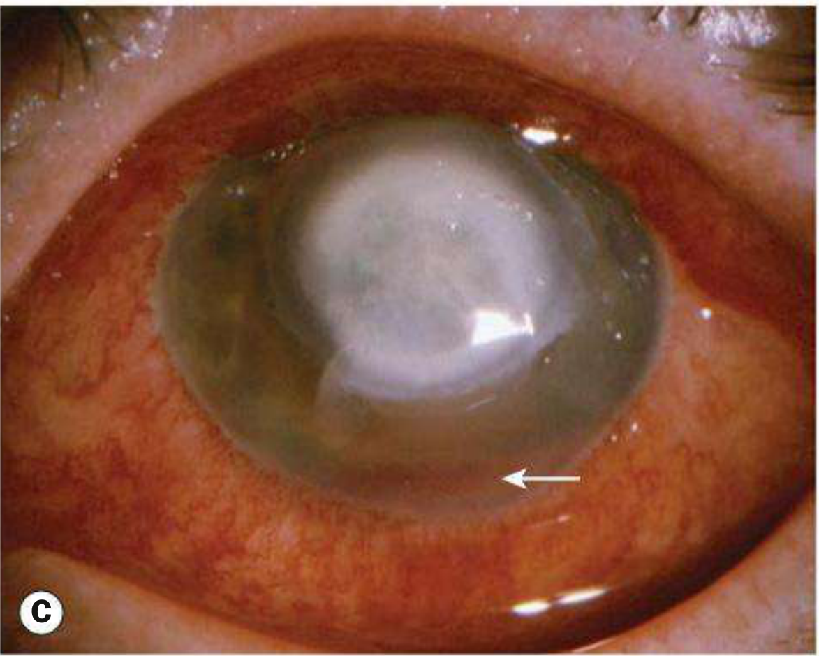
- Epithelial defect + underlying stromal infiltrate (white/yellow-white)
- Stromal oedema and folds in Descemet's membrane
- Anterior uveitis
- Hypopyon (pus in anterior chamber) - seen in moderate-severe cases
- Posterior synechiae
- Chemosis and eyelid swelling in severe cases
- Pseudomonas - can cause very large, rapidly spreading ulcer → descemetocele → perforation
Investigations
- Corneal scraping - gold standard (mandatory for ulcers >2 mm, central, deep stromal, chronic)
- Gram stain - identifies bacteria and fungi
- Culture on blood agar, chocolate agar, Sabouraud agar (incubated at 37°C)
- Brain-Heart Infusion (BHI) broth for enrichment
- Stains used: Gram, Giemsa, Ziehl-Neelsen (for Mycobacterium), Calcofluor white
- Sensitivity testing to guide antibiotic choice
Management
- Stop contact lens wear immediately (mandatory)
- Hospital admission for aggressive disease or poor compliance
- Clear eye shield if significant thinning/perforation risk
- Empirical broad-spectrum topical antibiotics - start before culture results
- Fluoroquinolones (Ciprofloxacin 0.3%, Ofloxacin 0.3%, Moxifloxacin 0.5%) - drug of choice for monotherapy; covers most pathogens
- Fortified antibiotics for severe cases: Fortified Tobramycin (1.3%) + Cefazolin (5%) - dual therapy
- Dosing: Hourly day and night for 24-48 hours, then tapered
- Cycloplegics (Atropine 1%) - for pain relief and to prevent posterior synechiae
- Topical steroids - only after infection is controlled and with caution
- Corneal transplant (Penetrating Keratoplasty) - for perforations or non-healing ulcers
- Tissue glue + bandage contact lens - for small perforations
- Conjunctival flap - for peripheral/chronic non-healing ulcers
Complications
- Corneal perforation - especially Pseudomonas
- Hypopyon (sterile pus in AC)
- Endophthalmitis (rare without perforation)
- Corneal scarring - permanent opacity, irregular astigmatism
- Corneal vascularization
- Descemetocele formation
- Iris prolapse after perforation
- Phthisis bulbi (shrunken, non-functional eye) - end stage
2. VIRAL CORNEAL ULCER (Herpes Simplex Keratitis)
Most common infectious cause of corneal blindness in developed countries. HSV-1 is the usual culprit.
Etiology
- Herpes Simplex Virus type 1 (HSV-1) - causes disease above the waist (face, lips, eyes)
- HSV-2 - genital herpes; rarely involves the eye (neonatal conjunctivitis, sexual transmission)
- Virus resides latent in trigeminal ganglion after primary infection
- Reactivated by: fever, UV light, stress, immunosuppression, menstruation, trauma
- Spread: droplet transmission or direct contact; common in poor hygiene/crowding
Types of Herpes Simplex Keratitis
| Type | Layer Involved | Key Feature |
|---|---|---|
| Epithelial (Dendritic) | Epithelium | Branching dendritic ulcer - pathognomonic |
| Disciform (Stromal) | Stroma + endothelium | Disc-shaped stromal oedema - immune-mediated |
| Necrotizing Stromal | Deep stroma | Active viral replication; stromal melting |
| Neurotrophic | Epithelium | Non-healing defect due to corneal anaesthesia |
Clinical Features
Epithelial HSK (Most Common)
- Foreign body sensation, watering, mild pain
- Reduced corneal sensation (anaesthesia - characteristic!)
- Blurred vision
- History of cold sores / lip blisters common
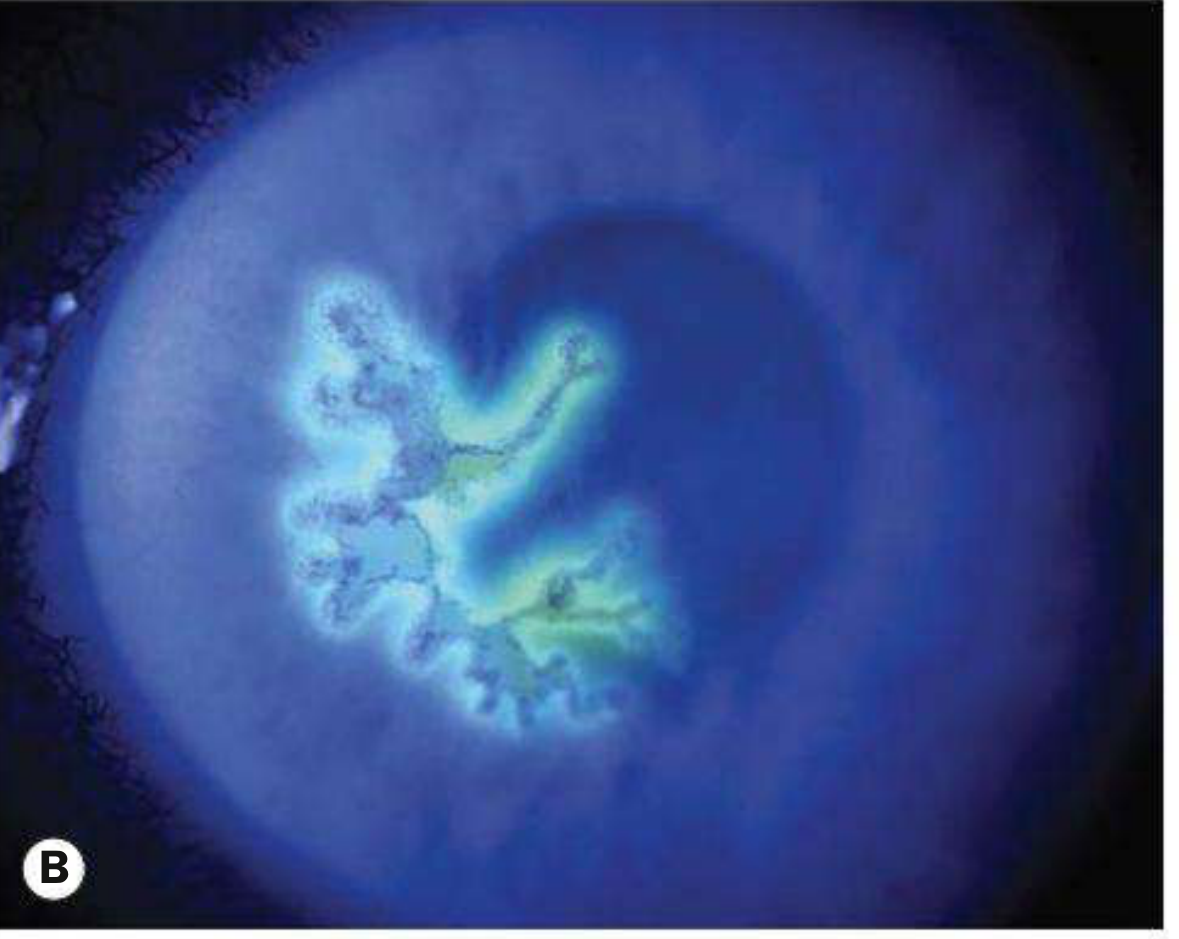
- Dendritic ulcer - branching, tree-like (dendrite = branch in Greek) ulcer with terminal bulbs at the ends
- Stains brilliantly with fluorescein (the ulcer bed stains green)
- Stains at margins with rose Bengal (the infected but living cells at edges stain red)
- May enlarge into a geographic ulcer (large, amoeboid shaped)
- Reduced/absent corneal sensation
Disciform (Stromal) HSK
- Central disc-shaped area of stromal oedema - hazy, thickened cornea
- Overlying epithelial oedema (bullae)
- Keratic precipitates (KP) directly under the oedematous area
- Raised IOP common (trabeculitis)
- Mild anterior uveitis
Investigations
- Clinical diagnosis usually (dendritic ulcer is pathognomonic)
- Corneal scraping in viral transport medium → PCR (most sensitive - up to 90%) or culture
- Giemsa stain → multinucleated giant cells
- HSV serology - only rises on primary infection
- Immunocytochemistry available
Management
Epithelial HSK:
- Topical antivirals:
- Aciclovir 3% eye ointment - 5 times daily for 14 days (first choice)
- Ganciclovir 0.15% gel - 5 times daily
- Trifluridine (vidarabine) - alternatives
- Oral antivirals - for immunocompromised, children, severe disease:
- Aciclovir 200-400 mg 5 times daily for 5-10 days
- Famciclovir or Valaciclovir (better tolerated, less frequent dosing)
- Debridement - wiping the corneal surface with a sterile swab, 2 mm beyond ulcer edge; removes virus-laden cells
- Cycloplegics (Homatropine 1%) for comfort
- Topical antibiotic prophylaxis (some practitioners)
⚠️ IMPORTANT RULE: NEVER use topical steroids in epithelial HSK - this dramatically worsens the ulceration and can cause corneal perforation!
Disciform HSK:
- Topical steroids (prednisolone) + topical antivirals together (steroids alone would allow viral replication)
- Long-term oral aciclovir 400 mg twice daily - prophylaxis to prevent recurrences (HEDS trial)
Recurrence prevention:
- Long-term oral aciclovir significantly reduces recurrence rate
Complications
- Corneal scarring - gets worse with each recurrence
- Corneal vascularization and lipid deposition
- Corneal anaesthesia (neurotrophic keratopathy)
- Irregular astigmatism
- Glaucoma (secondary, from trabeculitis or steroid use)
- Cataract (from chronic inflammation or steroids)
- Corneal perforation - in necrotizing form
- Amblyopia in children
- Final visual acuity <6/60 in ~10% of patients
3. FUNGAL CORNEAL ULCER (Fungal Keratitis / Keratomycosis)
The most common cause of corneal blindness in tropical and low-income countries. Often misdiagnosed as bacterial initially!
Etiology
| Type | Organisms | Climate |
|---|---|---|
| Filamentous (moulds) | Fusarium spp., Aspergillus spp. | Tropical (most common there) |
| Yeasts | Candida spp. | Temperate climates |
- Filamentous fungi: multicellular, form hyphae (tube-like projections); aggressive course
- Candida: ovoid, unicellular, reproduce by budding
Risk Factors (Predisposing Conditions)
- Trauma with plant material / agricultural tools - classic history for filamentous fungi (farmer scratched by paddy/thorn)
- Topical corticosteroid use - very important (suppresses immunity, allows fungi to proliferate)
- Chronic ocular surface disease
- Contact lens wear
- Previous corneal transplant
- Systemic immunosuppression, diabetes
Clinical Features
Key point: Diagnosis is often delayed because it looks like bacterial keratitis initially! Have a high index of suspicion, especially with history of plant trauma + steroid use + rural/tropical setting.
- Gradual onset of pain (slower than bacterial)
- Grittiness, photophobia
- Blurred vision
- Watery or mucopurulent discharge
- Yellow-white, densely suppurative infiltrate (looks like bacterial)
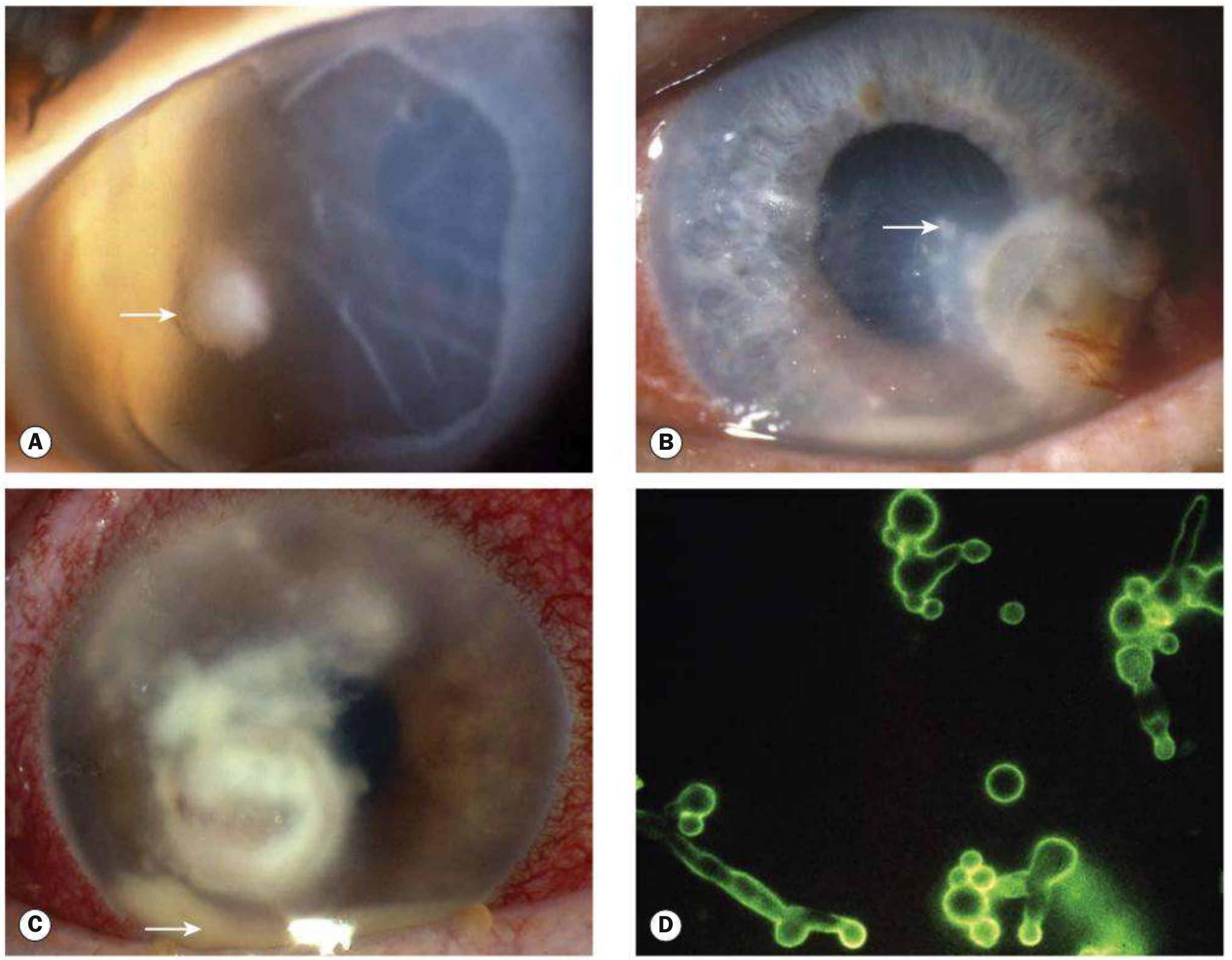
- Grey or yellow-white stromal infiltrate with indistinct, fluffy (feathery) margins - very characteristic!
- Satellite lesions (separate smaller infiltrates around the main one) - hallmark sign
- Feathery/branch-like extensions of infiltrate
- Ring-shaped infiltrate
- Rapid progression with necrosis and corneal thinning
- Epithelial defect may be small or absent initially
- Anterior uveitis, hypopyon, endothelial plaque, raised IOP
- Fungi can penetrate intact Descemet membrane → endophthalmitis without obvious perforation (unique to fungi!)
Investigations
- Corneal scraping - must be taken before starting treatment
- KOH (Potassium Hydroxide) preparation - rapid, highly sensitive; shows fungal elements under microscope
- Gram and Giemsa stain - ~50% sensitive for fungi
- Calcofluor white (fluorescent stain) - excellent for fungi and Acanthamoeba
- Periodic Acid-Schiff (PAS), Methenamine silver stains
- Culture on Sabouraud Dextrose Agar (specific for fungi; also grows on blood agar)
- PCR - up to 90% sensitive; investigation of choice in many centres
- Corneal biopsy - if no growth after 1 week of scraping and no improvement after 3-4 days of treatment (fungi proliferate just anterior to Descemet's - deep biopsy may be needed)
- Confocal microscopy - can identify organisms in vivo; not widely available
Management
- Hospital admission usually required
- Stop topical steroids (CRITICAL - they will make it much worse)
- Remove epithelium over the lesion to enhance antifungal penetration
- Regularly remove mucus and necrotic tissue with a spatula
- Improvement is much slower than bacterial - patient and doctor need patience
| Organism | Drug of Choice | Alternatives |
|---|---|---|
| Candida | Amphotericin B 0.15% | Econazole 1%, Natamycin 5%, Fluconazole 2%, Clotrimazole 1%, Voriconazole |
| Filamentous | Natamycin 5% (best for filamentous, especially Fusarium) | Voriconazole 1%, Econazole 1%, Amphotericin B |
- Oral voriconazole or itraconazole for severe/deep cases
- IV amphotericin B - for severe systemic spread
- Penetrating Keratoplasty (PKP) - for non-responding or perforated cases
- Anterior chamber washout for hypopyon
- Evisceration - in end-stage, blinded, painful eye
Complications
- Corneal perforation - very common in fungal keratitis
- Endophthalmitis - can occur even without perforation (unique feature - fungi penetrate Descemet's)
- Severe corneal scarring and opacity
- Secondary glaucoma
- Loss of eye (evisceration/enucleation) - poor prognosis overall
Quick Comparison Table - The Cheat Sheet!
| Feature | Bacterial | Viral (HSK) | Fungal |
|---|---|---|---|
| Onset | Acute / rapid | Acute (epithelial) | Gradual / insidious |
| Pain | Severe | Mild-moderate | Moderate (gradual) |
| Classic Appearance | White/yellow dense infiltrate | Dendritic ulcer (branching) | Fluffy margins + satellite lesions |
| Pathognomonic sign | Hypopyon | Dendrite with terminal bulbs | Satellite lesions + feathery edges |
| Corneal sensation | Normal | REDUCED / absent | Normal (initially) |
| Common setting | Contact lens wearer | Stress, fever, immunocompromised | Farmer, plant trauma, steroid user |
| Key stain/test | Gram stain, culture | Fluorescein (dendrite), PCR | KOH prep, Calcofluor white |
| Treatment | Fluoroquinolone drops | Aciclovir (NEVER steroids alone) | Natamycin / Amphotericin B |
| Steroids | Cautious use after infection controlled | CONTRAINDICATED in epithelial | CONTRAINDICATED (makes it worse) |
| Prognosis | Good if treated early | Recurrent; can scar over time | Often poor; perforation common |
One-Line Memory Tricks
- Bacterial = Contact lens + Quick onset + Dense white infiltrate + Gram stain
- Viral (HSK) = Dendrite + Terminal bulbs + Reduced sensation + NEVER steroids alone
- Fungal = Farmer + Plant trauma + Fluffy/feathery + Satellites + KOH prep + NEVER steroids
Su ut 12/15पूयालसे शोणितमोक्षणं च हितं तथा एव अपि उपनाहनं च कृत्स्नोविधिश्वेक्षण पाकघाती यथाविधानं meaning of this word to word
Sushruta Uttaratantra chapter 12 verse 15 Pooyalasa Shonitamokshanam shloka Sanskrit meaning
https://www.wisdomlib.org/hinduism/book/sushruta-samhita-vol…
Sushruta Uttaratantra chapter 12 sandhi gata roga Pooyalasa Upanaha treatment shloka translation
https://www.wisdomlib.org/hinduism/book/sushruta-samhita-vol…
Su. Ut. 12/15 - Word-to-Word Meaning
The Shloka (Devanagari Reconstructed):
पूयालसे शोणितमोक्षणं च हितं तथा एव अपि उपनाहनं च कृत्स्नो विधिश्च एक्षण पाकघाती यथाविधानं
Word-to-Word (Pada-Artha) Breakdown
| Sanskrit Word | Sandhi Split | Grammatical Form | Word Meaning |
|---|---|---|---|
| पूयालसे | Pūyālase | Saptami Vibhakti (Locative case) - "in the case of" | In Puyalasa (the disease of the eye junction - dacryocystitis / suppurative condition of eye canthus) |
| शोणितमोक्षणं | Śoṇita + mokṣaṇam | Noun, Nominative | Bloodletting / Venesection - "Shoṇita" = blood; "Mokṣaṇa" = releasing/letting out |
| च | ca | Indeclinable particle | And / also |
| हितं | Hitam | Adjective, Nominative | Beneficial / indicated / conducive (i.e., the treatment is beneficial) |
| तथा | tathā | Indeclinable adverb | Similarly / likewise / in the same way |
| एव | eva | Indeclinable (emphatic particle) | Indeed / also / even (gives emphasis) |
| अपि | api | Indeclinable particle | Also / too / as well |
| उपनाहनं | Upanāhanam | Noun, Nominative | Upanaha = poulticing / warm fomentation application / medicated poultice tied over the affected part |
| च | ca | Indeclinable | And |
| कृत्स्नः | Kṛtsnaḥ | Adjective | All / entire / whole / complete |
| विधिः | Vidhiḥ | Noun | Treatment regimen / procedure / method / rule |
| च | ca | Indeclinable | And / also |
| एक्षण (=अक्षिपाक) | Akṣi + Pāka | Compound noun | Akshipaka = inflammation/suppuration of the eye ("Akshi" = eye; "Paka" = suppuration/cooking/maturation of inflammation) |
| पाकघाती | Pāka + ghātī | Compound adjective | That which destroys/cures the paka (suppuration) - "Ghāti" = destroyer/one that kills |
| यथाविधानं | Yathā + vidhānam | Indeclinable compound | According to the prescribed method / as per the rules / as directed |
Complete Translation (Word-to-Word Order):
| Pada | Meaning |
|---|---|
| पूयालसे | In the case of Puyalasa (eye disease of the canthus) |
| शोणितमोक्षणं च | bloodletting (Raktamokshana) also |
| हितं | is beneficial |
| तथा एव अपि | similarly, indeed, also |
| उपनाहनं च | poulticing (Upanaha) too |
| कृत्स्नः विधिः च | and the entire / complete treatment regimen |
| अक्षिपाकघाती | that destroys/cures Akshipaka (eye suppuration) |
| यथाविधानं | according to proper procedure / as prescribed |
Full Meaning in Simple English:
"In the case of Puyalasa, bloodletting (Raktamokshana) is beneficial, and similarly, poulticing (Upanaha) is also indicated. Furthermore, the entire treatment regimen that is used to cure Akshipaka (suppurative inflammation of the eye) should also be employed here, following the prescribed method."
Clinical Meaning for BAMS:
| Treatment | Sanskrit Term | What it Means Practically |
|---|---|---|
| Shonitamokshana | Bloodletting | Leech application or venesection at the inner canthus area to drain vitiated Rakta |
| Upanaha | Poultice | Warm medicated paste tied/applied over the swollen lacrimal sac area to ripen the pus and reduce inflammation |
| Akshipaka Vidhana (complete regimen) | All treatments of Akshipaka | Sneha (oleation), Sveda (fomentation), eye washes (Seka), Aschyotana (eye drops), Nasya (nasal drops), Putapaka - all applied to Puyalasa as well |
Memory tip for the exam:
- Raktamokshana (bloodletting)
- Upanaha (poultice)
- Complete Akshipaka treatment protocol - as per rules