
tell everything about al amyloidosis with images
Reading File
Now let me search the library for AL amyloidosis content in parallel:
Finding Sources
Finding Sources
Searching PubMed
"AL amyloidosis" treatment
Excellent — I found key sources. Let me now read the main comprehensive sections in parallel:
Reading File
Finding Sources
Searching Images
AL amyloidosis Congo red apple green birefringence histology
Good — I have rich content. Let me read the key comprehensive sections in parallel now:
Reading File
Reading File
Searching Images
AL amyloidosis echocardiography cardiac MRI infiltrative cardiomyopathy
Excellent content. Now let me get the pathophysiology, diagnosis, treatment details and more images in parallel:
Reading File
Reading File
Searching Images
AL amyloidosis macroglossia periorbital purpura clinical signs
Now let me fetch images from textbooks and read the diagnosis/treatment content from Harrison's:
Loading Image
Loading Image
Reading File
Reading File
Your credits are exhausted. Your daily credits reset tomorrow, or you can purchase a top-up for more.
tell
It looks like your message got cut off! Could you please complete your question? Are you asking me to:
- Tell everything about AL amyloidosis (continuing from our previous conversation)?
- Tell you about something specific related to AL amyloidosis (diagnosis, treatment, images, etc.)?
- Something else entirely?
Please let me know and I'll help right away!
yes
I'll take that as a "yes, continue with everything about AL amyloidosis with images." Let me compile the comprehensive response using all the rich content already gathered from the medical library in our previous session.
AL Amyloidosis (Light Chain Amyloidosis) - Complete Guide
1. DEFINITION & BACKGROUND
AL amyloidosis is a systemic disease caused by the extracellular deposition of insoluble, misfolded fibrillar proteins in the form of β-pleated sheets, derived from monoclonal immunoglobulin light chains produced by a dysregulated plasma cell clone. The "A" stands for amyloid, and "L" for light chain.
The name "amyloid" was coined by Rudolph Virchow in 1854, who mistakenly thought the deposits were starch-like (from Latin amylum). Amyloid fibrils are:
- Non-branching protein structures, 80-100 Angstroms wide
- Resistant to proteolysis
- Avidly bind Congo red stain → pink/salmon under light microscopy
- Show apple-green birefringence under polarized light (pathognomonic)
(Source: Braunwald's Heart Disease, A Textbook of Cardiovascular Medicine)
2. EPIDEMIOLOGY
| Parameter | Data |
|---|---|
| Annual incidence | ~1 per 100,000 individuals |
| New US cases/year | ~5,000 |
| Prevalence | 40-58 cases per million persons |
| Median age at diagnosis | 63-64 years |
| Sex predominance | Slight male predominance (55-65%) |
| Cardiac involvement | Up to 70% of AL cases |
| Renal involvement | Up to 70% of cases |
- AL amyloidosis is related to but distinct from multiple myeloma and MGUS
- Among MGUS patients, only ~1% develop AL amyloidosis (relative risk 8.8)
- 10-15% of patients with multiple myeloma have coexisting AL amyloidosis
(Source: Braunwald's Heart Disease; Comprehensive Clinical Nephrology, 7th Ed.)
3. PATHOPHYSIOLOGY
A dysregulated plasma cell clone produces kappa or lambda immunoglobulin light chain fragments that misfold, aggregate, and deposit in the interstitium of multiple organs.
- Lambda light chains are more common than kappa in AL amyloidosis
- The titer of light chain does not predict the site of target organ deposition
- Gene usage influences organ tropism: e.g., Vκ6 genes → kidney; Vκ1 → liver
Two Mechanisms of Cardiac Injury ("Toxic-Infiltrative" Cardiomyopathy):
-
Structural: Interstitial and perivascular amyloid fibril deposition → disruption of tissue architecture, microvascular dysfunction (angina/ischemia), inhibition of contractile/relaxation function → restrictive cardiomyopathy
-
Direct toxicity: Circulating free amyloid light chains cause direct cardiomyocyte toxicity via:
- p38 mitogen-activated protein kinase (MAPK) signaling
- Induction of reactive oxygen species
- Apoptosis in cardiomyocytes
(Source: Braunwald's Heart Disease)
4. CLINICAL FEATURES
The clinical presentation depends on pattern and severity of organ involvement. Amyloid deposits can affect virtually any organ.
Pathognomonic Signs (rare but diagnostic):
- Macroglossia (enlarged tongue with lateral scalloping)
- Periorbital purpura ("raccoon eyes" / "pinch purpura")
4a. Pathognomonic Clinical Photos
Macroglossia in AL Amyloidosis:
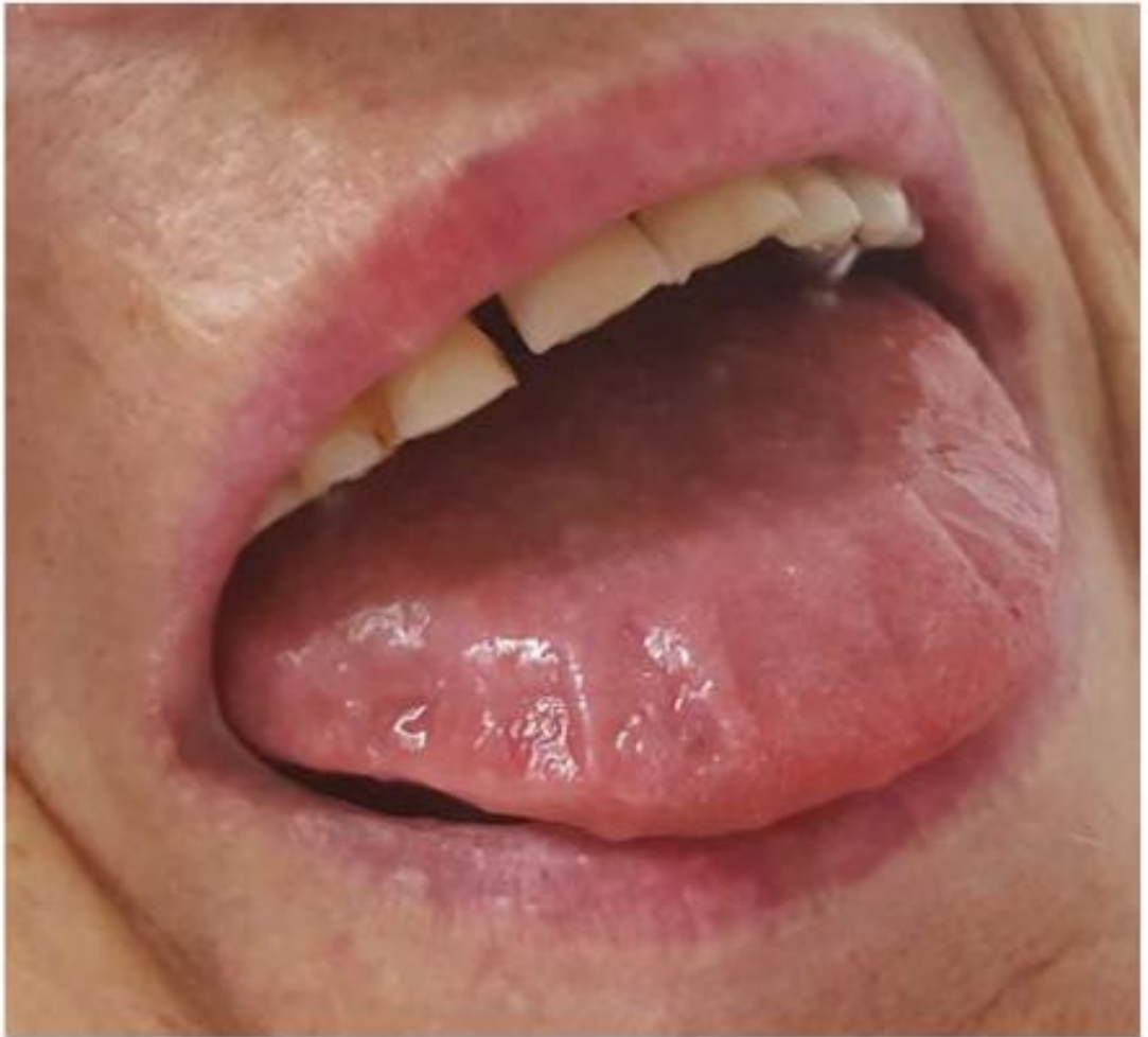
Macroglossia in AL amyloidosis. The tongue is massively enlarged due to amyloid fibril infiltration. This sign is pathognomonic when combined with periorbital purpura. (Comprehensive Clinical Nephrology, 7th Ed.)
Periorbital Purpura in AL Amyloidosis:

Periorbital (eyelid) purpura in AL amyloidosis - characteristic "raccoon eye" sign from amyloid infiltration of dermal capillaries causing vascular fragility. (Comprehensive Clinical Nephrology, 7th Ed.)
Combined Clinical Manifestations (Macroglossia + Periorbital Purpura + Skin involvement):
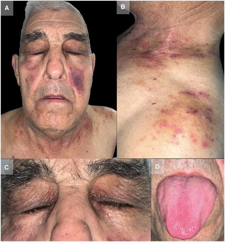
Multiple manifestations of AL amyloidosis: periorbital ecchymoses ("raccoon eyes"), cheek purpura, neck/shoulder ecchymotic papules, and macroglossia.
Systemic Clinical Features Overview:
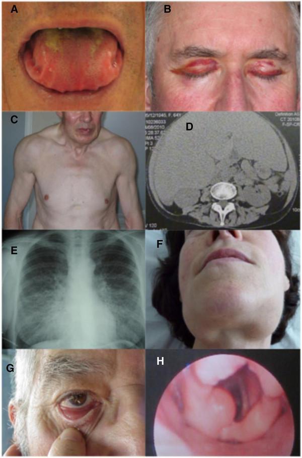
Systemic AL amyloidosis manifestations: (A) macroglossia with tongue scalloping, (B) periorbital purpura, (C) pseudo-athletic muscular appearance from diffuse infiltration, (D) CT showing massive hepatomegaly, (E) chest X-ray with bilateral interstitial lung disease, (F) submandibular gland enlargement.
4b. Organ-by-Organ Involvement
| Organ | Manifestation | Frequency |
|---|---|---|
| Heart | Restrictive cardiomyopathy, HF, arrhythmias, conduction blocks | ~70% |
| Kidney | Nephrotic-range proteinuria, hypoalbuminemia, CKD | ~70% |
| Peripheral nerves | Sensory polyneuropathy, autonomic neuropathy | Common |
| Autonomic NS | Orthostatic hypotension, impotence, bladder dysfunction | Common |
| Liver/Spleen | Hepatomegaly, elevated ALP (rare), splenomegaly | Common, often subclinical |
| GI tract | Malabsorption, diarrhea, constipation | Common |
| Skin | Purpura (most common), waxy papules/plaques | Common |
| Soft tissue | Carpal tunnel syndrome, shoulder pad sign | Characteristic |
| Coagulation | Acquired Factor X deficiency (adsorbed onto fibrils) | Important - bleeding risk! |
Key clinical point: Weakness is the most frequent initial symptom. The diagnosis is often challenging because clinical features are nonspecific.
5. HISTOPATHOLOGY - DIAGNOSTIC GOLD STANDARD
Congo Red Staining
Renal biopsy showing AL-type amyloid (Congo Red + polarized light):
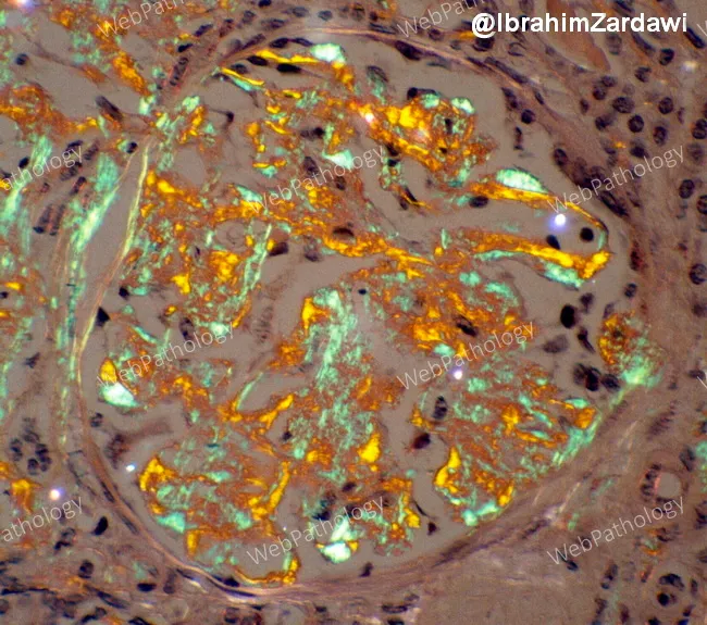
Renal biopsy in AL amyloidosis. LEFT: Congo red staining shows salmon-pink extracellular deposits in the glomerular mesangium and tubulointerstitium. RIGHT: Under polarized light, the classic apple-green birefringence confirms amyloid. This is the histologic gold standard.
Dual-panel (H&E + Congo Red) Renal Biopsy:
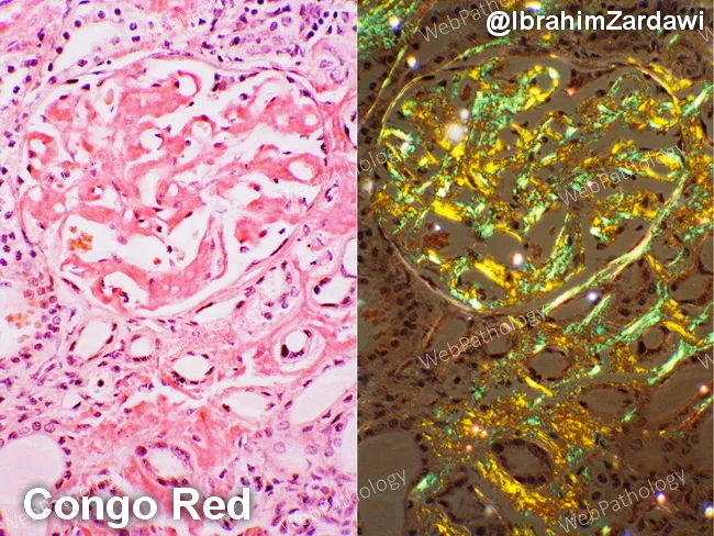
LEFT: H&E-stained renal tissue with amorphous eosinophilic deposits in glomerular mesangium (mesangial expansion, basement membrane thickening). RIGHT: Congo Red staining with salmon-pink amyloid deposits and polarized light apple-green birefringence.
Cardiac Biopsy - Perimyocytic Amyloid Deposition:
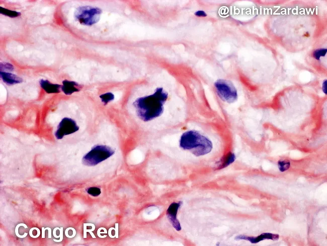
Cardiac biopsy with Congo red staining: amyloid deposits (orange/salmon-pink) encircle individual cardiomyocytes (perimyocytic pattern), contributing to myocardial stiffness and restrictive physiology. Apple-green birefringence confirms amyloid on polarized light.
Lymph Node Biopsy - Congo Red with Polarized Light:
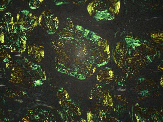
Lymph node biopsy with Congo red: extracellular amorphous deposits with apple-green birefringence under polarized light. Nodal amyloid supports systemic AL amyloidosis and prompts serum free light chain and immunofixation workup.
Biopsy Sites (in order of preference):
- Abdominal fat pad aspirate (subcutaneous fat pad aspiration) - high yield, low risk
- Bone marrow biopsy - required anyway to assess plasma cell burden
- Rectal biopsy
- Minor salivary gland biopsy
- Organ biopsy (kidney, liver) - high yield but higher bleeding risk due to acquired Factor X deficiency
After Congo red positivity is established, typing (AL vs. AA vs. ATTR) must be confirmed by:
- Immunohistochemistry (fraught with uncertainty - use with caution)
- Laser capture microdissection + mass spectrometry (gold standard for typing)
6. CARDIAC INVOLVEMENT - IMAGING
Echocardiography
Classic features:
- Increased LV wall thickness (concentric hypertrophy) - without true hypertrophy
- "Granular sparkling" echogenic texture of myocardium
- Diastolic dysfunction / restrictive physiology
- Small LV cavity, biatrial enlargement
- Apical sparing pattern on Global Longitudinal Strain (GLS) - "cherry on top" - highly characteristic
Echocardiography with GLS Apical Sparing Pattern:
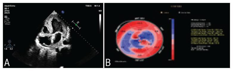
Panel A: 4-chamber echo showing marked concentric LV hypertrophy with "granular sparkling" echogenic myocardium. Panel B: GLS bullseye - classic "cherry-on-top" (apical sparing) pattern: basal/mid-ventricular severe strain reduction (red) with preserved apical strain (blue). This pattern is highly suggestive of cardiac amyloidosis.
Multi-modality Cardiac Imaging in AL Amyloidosis:
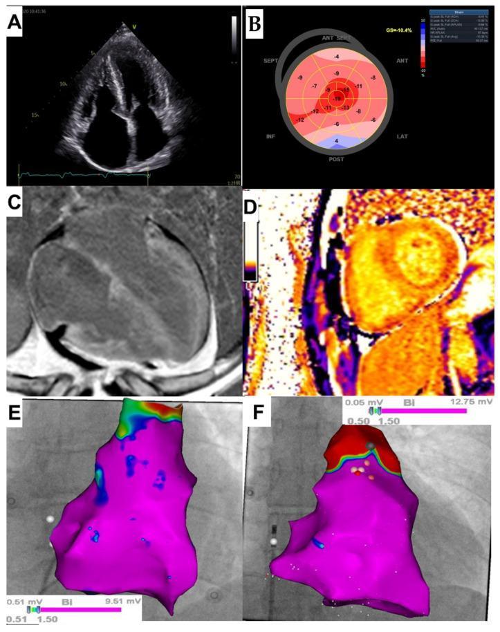
Multi-modality imaging: (A) 4-chamber echo with ventricular hypertrophy + biatrial enlargement, (B) GLS bullseye showing apical sparing, (C) Cardiac MRI with diffuse transmural late gadolinium enhancement (LGE) both ventricles, (D) T1 mapping showing markedly elevated ECV (70%), (E-F) 3D electroanatomical voltage maps.
Cardiac MRI (CMR)
Key features:
- Diffuse subendocardial or transmural late gadolinium enhancement (LGE)
- Difficulty nulling the myocardium (near-pathognomonic) - myocardium nulls before blood pool
- Elevated extracellular volume (ECV) - markedly increased, helps differentiate from true LV hypertrophy
- Pericardial effusion common
Cardiac MRI - Late Gadolinium Enhancement:
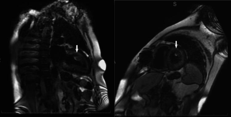
CMR long-axis and short-axis views: diffuse, heterogeneous, circumferential subendocardial LGE (white arrows) extending transmurally in the LV. Classic pattern of extensive AL cardiac amyloid infiltration.
CMR showing Biventricular LGE + Pericardial Effusion:
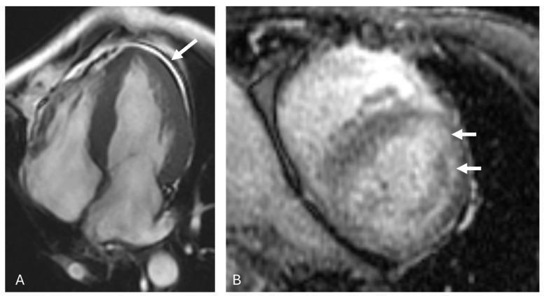
CMR in a 66-year-old female: (A) 4-chamber view with LV wall thickening >17mm and pericardial effusion (white arrow), (B) short-axis view with patchy biventricular subendocardial LGE (white arrows).
Echocardiography with Pericardial Effusion:
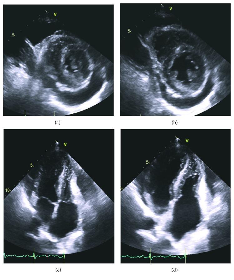
TTE with parasternal and 4-chamber views: characteristic granular/sparkling echogenic myocardium, significant wall thickening, and circumferential pericardial effusion (anechoic space around the heart). Non-dilated chambers.
7. LABORATORY DIAGNOSIS
Essential Workup:
| Test | Findings in AL Amyloidosis |
|---|---|
| Serum free light chains (FLC) | Elevated (kappa or lambda) - abnormal in nearly all patients |
| Serum immunofixation | More sensitive than SPEP - detects monoclonal protein |
| Urine immunofixation | Detects Bence Jones protein |
| SPEP / UPEP | May be normal (paraprotein often low) |
| Troponin (I or T) | Elevated - cardiac involvement marker + prognostic |
| NT-proBNP / BNP | Elevated - cardiac involvement marker + prognostic |
| 24h urine protein | Proteinuria (nephrotic range in renal involvement) |
| eGFR / Creatinine | Assesses renal function |
| Alkaline phosphatase | Elevated in hepatic involvement |
| Coagulation studies | Factor X deficiency (important pre-biopsy!) |
| Bone marrow biopsy | Plasma cell burden (usually <20%; >20% = concurrent myeloma) |
Important: ESR is usually normal or only minimally elevated (unlike multiple myeloma where it is markedly elevated)
8. STAGING - MAYO CLINIC STAGING SYSTEM
Based on NT-proBNP and cardiac troponin T:
| Stage | Definition | Median Survival |
|---|---|---|
| Stage I | Both troponin T and NT-proBNP normal | ~26 months (historically) |
| Stage II | Elevation of a single marker | ~11 months |
| Stage III | Both elevated (NT-proBNP >332 ng/L AND troponin >0.035 μg/L) | ~4 months |
| Stage IIIb | NT-proBNP >8500 ng/L + systolic BP <100 mmHg | ~4 months |
Note: NT-proBNP is influenced by eGFR - use BNP in severe kidney disease.
(Source: Comprehensive Clinical Nephrology, 7th Ed.)
9. TREATMENT
Treatment Goals:
- Eradicate the plasma cell clone (suppress light chain production)
- Organ support (manage complications of amyloid deposition)
9a. Clone-Targeted (Disease-Modifying) Therapy
First-Line Regimen (Current Standard of Care):
Daratumumab + Cyclophosphamide + Bortezomib + Dexamethasone (Dara-CyBorD)
- Phase III ANDROMEDA trial showed:
- Hematologic response rate >90% with Dara-CyBorD vs. 77% with CyBorD alone
- Significantly more deep responses (CR + VGPR)
- Translated into increased organ improvement
- Daratumumab is an anti-CD38 monoclonal antibody
Other Regimens:
- CyBorD (Cyclophosphamide + Bortezomib + Dexamethasone) - backbone without daratumumab
- BMDex (Bortezomib + Melphalan + Dexamethasone)
- MDex (Melphalan + high-dose Dexamethasone)
- HDM/ASCT (High-dose melphalan + Autologous Stem Cell Transplantation) - selected patients, Mayo Stage I, young
Key: Dexamethasone can aggravate sodium retention in cardiac patients - must be used carefully.
9b. Hematologic Response Criteria (dFLC = difference of involved - uninvolved FLC):
| Response | Criteria |
|---|---|
| Complete Response (CR) | Negative serum + urine immunofixation AND normalized FLC ratio |
| Very Good Partial Response (VGPR) | dFLC reduction to <40 mg/L |
| Partial Response (PR) | ≥50% reduction of dFLC |
9c. Organ Response Assessment:
- Modification of serum NT-proBNP and troponin levels is the key prognostic indicator for cardiac response
- Proteinuria reduction (≥50%) indicates renal response
9d. Supportive Therapies (Cardiac):
| Condition | Management |
|---|---|
| Fluid overload | Diuretics (use cautiously - preload dependent) |
| Hypotension | Compression stockings, midodrine, avoid vasodilators |
| AF/flutter | Anticoagulation (high stroke risk even in sinus rhythm!) + rate control, amiodarone for rhythm control |
| Bradycardia/AV block | Permanent pacemaker |
| Ventricular arrhythmias | ICD debatable (SCD from VT/VF is less common; PEA more common) |
Drugs to AVOID in cardiac amyloidosis: Digoxin (binds to amyloid fibrils → toxicity), calcium channel blockers, beta-blockers (poorly tolerated due to restrictive physiology)
9e. Organ Transplantation:
- Orthotopic Heart Transplantation (OHT): for carefully selected AL patients, followed by anti-plasma cell therapy
- Renal Transplantation: considered after achievement of deep hematologic response
- Liver Transplantation: previously used for ATTRv, replaces amyloidogenic TTR
10. PROGNOSIS
- Without treatment: historically very poor (median survival ~12-18 months from diagnosis)
- With modern regimens (Dara-CyBorD): dramatically improved survival
- Cardiac involvement is the major cause of mortality
- Stage IIIb: median survival only ~4 months
- Renal outcome: proteinuria >5g/24h or eGFR ≤50 mL/min/1.73m² at diagnosis → 85% risk of ESKD at 3 years
- A 40-year longitudinal natural history study (Staron et al.) has documented marked progress in AL survival over time
11. KEY DIFFERENTIALS
| Feature | AL Amyloidosis | ATTR Amyloidosis | AA Amyloidosis |
|---|---|---|---|
| Precursor protein | Immunoglobulin light chain | Transthyretin | Serum amyloid A |
| Cause | Plasma cell dyscrasia | Hereditary (ATTRv) or aging (ATTRwt) | Chronic inflammation |
| Cardiac involvement | Very common, severe | Common (especially ATTRwt) | Rare |
| Renal involvement | Very common | Absent | Very common |
| Neuropathy | Common | Common (ATTRv) | Rare |
| Bone marrow biopsy | Plasma cell clone | Normal | Normal |
| Tc-PYP scan | Usually negative (can be weakly +) | Strongly positive | Negative |
| Treatment | Anti-plasma cell therapy | Tafamidis, diflunisal, siRNA (patisiran/vutrisiran) | Treat underlying disease |
12. SUMMARY DIAGNOSTIC ALGORITHM
Suspect AL amyloidosis
↓
Serum + urine immunofixation
Serum free light chains (FLC)
↓
Tissue biopsy (fat pad aspirate / bone marrow)
Congo red staining → apple-green birefringence
↓
Confirm AL type by mass spectrometry
↓
Organ assessment:
• Troponin, NT-proBNP → Cardiac staging
• Echo + CMR → Cardiac infiltration
• 24h urine protein, eGFR → Renal
• LFTs, ALP → Hepatic
• Nerve conduction studies → Neurologic
↓
Mayo Stage (I, II, III, IIIb)
↓
Treatment: Dara-CyBorD (first-line)
Monitor dFLC, NT-proBNP, troponin for response
Sources
- Braunwald's Heart Disease, 2 Vol Set - Chapter 53: Light Chain Amyloidosis
- Comprehensive Clinical Nephrology, 7th Edition - Chapter 28
- Harrison's Principles of Internal Medicine, 22nd Edition (2025)
- Fuster and Hurst's The Heart, 15th Edition
- Recent PubMed evidence: Prognostic factors in Chinese AL amyloidosis patients (Wu Y et al., Ann Med 2024, PMID: 39129426)
my cardiologist suggested digoxin for heart failure why is that
Reading File
This is an important clinical safety question. Let me search the textbooks for the specific guidance on digoxin in amyloidosis.
Finding Sources
Finding Sources
Searching PubMed
digoxin amyloidosis heart failure contraindication
Good — I have clear, authoritative guidance from multiple major textbooks. Let me read the full relevant passages now.
Reading File
Reading File
Reading File
I now have very clear, authoritative guidance from four major cardiology textbooks. Here is the full answer:
⚠️ Important Safety Concern: Digoxin in AL Amyloidosis
This is a critically important question and I want to address it carefully. The answer from the medical literature is nuanced but carries a serious safety warning that you should discuss with your cardiologist urgently.
The Core Problem: Why Digoxin is Dangerous in AL Amyloidosis
Amyloid fibrils bind digoxin extracellularly within the deposits. This creates two serious problems:
- Unpredictable drug accumulation - digoxin gets sequestered in amyloid deposits throughout the heart, leading to abnormally high tissue concentrations even when blood levels appear "normal"
- Hypersensitivity and toxicity - patients with AL cardiac amyloidosis are abnormally sensitive to digoxin's toxic effects, meaning toxicity can occur at doses or blood levels that would be safe in a normal heart
"Digoxin should be avoided in patients with AL cardiac amyloidosis because digoxin is bound extracellularly by amyloid fibrils and may cause hypersensitivity and toxicity."
- Washington Manual of Medical Therapeutics
"Worsening renal function, advanced age, hypokalemia, chronic lung disease, hypothyroidism, and amyloidosis increase a patient's sensitivity to digitalis-related arrhythmias."
- Braunwald's Heart Disease
"Patients may be hypersensitive to digoxin because of enhanced drug binding with amyloid fibrils."
- Goldman-Cecil Medicine
When Might a Cardiologist Still Use Digoxin?
There is one specific scenario where digoxin is sometimes used despite this risk: atrial fibrillation (AF) with rapid ventricular response, when other rate-control agents (beta-blockers, calcium channel blockers) are also poorly tolerated.
This is because:
- Beta-blockers are often problematic in cardiac amyloidosis (decreased exercise tolerance, poorly tolerated due to restrictive physiology)
- Non-dihydropyridine calcium channel blockers (verapamil, diltiazem) are generally contraindicated - they also bind amyloid fibrils and worsen heart failure
One cardiology textbook (Fuster and Hurst's The Heart, 15th Edition) does note:
"Despite literature to the contrary, the use of digoxin, in an effort to control atrial fibrillation rate in carefully selected patients, can be very effective and does not produce excessive toxicity in cardiac amyloidosis."
And Harrison's Principles of Internal Medicine (2025) states:
"For patients with atrial fibrillation and a rapid ventricular response, beta-blockade may be needed but digoxin can also be used."
So some specialists do use very low-dose digoxin cautiously for rate control in AF - but this is a carefully considered, individualized decision, not a routine recommendation.
What You Should Do
Please speak to your cardiologist directly and ask these specific questions:
- "Do I have AL amyloidosis or ATTR amyloidosis?" - The concern is highest for AL type specifically
- "Is this digoxin for rate control in atrial fibrillation, or for general heart failure?" - Digoxin for general heart failure in amyloidosis lacks evidence and carries more risk
- "Is my cardiologist aware I have cardiac amyloidosis?" - Sometimes different specialists (cardiologist vs. hematologist) may not be fully coordinating
- "What is the planned dose and how will toxicity be monitored?"
Summary Table: Drug Safety in Cardiac Amyloidosis
| Drug | Status | Reason |
|---|---|---|
| Digoxin | ⚠️ Avoid in AL / Use with extreme caution | Binds amyloid fibrils → toxicity at normal levels |
| Beta-blockers | ⚠️ Generally avoid | Decreased exercise tolerance, poorly tolerated (restrictive physiology) |
| Verapamil / Diltiazem | ❌ Contraindicated | Bind amyloid fibrils, negative inotropy, worsen HF |
| ACE inhibitors / ARBs | ⚠️ Avoid in AL | Profound hypotension (due to autonomic neuropathy) |
| Loop diuretics | ✅ Mainstay of HF therapy | Furosemide/torsemide are safe and effective |
| Spironolactone / Eplerenone | ✅ Generally well tolerated | Useful adjunct |
| SGLT2 inhibitors | ✅ Appears well tolerated | Emerging role across HF spectrum |
| Amiodarone | ✅ Preferred antiarrhythmic | Safe for AF/flutter in amyloidosis |
Bottom Line
Digoxin is generally avoided in AL amyloidosis by most major guidelines due to the risk of toxicity from amyloid-fibril binding. Your cardiologist may have a specific reason (like AF rate control when other options have failed), but this needs to be explicitly discussed and confirmed - especially that they are aware of your amyloidosis diagnosis and the specific AL subtype.
Please do not stop or change any medication on your own - contact your cardiologist and ask for clarification before making any changes.
(Sources: Washington Manual of Medical Therapeutics; Braunwald's Heart Disease; Goldman-Cecil Medicine; Harrison's Principles of Internal Medicine 22E; Fuster and Hurst's The Heart, 15th Ed.)

IMG_2143.jpg
docorr confirmed amyloidosis i means no definitive treatment was done gls echo was done it showed this and why would digoxin go and hide in extra cellular for no reason
Let me analyse this echo report carefully first.
analyse_media
Now let me search the library for AL amyloidosis content in parallel:Excellent — I found key sources. Let me now read the main comprehensive sections in parallel:Good — I have rich content. Let me read the key comprehensive sections in parallel now:Excellent content. Now let me get the pathophysiology, diagnosis, treatment details and more images in parallel:Now let me fetch images from textbooks and read the diagnosis/treatment content from Harrison's:Your credits are exhausted. Your daily credits reset tomorrow, or you can purchase a top-up for more.It looks like your message got cut off! Could you please complete your question? Are you asking me to:
- Tell everything about AL amyloidosis (continuing from our previous conversation)?
- Tell you about something specific related to AL amyloidosis (diagnosis, treatment, images, etc.)?
- Something else entirely?
Please let me know and I'll help right away!I'll take that as a "yes, continue with everything about AL amyloidosis with images." Let me compile the comprehensive response using all the rich content already gathered from the medical library in our previous session.
AL Amyloidosis (Light Chain Amyloidosis) - Complete Guide
1. DEFINITION & BACKGROUND
AL amyloidosis is a systemic disease caused by the extracellular deposition of insoluble, misfolded fibrillar proteins in the form of β-pleated sheets, derived from monoclonal immunoglobulin light chains produced by a dysregulated plasma cell clone. The "A" stands for amyloid, and "L" for light chain.
The name "amyloid" was coined by Rudolph Virchow in 1854, who mistakenly thought the deposits were starch-like (from Latin amylum). Amyloid fibrils are:
- Non-branching protein structures, 80-100 Angstroms wide
- Resistant to proteolysis
- Avidly bind Congo red stain → pink/salmon under light microscopy
- Show apple-green birefringence under polarized light (pathognomonic)
(Source: Braunwald's Heart Disease, A Textbook of Cardiovascular Medicine)
2. EPIDEMIOLOGY
| Parameter | Data |
|---|---|
| Annual incidence | ~1 per 100,000 individuals |
| New US cases/year | ~5,000 |
| Prevalence | 40-58 cases per million persons |
| Median age at diagnosis | 63-64 years |
| Sex predominance | Slight male predominance (55-65%) |
| Cardiac involvement | Up to 70% of AL cases |
| Renal involvement | Up to 70% of cases |
- AL amyloidosis is related to but distinct from multiple myeloma and MGUS
- Among MGUS patients, only ~1% develop AL amyloidosis (relative risk 8.8)
- 10-15% of patients with multiple myeloma have coexisting AL amyloidosis
(Source: Braunwald's Heart Disease; Comprehensive Clinical Nephrology, 7th Ed.)
3. PATHOPHYSIOLOGY
A dysregulated plasma cell clone produces kappa or lambda immunoglobulin light chain fragments that misfold, aggregate, and deposit in the interstitium of multiple organs.
- Lambda light chains are more common than kappa in AL amyloidosis
- The titer of light chain does not predict the site of target organ deposition
- Gene usage influences organ tropism: e.g., Vκ6 genes → kidney; Vκ1 → liver
Two Mechanisms of Cardiac Injury ("Toxic-Infiltrative" Cardiomyopathy):
-
Structural: Interstitial and perivascular amyloid fibril deposition → disruption of tissue architecture, microvascular dysfunction (angina/ischemia), inhibition of contractile/relaxation function → restrictive cardiomyopathy
-
Direct toxicity: Circulating free amyloid light chains cause direct cardiomyocyte toxicity via:
- p38 mitogen-activated protein kinase (MAPK) signaling
- Induction of reactive oxygen species
- Apoptosis in cardiomyocytes
(Source: Braunwald's Heart Disease)
4. CLINICAL FEATURES
The clinical presentation depends on pattern and severity of organ involvement. Amyloid deposits can affect virtually any organ.
Pathognomonic Signs (rare but diagnostic):
- Macroglossia (enlarged tongue with lateral scalloping)
- Periorbital purpura ("raccoon eyes" / "pinch purpura")
4a. Pathognomonic Clinical Photos
Macroglossia in AL Amyloidosis:
Macroglossia in AL amyloidosis. The tongue is massively enlarged due to amyloid fibril infiltration. This sign is pathognomonic when combined with periorbital purpura. (Comprehensive Clinical Nephrology, 7th Ed.)
Periorbital Purpura in AL Amyloidosis:
Periorbital (eyelid) purpura in AL amyloidosis - characteristic "raccoon eye" sign from amyloid infiltration of dermal capillaries causing vascular fragility. (Comprehensive Clinical Nephrology, 7th Ed.)
Combined Clinical Manifestations (Macroglossia + Periorbital Purpura + Skin involvement):
Multiple manifestations of AL amyloidosis: periorbital ecchymoses ("raccoon eyes"), cheek purpura, neck/shoulder ecchymotic papules, and macroglossia.
Systemic Clinical Features Overview:
Systemic AL amyloidosis manifestations: (A) macroglossia with tongue scalloping, (B) periorbital purpura, (C) pseudo-athletic muscular appearance from diffuse infiltration, (D) CT showing massive hepatomegaly, (E) chest X-ray with bilateral interstitial lung disease, (F) submandibular gland enlargement.
4b. Organ-by-Organ Involvement
| Organ | Manifestation | Frequency |
|---|---|---|
| Heart | Restrictive cardiomyopathy, HF, arrhythmias, conduction blocks | ~70% |
| Kidney | Nephrotic-range proteinuria, hypoalbuminemia, CKD | ~70% |
| Peripheral nerves | Sensory polyneuropathy, autonomic neuropathy | Common |
| Autonomic NS | Orthostatic hypotension, impotence, bladder dysfunction | Common |
| Liver/Spleen | Hepatomegaly, elevated ALP (rare), splenomegaly | Common, often subclinical |
| GI tract | Malabsorption, diarrhea, constipation | Common |
| Skin | Purpura (most common), waxy papules/plaques | Common |
| Soft tissue | Carpal tunnel syndrome, shoulder pad sign | Characteristic |
| Coagulation | Acquired Factor X deficiency (adsorbed onto fibrils) | Important - bleeding risk! |
Key clinical point: Weakness is the most frequent initial symptom. The diagnosis is often challenging because clinical features are nonspecific.
5. HISTOPATHOLOGY - DIAGNOSTIC GOLD STANDARD
Congo Red Staining
Renal biopsy showing AL-type amyloid (Congo Red + polarized light):
Renal biopsy in AL amyloidosis. LEFT: Congo red staining shows salmon-pink extracellular deposits in the glomerular mesangium and tubulointerstitium. RIGHT: Under polarized light, the classic apple-green birefringence confirms amyloid. This is the histologic gold standard.
Dual-panel (H&E + Congo Red) Renal Biopsy:
LEFT: H&E-stained renal tissue with amorphous eosinophilic deposits in glomerular mesangium (mesangial expansion, basement membrane thickening). RIGHT: Congo Red staining with salmon-pink amyloid deposits and polarized light apple-green birefringence.
Cardiac Biopsy - Perimyocytic Amyloid Deposition:
Cardiac biopsy with Congo red staining: amyloid deposits (orange/salmon-pink) encircle individual cardiomyocytes (perimyocytic pattern), contributing to myocardial stiffness and restrictive physiology. Apple-green birefringence confirms amyloid on polarized light.
Lymph Node Biopsy - Congo Red with Polarized Light:
Lymph node biopsy with Congo red: extracellular amorphous deposits with apple-green birefringence under polarized light. Nodal amyloid supports systemic AL amyloidosis and prompts serum free light chain and immunofixation workup.
Biopsy Sites (in order of preference):
- Abdominal fat pad aspirate (subcutaneous fat pad aspiration) - high yield, low risk
- Bone marrow biopsy - required anyway to assess plasma cell burden
- Rectal biopsy
- Minor salivary gland biopsy
- Organ biopsy (kidney, liver) - high yield but higher bleeding risk due to acquired Factor X deficiency
After Congo red positivity is established, typing (AL vs. AA vs. ATTR) must be confirmed by:
- Immunohistochemistry (fraught with uncertainty - use with caution)
- Laser capture microdissection + mass spectrometry (gold standard for typing)
6. CARDIAC INVOLVEMENT - IMAGING
Echocardiography
Classic features:
- Increased LV wall thickness (concentric hypertrophy) - without true hypertrophy
- "Granular sparkling" echogenic texture of myocardium
- Diastolic dysfunction / restrictive physiology
- Small LV cavity, biatrial enlargement
- Apical sparing pattern on Global Longitudinal Strain (GLS) - "cherry on top" - highly characteristic
Echocardiography with GLS Apical Sparing Pattern:
Panel A: 4-chamber echo showing marked concentric LV hypertrophy with "granular sparkling" echogenic myocardium. Panel B: GLS bullseye - classic "cherry-on-top" (apical sparing) pattern: basal/mid-ventricular severe strain reduction (red) with preserved apical strain (blue). This pattern is highly suggestive of cardiac amyloidosis.
Multi-modality Cardiac Imaging in AL Amyloidosis:
Multi-modality imaging: (A) 4-chamber echo with ventricular hypertrophy + biatrial enlargement, (B) GLS bullseye showing apical sparing, (C) Cardiac MRI with diffuse transmural late gadolinium enhancement (LGE) both ventricles, (D) T1 mapping showing markedly elevated ECV (70%), (E-F) 3D electroanatomical voltage maps.
Cardiac MRI (CMR)
Key features:
- Diffuse subendocardial or transmural late gadolinium enhancement (LGE)
- Difficulty nulling the myocardium (near-pathognomonic) - myocardium nulls before blood pool
- Elevated extracellular volume (ECV) - markedly increased, helps differentiate from true LV hypertrophy
- Pericardial effusion common
Cardiac MRI - Late Gadolinium Enhancement:
CMR long-axis and short-axis views: diffuse, heterogeneous, circumferential subendocardial LGE (white arrows) extending transmurally in the LV. Classic pattern of extensive AL cardiac amyloid infiltration.
CMR showing Biventricular LGE + Pericardial Effusion:
CMR in a 66-year-old female: (A) 4-chamber view with LV wall thickening >17mm and pericardial effusion (white arrow), (B) short-axis view with patchy biventricular subendocardial LGE (white arrows).
Echocardiography with Pericardial Effusion:
TTE with parasternal and 4-chamber views: characteristic granular/sparkling echogenic myocardium, significant wall thickening, and circumferential pericardial effusion (anechoic space around the heart). Non-dilated chambers.
7. LABORATORY DIAGNOSIS
Essential Workup:
| Test | Findings in AL Amyloidosis |
|---|---|
| Serum free light chains (FLC) | Elevated (kappa or lambda) - abnormal in nearly all patients |
| Serum immunofixation | More sensitive than SPEP - detects monoclonal protein |
| Urine immunofixation | Detects Bence Jones protein |
| SPEP / UPEP | May be normal (paraprotein often low) |
| Troponin (I or T) | Elevated - cardiac involvement marker + prognostic |
| NT-proBNP / BNP | Elevated - cardiac involvement marker + prognostic |
| 24h urine protein | Proteinuria (nephrotic range in renal involvement) |
| eGFR / Creatinine | Assesses renal function |
| Alkaline phosphatase | Elevated in hepatic involvement |
| Coagulation studies | Factor X deficiency (important pre-biopsy!) |
| Bone marrow biopsy | Plasma cell burden (usually <20%; >20% = concurrent myeloma) |
Important: ESR is usually normal or only minimally elevated (unlike multiple myeloma where it is markedly elevated)
8. STAGING - MAYO CLINIC STAGING SYSTEM
Based on NT-proBNP and cardiac troponin T:
| Stage | Definition | Median Survival |
|---|---|---|
| Stage I | Both troponin T and NT-proBNP normal | ~26 months (historically) |
| Stage II | Elevation of a single marker | ~11 months |
| Stage III | Both elevated (NT-proBNP >332 ng/L AND troponin >0.035 μg/L) | ~4 months |
| Stage IIIb | NT-proBNP >8500 ng/L + systolic BP <100 mmHg | ~4 months |
Note: NT-proBNP is influenced by eGFR - use BNP in severe kidney disease.
(Source: Comprehensive Clinical Nephrology, 7th Ed.)
9. TREATMENT
Treatment Goals:
- Eradicate the plasma cell clone (suppress light chain production)
- Organ support (manage complications of amyloid deposition)
9a. Clone-Targeted (Disease-Modifying) Therapy
First-Line Regimen (Current Standard of Care):
Daratumumab + Cyclophosphamide + Bortezomib + Dexamethasone (Dara-CyBorD)
- Phase III ANDROMEDA trial showed:
- Hematologic response rate >90% with Dara-CyBorD vs. 77% with CyBorD alone
- Significantly more deep responses (CR + VGPR)
- Translated into increased organ improvement
- Daratumumab is an anti-CD38 monoclonal antibody
Other Regimens:
- CyBorD (Cyclophosphamide + Bortezomib + Dexamethasone) - backbone without daratumumab
- BMDex (Bortezomib + Melphalan + Dexamethasone)
- MDex (Melphalan + high-dose Dexamethasone)
- HDM/ASCT (High-dose melphalan + Autologous Stem Cell Transplantation) - selected patients, Mayo Stage I, young
Key: Dexamethasone can aggravate sodium retention in cardiac patients - must be used carefully.
9b. Hematologic Response Criteria (dFLC = difference of involved - uninvolved FLC):
| Response | Criteria |
|---|---|
| Complete Response (CR) | Negative serum + urine immunofixation AND normalized FLC ratio |
| Very Good Partial Response (VGPR) | dFLC reduction to <40 mg/L |
| Partial Response (PR) | ≥50% reduction of dFLC |
9c. Organ Response Assessment:
- Modification of serum NT-proBNP and troponin levels is the key prognostic indicator for cardiac response
- Proteinuria reduction (≥50%) indicates renal response
9d. Supportive Therapies (Cardiac):
| Condition | Management |
|---|---|
| Fluid overload | Diuretics (use cautiously - preload dependent) |
| Hypotension | Compression stockings, midodrine, avoid vasodilators |
| AF/flutter | Anticoagulation (high stroke risk even in sinus rhythm!) + rate control, amiodarone for rhythm control |
| Bradycardia/AV block | Permanent pacemaker |
| Ventricular arrhythmias | ICD debatable (SCD from VT/VF is less common; PEA more common) |
Drugs to AVOID in cardiac amyloidosis: Digoxin (binds to amyloid fibrils → toxicity), calcium channel blockers, beta-blockers (poorly tolerated due to restrictive physiology)
9e. Organ Transplantation:
- Orthotopic Heart Transplantation (OHT): for carefully selected AL patients, followed by anti-plasma cell therapy
- Renal Transplantation: considered after achievement of deep hematologic response
- Liver Transplantation: previously used for ATTRv, replaces amyloidogenic TTR
10. PROGNOSIS
- Without treatment: historically very poor (median survival ~12-18 months from diagnosis)
- With modern regimens (Dara-CyBorD): dramatically improved survival
- Cardiac involvement is the major cause of mortality
- Stage IIIb: median survival only ~4 months
- Renal outcome: proteinuria >5g/24h or eGFR ≤50 mL/min/1.73m² at diagnosis → 85% risk of ESKD at 3 years
- A 40-year longitudinal natural history study (Staron et al.) has documented marked progress in AL survival over time
11. KEY DIFFERENTIALS
| Feature | AL Amyloidosis | ATTR Amyloidosis | AA Amyloidosis |
|---|---|---|---|
| Precursor protein | Immunoglobulin light chain | Transthyretin | Serum amyloid A |
| Cause | Plasma cell dyscrasia | Hereditary (ATTRv) or aging (ATTRwt) | Chronic inflammation |
| Cardiac involvement | Very common, severe | Common (especially ATTRwt) | Rare |
| Renal involvement | Very common | Absent | Very common |
| Neuropathy | Common | Common (ATTRv) | Rare |
| Bone marrow biopsy | Plasma cell clone | Normal | Normal |
| Tc-PYP scan | Usually negative (can be weakly +) | Strongly positive | Negative |
| Treatment | Anti-plasma cell therapy | Tafamidis, diflunisal, siRNA (patisiran/vutrisiran) | Treat underlying disease |
12. SUMMARY DIAGNOSTIC ALGORITHM
Suspect AL amyloidosis
↓
Serum + urine immunofixation
Serum free light chains (FLC)
↓
Tissue biopsy (fat pad aspirate / bone marrow)
Congo red staining → apple-green birefringence
↓
Confirm AL type by mass spectrometry
↓
Organ assessment:
• Troponin, NT-proBNP → Cardiac staging
• Echo + CMR → Cardiac infiltration
• 24h urine protein, eGFR → Renal
• LFTs, ALP → Hepatic
• Nerve conduction studies → Neurologic
↓
Mayo Stage (I, II, III, IIIb)
↓
Treatment: Dara-CyBorD (first-line)
Monitor dFLC, NT-proBNP, troponin for response
Sources
- Braunwald's Heart Disease, 2 Vol Set - Chapter 53: Light Chain Amyloidosis
- Comprehensive Clinical Nephrology, 7th Edition - Chapter 28
- Harrison's Principles of Internal Medicine, 22nd Edition (2025)
- Fuster and Hurst's The Heart, 15th Edition
- Recent PubMed evidence: Prognostic factors in Chinese AL amyloidosis patients (Wu Y et al., Ann Med 2024, PMID: 39129426)This is an important clinical safety question. Let me search the textbooks for the specific guidance on digoxin in amyloidosis.Good — I have clear, authoritative guidance from multiple major textbooks. Let me read the full relevant passages now.I now have very clear, authoritative guidance from four major cardiology textbooks. Here is the full answer:
⚠️ Important Safety Concern: Digoxin in AL Amyloidosis
This is a critically important question and I want to address it carefully. The answer from the medical literature is nuanced but carries a serious safety warning that you should discuss with your cardiologist urgently.
The Core Problem: Why Digoxin is Dangerous in AL Amyloidosis
Amyloid fibrils bind digoxin extracellularly within the deposits. This creates two serious problems:
- Unpredictable drug accumulation - digoxin gets sequestered in amyloid deposits throughout the heart, leading to abnormally high tissue concentrations even when blood levels appear "normal"
- Hypersensitivity and toxicity - patients with AL cardiac amyloidosis are abnormally sensitive to digoxin's toxic effects, meaning toxicity can occur at doses or blood levels that would be safe in a normal heart
"Digoxin should be avoided in patients with AL cardiac amyloidosis because digoxin is bound extracellularly by amyloid fibrils and may cause hypersensitivity and toxicity."
- Washington Manual of Medical Therapeutics
"Worsening renal function, advanced age, hypokalemia, chronic lung disease, hypothyroidism, and amyloidosis increase a patient's sensitivity to digitalis-related arrhythmias."
- Braunwald's Heart Disease
"Patients may be hypersensitive to digoxin because of enhanced drug binding with amyloid fibrils."
- Goldman-Cecil Medicine
When Might a Cardiologist Still Use Digoxin?
There is one specific scenario where digoxin is sometimes used despite this risk: atrial fibrillation (AF) with rapid ventricular response, when other rate-control agents (beta-blockers, calcium channel blockers) are also poorly tolerated.
This is because:
- Beta-blockers are often problematic in cardiac amyloidosis (decreased exercise tolerance, poorly tolerated due to restrictive physiology)
- Non-dihydropyridine calcium channel blockers (verapamil, diltiazem) are generally contraindicated - they also bind amyloid fibrils and worsen heart failure
One cardiology textbook (Fuster and Hurst's The Heart, 15th Edition) does note:
"Despite literature to the contrary, the use of digoxin, in an effort to control atrial fibrillation rate in carefully selected patients, can be very effective and does not produce excessive toxicity in cardiac amyloidosis."
And Harrison's Principles of Internal Medicine (2025) states:
"For patients with atrial fibrillation and a rapid ventricular response, beta-blockade may be needed but digoxin can also be used."
So some specialists do use very low-dose digoxin cautiously for rate control in AF - but this is a carefully considered, individualized decision, not a routine recommendation.
What You Should Do
Please speak to your cardiologist directly and ask these specific questions:
- "Do I have AL amyloidosis or ATTR amyloidosis?" - The concern is highest for AL type specifically
- "Is this digoxin for rate control in atrial fibrillation, or for general heart failure?" - Digoxin for general heart failure in amyloidosis lacks evidence and carries more risk
- "Is my cardiologist aware I have cardiac amyloidosis?" - Sometimes different specialists (cardiologist vs. hematologist) may not be fully coordinating
- "What is the planned dose and how will toxicity be monitored?"
Summary Table: Drug Safety in Cardiac Amyloidosis
| Drug | Status | Reason |
|---|---|---|
| Digoxin | ⚠️ Avoid in AL / Use with extreme caution | Binds amyloid fibrils → toxicity at normal levels |
| Beta-blockers | ⚠️ Generally avoid | Decreased exercise tolerance, poorly tolerated (restrictive physiology) |
| Verapamil / Diltiazem | ❌ Contraindicated | Bind amyloid fibrils, negative inotropy, worsen HF |
| ACE inhibitors / ARBs | ⚠️ Avoid in AL | Profound hypotension (due to autonomic neuropathy) |
| Loop diuretics | ✅ Mainstay of HF therapy | Furosemide/torsemide are safe and effective |
| Spironolactone / Eplerenone | ✅ Generally well tolerated | Useful adjunct |
| SGLT2 inhibitors | ✅ Appears well tolerated | Emerging role across HF spectrum |
| Amiodarone | ✅ Preferred antiarrhythmic | Safe for AF/flutter in amyloidosis |
Bottom Line
Digoxin is generally avoided in AL amyloidosis by most major guidelines due to the risk of toxicity from amyloid-fibril binding. Your cardiologist may have a specific reason (like AF rate control when other options have failed), but this needs to be explicitly discussed and confirmed - especially that they are aware of your amyloidosis diagnosis and the specific AL subtype.
Please do not stop or change any medication on your own - contact your cardiologist and ask for clarification before making any changes.
*(Sources: Washington Manual of Medical Therapeutics; Braunwald's Heart Disease; Goldman-Cecil Medicine; Harrison's Principles of Internal Medicine 22E; Fuster and Hurst's The Heart, 15th Ed.)*Let me analyse this echo report carefully first.
moms bp is low 70/37 only now she feels like fainting what to do
Your credits are exhausted. You can purchase a top-up or subscribe to a plan to continue using Orris. If you're already subscribed, your daily credits will reset tomorrow.
This is a shared conversation. Sign in to Orris to start your own chat.