
Duran venous sinuses
Reading File
The dural venous sinuses are large venous channels within the dura mater that drain blood from the brain. They have no valves and receive blood from cerebral veins, diploic veins, and emissary veins before ultimately draining into the internal jugular veins.
Searching sources...
Finding Sources
Finding Sources
Reading File
Reading File
Reading File
Loading Image
Reading File
Loading Image
Reading File
Here is a complete account of the dural venous sinuses, drawn from Gray's Anatomy for Students and the Color Atlas of Human Anatomy.
Dural Venous Sinuses
Structure
The dural venous sinuses are endothelial-lined, valveless spaces formed between the outer periosteal and inner meningeal layers of the dura mater. Their walls are rigid (unlike peripheral veins), being composed of cranial periosteum and dura. Because they have no valves, blood can flow in either direction.
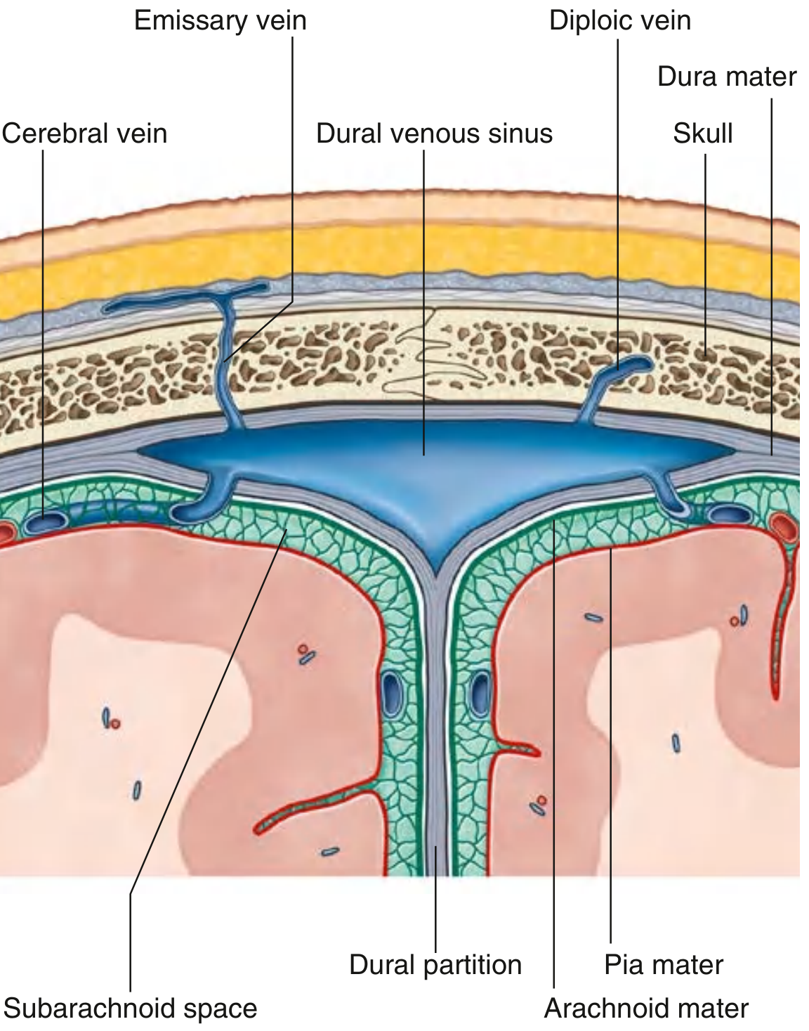
Tributaries (What drains into them)
| Source | Notes |
|---|---|
| Cerebral veins | Superficial veins drain directly; deep veins drain via the great cerebral vein |
| Cerebellar veins | From the superior and inferior surfaces of the cerebellum |
| Brainstem veins | Various small tributaries |
| Diploic veins | Run between the inner and outer tables of the skull vault |
| Emissary veins | Connect extracranial veins to the sinuses - valveless, clinically important as infection conduits |
| Arachnoid granulations | CSF re-enters venous circulation primarily via granulations projecting into the superior sagittal sinus |
Individual Sinuses
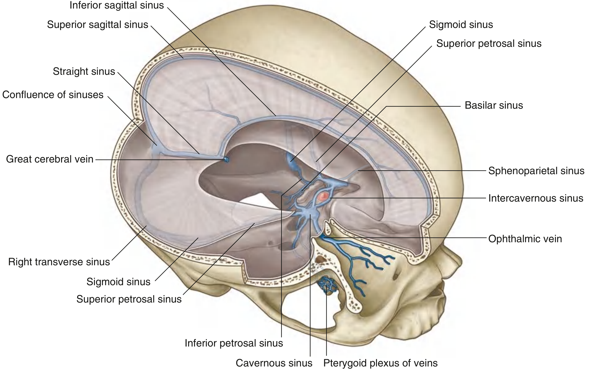
1. Superior Sagittal Sinus
- Runs in the superior border of the falx cerebri
- Begins anteriorly at the foramen cecum (may receive a small emissary vein from the nasal cavity)
- Ends posteriorly at the confluence of sinuses, usually bending right into the right transverse sinus
- Has lateral extensions called lateral lacunae containing arachnoid granulations
- Receives: superior cerebral veins, diploic veins, emissary veins, CSF
2. Inferior Sagittal Sinus
- Runs in the inferior (free) margin of the falx cerebri
- Receives a few cerebral veins and veins from the falx cerebri
- Ends at the anterior edge of the tentorium cerebelli, where it joins the great cerebral vein to form the straight sinus
3. Straight Sinus
- Runs along the junction of the falx cerebri and tentorium cerebelli
- Formed by the union of the inferior sagittal sinus + great cerebral vein (of Galen)
- Ends at the confluence of sinuses, usually bending left into the left transverse sinus
- Receives: posterior cerebral veins, superior cerebellar veins, falx veins
4. Confluence of Sinuses (Torcular Herophili)
- A dilated space at the internal occipital protuberance
- Receives the superior sagittal, straight, and occipital sinuses
- Drains into the right and left transverse sinuses
5. Transverse Sinuses (paired)
- Extend horizontally from the confluence of sinuses along the posterior and lateral attachments of the tentorium cerebelli
- Right transverse sinus usually drains the superior sagittal sinus; left drains the straight sinus
- Also receive the superior petrosal sinus, inferior cerebral and cerebellar veins, diploic and emissary veins
6. Sigmoid Sinuses (paired)
- Continuation of the transverse sinuses, turning inferiorly in an S-shaped course
- Groove the parietal, temporal, and occipital bones
- End at the jugular foramen where they become the internal jugular veins
- Receive cerebral, cerebellar, diploic, and emissary veins
7. Occipital Sinus
- Lies in the falx cerebelli against the occipital bone
- Begins at the foramen magnum (communicates with the marginal sinus and vertebral venous plexus)
- Drains into the confluence of sinuses
8. Cavernous Sinuses (paired) - Clinically Important
- Located on either side of the body of the sphenoid / sella turcica
- Receive blood from: cerebral veins, ophthalmic veins (from orbit), emissary veins (from pterygoid plexus)
- Connected to each other via intercavernous sinuses (anterior and posterior to the pituitary stalk)
- Drain via the superior petrosal sinus (to transverse sinus) and inferior petrosal sinus (to internal jugular vein)
Contents of the cavernous sinus:
- Running through the sinus: internal carotid artery (with sympathetic plexus), abducent nerve [CN VI]
- In the lateral wall (superior to inferior): CN III (oculomotor), CN IV (trochlear), CN V1 (ophthalmic), CN V2 (maxillary)
9. Sphenoparietal Sinuses
- Run along the inferior surface of the lesser wings of the sphenoid
- Drain into the anterior ends of the cavernous sinuses
- Receive diploic and meningeal veins
10. Superior Petrosal Sinuses (paired)
- Drain the cavernous sinuses posterolaterally along the superior margin of the petrous temporal bone
- Connect the cavernous sinus to the transverse/sigmoid sinus junction
- Also receive cerebral and cerebellar veins
11. Inferior Petrosal Sinuses (paired)
- Run posteroinferiorly in a groove between the petrous temporal bone and basal occipital bone
- Drain the cavernous sinuses directly into the internal jugular veins at the jugular foramen
- Receive cerebellar veins, veins of the internal ear and brainstem
12. Basilar Sinuses / Basilar Plexus
- A venous plexus lying on the clivus between the marginal and cavernous sinuses
- Connects the two inferior petrosal sinuses and communicates with the vertebral venous plexus
13. Marginal Sinus
- Encircles the foramen magnum
- Connects the dural sinuses with the vertebral venous plexuses
Final Drainage Pathway
Superior sagittal sinus + Straight sinus + Occipital sinus → Confluence of sinuses → Transverse sinuses → Sigmoid sinuses → Internal jugular veins
Cavernous sinus → (via superior petrosal sinus) → Transverse/sigmoid sinus → Internal jugular vein
- also → (via inferior petrosal sinus) → directly into internal jugular vein
Clinical Highlights
- Emissary veins - valveless, bidirectional; provide a route for extracranial infections (e.g., scalp cellulitis, mastoiditis) to reach the dural sinuses, leading to septic thrombosis
- Cavernous sinus thrombosis - can result from spread of facial infections (via ophthalmic veins); manifests with painful ophthalmoplegia (CN III, IV, VI) and chemosis
- Lateral/sigmoid sinus thrombosis - a known intracranial complication of otitis media/mastoiditis (the sigmoid sinus lies close to the mastoid air cells)
- Subdural hematoma - tearing of bridging cerebral veins as they cross the dura to enter the sinuses
- Superior sagittal sinus thrombosis - presents with headache, seizures, venous infarcts; associated with prothrombotic states, pregnancy, dehydration
- Gray's Anatomy for Students, pp. 1024-1027
- Color Atlas of Human Anatomy Vol. 2 (Internal Organs), pp. 134-135
This is a shared conversation. Sign in to Orris to start your own chat.