
Can you explain right atrium
Reading File
Now let me search the medical library for right atrium content in parallel.
Finding Sources
Finding Sources
Excellent - I have multiple great sources. Let me read the most comprehensive ones in parallel.
Reading File
Reading File
Reading File
I have rich content. Let me also fetch the anatomy diagram and read the physiology section.
Loading Image
Reading File
Reading File
I now have comprehensive information from multiple authoritative textbooks. Let me compile a thorough explanation.
The Right Atrium
The right atrium (RA) is the upper-right chamber of the heart, responsible for receiving deoxygenated blood from the systemic circulation and delivering it to the right ventricle. Here is a detailed breakdown:
Position and External Landmarks
In the anatomical position, the right atrium forms the right border of the heart and contributes to the right portion of the anterior surface. Externally, a shallow vertical groove called the sulcus terminalis cordis runs from the right side of the SVC opening down to the right side of the IVC opening, marking the division between the chamber's two internal compartments. This groove also serves as a macroscopic landmark for the sinoatrial (SA) node.
- Gray's Anatomy for Students, p. 232
Two Internal Compartments
The interior of the right atrium is functionally and embryologically divided into two continuous spaces:
1. Sinus of the Venae Cavae (Smooth-Walled Part)
- Located posteriorly
- Derived embryologically from the right horn of the sinus venosus
- Has smooth, thin walls
- Receives the SVC (superiorly) and IVC (inferiorly)
2. Atrium Proper (Rough-Walled Part) + Right Auricle
- Located anteriorly
- Derived from the embryonic primitive atrium
- Walls are covered by musculi pectinati (pectinate muscles) - ridges of cardiac muscle that fan out like the teeth of a comb
- Continuous anteriorly with the right auricle (an ear-shaped muscular pouch that overlaps the ascending aorta)
The internal boundary between these two spaces is the crista terminalis - a smooth muscular ridge that begins at the roof just anterior to the SVC opening and descends along the lateral wall to the anterior lip of the IVC. The pectinate muscles fan out from this ridge. Importantly, the crista terminalis is a frequent source of atrial tachycardia.
- Color Atlas of Human Anatomy Vol. 2, p. 43
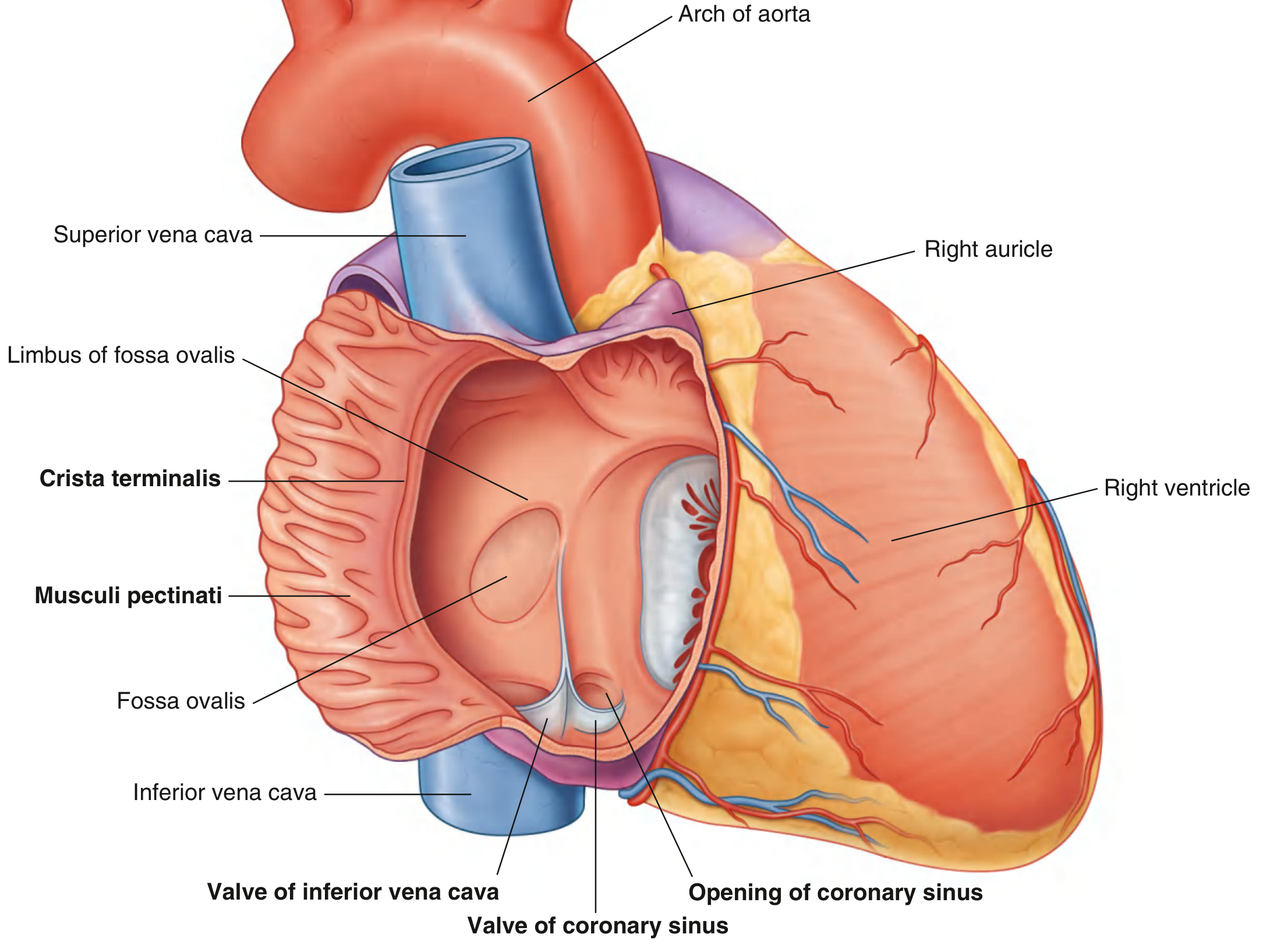
Inflow (Openings into the Right Atrium)
Blood enters the RA through three vessels:
| Vessel | Location | Valve |
|---|---|---|
| Superior vena cava (SVC) | Upper posterior wall, directed downward and anteriorly | No valve |
| Inferior vena cava (IVC) | Lower posterior wall | Valve of the IVC (Eustachian valve) - crescent-shaped; in fetal life, directs blood through the foramen ovale |
| Coronary sinus | Medial to the IVC opening, returns venous blood from the heart walls | Valve of the coronary sinus (Thebesian valve) |
Additionally, foramina of the venae cordis minimae (Thebesian foramina) are scattered along the walls - tiny openings from the smallest cardiac veins draining directly into the chamber.
Interatrial Septum
The RA is separated from the left atrium by the interatrial septum, which faces forward and to the right. A key landmark is the fossa ovalis - an oval depression in the septum, bordered by a prominent muscular rim called the limbus fossae ovalis. This is the remnant of the fetal foramen ovale, through which oxygenated placental blood passed from the RA to the left atrium. After birth, this closes in most individuals, though a patent foramen ovale (PFO) persists in ~25% of adults.
Outflow
From the right atrium, blood passes into the right ventricle through the right atrioventricular (tricuspid) orifice, which faces forward and medially. This opening is guarded by the tricuspid valve, which closes during ventricular systole to prevent backflow.
Physiology - Right Atrial Pressure
The normal right atrial pressure is approximately 0 mmHg (equal to atmospheric pressure). This is also called central venous pressure (CVP) since all systemic veins drain into it.
- Pressure rises to 20-30 mmHg in severe heart failure or after massive transfusion
- Pressure can fall to -3 to -5 mmHg with vigorous cardiac pumping or after severe hemorrhage
Right atrial pressure is regulated by a balance between:
- The heart's ability to pump blood into the pulmonary circulation
- The rate of venous return from peripheral vessels
Factors that raise right atrial pressure include: increased blood volume, increased venous tone, and arteriolar dilation (which accelerates venous return).
- Guyton and Hall Textbook of Medical Physiology
Embryological Origins (Summary)
| Structure | Embryological Origin |
|---|---|
| Sinus of venae cavae | Right horn of sinus venosus |
| Atrium proper + auricle | Embryonic primitive atrium |
| Valve of IVC, valve of coronary sinus | Valve of embryonic sinus venosus |
| Fossa ovalis | Foramen ovale (closes at birth) |
Clinical Significance
- Right atrial enlargement causes the right heart border to become more prominent and rounded on chest X-ray (Goldman-Cecil Medicine)
- Crista terminalis is a trigger zone for atrial tachycardia
- The sulcus terminalis is the external landmark for the SA node - the primary pacemaker of the heart
- The right auricle's thin, trabeculated walls are susceptible to thrombus formation in atrial fibrillation
- On echocardiography (TEE), the RA body is best seen in the four-chamber view; the auricle and caval entrances are best assessed at 90° with the probe rotated rightward (Textbook of Clinical Echocardiography)
This is a shared conversation. Sign in to Orris to start your own chat.