Dorsal slit circumcision in detail

Reading File
Finding Sources
Finding Sources
Searching Images

dorsal slit circumcision procedure technique
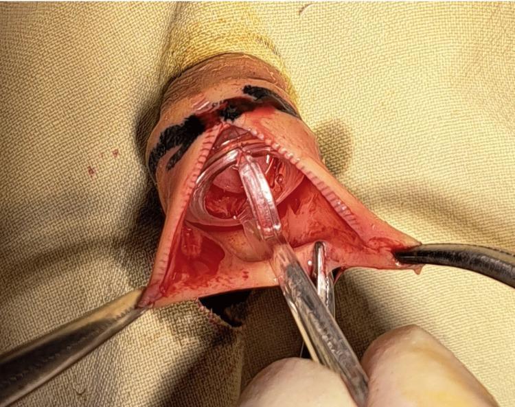
This clinical photograph captures a key intraoperative step of a neonatal circumcision using the Plastibell technique. The image shows the penis prepped and draped for surgery. A dorsal slit has been performed at the 12 o'clock position of the prepuce (foreskin), which is seen incised and retracted laterally. A clear plastic Plastibell device is inserted over the glans penis, with the distal handle protruding through the dorsal slit. Surgical instruments, including hemostats, are visible: one is used to hold and stabilize the edges of the incised foreskin, while another is positioned near the Plastibell ring to facilitate the procedure. The surgical field shows expected mild erythema and a small amount of blood consistent with acute surgical intervention. Black markings are visible on the skin, likely used for pre-surgical measurement and alignment. This image serves as an educational tool for demonstrating surgical landmarks, instrument placement, and device application in pediatric urological procedures.
This clinical photograph captures a key intraoperative step of a neonatal circumcision using the Plastibell technique. The image shows the penis prepped and draped for surgery. A dorsal slit has been performed at the 12 o'clock position of the prepuce (foreskin), which is seen incised and retracted laterally. A clear plastic Plastibell device is inserted over the glans penis, with the distal handle protruding through the dorsal slit. Surgical instruments, including hemostats, are visible: one is used to hold and stabilize the edges of the incised foreskin, while another is positioned near the Plastibell ring to facilitate the procedure. The surgical field shows expected mild erythema and a small amount of blood consistent with acute surgical intervention. Black markings are visible on the skin, likely used for pre-surgical measurement and alignment. This image serves as an educational tool for demonstrating surgical landmarks, instrument placement, and device application in pediatric urological procedures.

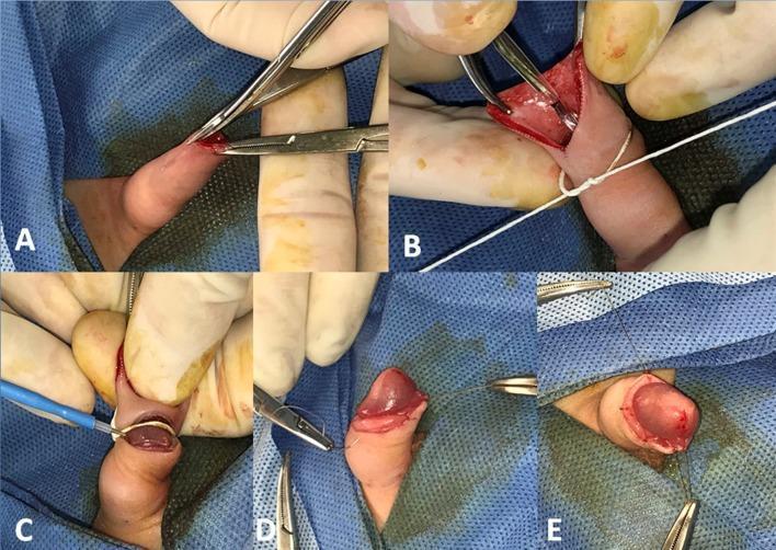
A series of five intraoperative clinical photographs (labelled A-E) documenting the surgical stages of a circumcision procedure using a modified Plastibell technique. Panel A illustrates the creation of a dorsal slit using surgical scissors while the foreskin is stabilized with a straight clamp. Panel B shows the placement of the Plastibell device over the glans with a circumferential ligature tied securely in the groove. Panel C demonstrates the division of the prepuce on the proximal side of the string using a needle-tip Bovie electrocautery tool. Panels D and E show the final reconstructive phase, where the skin-mucosal interface is re-approximated using simple interrupted sutures (chromic gut) to achieve meticulous hemostasis and final closure. The series highlights specific surgical instrumentation including forceps, needle holders, and electrocautery within a sterile field, intended for educational instruction on urological surgical techniques.
A series of five intraoperative clinical photographs (labelled A-E) documenting the surgical stages of a circumcision procedure using a modified Plastibell technique. Panel A illustrates the creation of a dorsal slit using surgical scissors while the foreskin is stabilized with a straight clamp. Panel B shows the placement of the Plastibell device over the glans with a circumferential ligature tied securely in the groove. Panel C demonstrates the division of the prepuce on the proximal side of the string using a needle-tip Bovie electrocautery tool. Panels D and E show the final reconstructive phase, where the skin-mucosal interface is re-approximated using simple interrupted sutures (chromic gut) to achieve meticulous hemostasis and final closure. The series highlights specific surgical instrumentation including forceps, needle holders, and electrocautery within a sterile field, intended for educational instruction on urological surgical techniques.

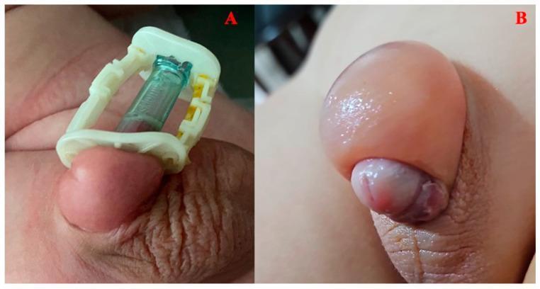
Clinical photograph comparing two different postoperative outcomes following circumcision in pediatric patients. Image A displays the Alisklamp (AK) technique, featuring a plastic disposable circumcision device in situ. The penile tissue shows mild, localized edema with normal skin texture and healthy pink coloration. Image B illustrates a complication following the dorsal slit (DS) technique, showing severe, circumferential penile edema. The prepuce and surrounding shaft tissue are significantly distended, appearing tense, shiny, and semi-translucent. The glans penis is visible but appears partially constricted at the base due to the surrounding swelling, with focal areas of purplish discoloration suggestive of venous congestion. This comparison serves as an educational tool for identifying varying degrees of postoperative edema and assessing the visual safety profile of different surgical circumcision modalities in urological practice.
Clinical photograph comparing two different postoperative outcomes following circumcision in pediatric patients. Image A displays the Alisklamp (AK) technique, featuring a plastic disposable circumcision device in situ. The penile tissue shows mild, localized edema with normal skin texture and healthy pink coloration. Image B illustrates a complication following the dorsal slit (DS) technique, showing severe, circumferential penile edema. The prepuce and surrounding shaft tissue are significantly distended, appearing tense, shiny, and semi-translucent. The glans penis is visible but appears partially constricted at the base due to the surrounding swelling, with focal areas of purplish discoloration suggestive of venous congestion. This comparison serves as an educational tool for identifying varying degrees of postoperative edema and assessing the visual safety profile of different surgical circumcision modalities in urological practice.

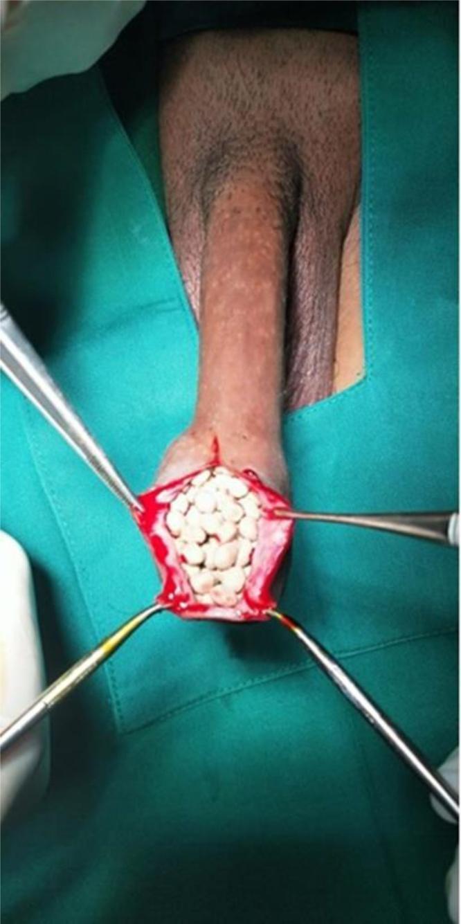
This clinical photograph captures an intraoperative view of a dorsal slit circumcision performed on a patient with chronic phimosis. The image focuses on the distal aspect of the penis, where a surgical dorsal incision has been made in the preputial skin. Four surgical retractors are arranged radially to hold the incision open, revealing a large collection of tightly packed, multiple white calculi (preputial stones) within the preputial cavity. The stones vary in size and shape, exhibiting a smooth, rounded morphology typical of long-term lithiasis. The incised edges of the prepuce appear erythematous, indicative of chronic inflammation or surgical trauma. This visual demonstrates a rare complication of severe phimosis and poor local hygiene, where trapped smegma and urinary salts provide a nidus for preputial stone formation. The image is intended for medical education in urology, illustrating the surgical management of preputial lithiasis and its gross clinical presentation.

This clinical photograph captures an intraoperative view of a dorsal slit circumcision performed on a patient with chronic phimosis. The image focuses on the distal aspect of the penis, where a surgical dorsal incision has been made in the preputial skin. Four surgical retractors are arranged radially to hold the incision open, revealing a large collection of tightly packed, multiple white calculi (preputial stones) within the preputial cavity. The stones vary in size and shape, exhibiting a smooth, rounded morphology typical of long-term lithiasis. The incised edges of the prepuce appear erythematous, indicative of chronic inflammation or surgical trauma. This visual demonstrates a rare complication of severe phimosis and poor local hygiene, where trapped smegma and urinary salts provide a nidus for preputial stone formation. The image is intended for medical education in urology, illustrating the surgical management of preputial lithiasis and its gross clinical presentation.

Reading File
Reading File
Reading File
Reading File
Reading File
Loading Image
I now have comprehensive information from multiple authoritative sources. Let me compile a detailed, well-structured answer.

Dorsal Slit Circumcision - Detailed Overview

Definition and Concept

The dorsal slit is a longitudinal midline incision made through the dorsal (12 o'clock) aspect of the prepuce, extending from its tip proximally toward the coronal sulcus. Unlike full circumcision, the foreskin is not excised - it is merely divided, widening the preputial opening. It can serve as a standalone definitive procedure or as a preliminary step in formal circumcision.
  • Hinman's Atlas of Urologic Surgery (p. 878): "The dorsal slit procedure is useful as a standalone operation for phimosis and paraphimosis or as an adjunct to other operations that require preliminary exposure of the glans. For families and patients who need to resolve phimosis but who do not wish to appear circumcised, the dorsal slit is a useful alternative."

Indications

Clinical SettingRationale
Phimosis (tight foreskin)Widens the preputial ring without full excision
Paraphimosis (irreducible foreskin)Emergency - releases the constricting band when manual reduction fails
Urethral catheterization accessAny situation where the meatus cannot be accessed
As an adjunctPreliminary step in formal circumcision or other penile operations
  • Roberts and Hedges' Clinical Procedures in Emergency Medicine: "Dorsal slit of the foreskin is performed in any emergency situation to gain access to the urethral meatus for urethral catheterization... Elective circumcision rather than dorsal slit of the foreskin is the definitive procedure of choice in nonemergency situations."
  • CPT Code: 54160 (Dorsal slit for phimosis); 54161 (Circumcision, surgical excision other than clamp device, or dorsal slit, other than newborn)
Contraindications: None in the emergency setting. In elective settings, the procedure should not be undertaken in the presence of hypospadias, penile curvature, or buried penis.

Anatomy Relevant to the Procedure

  • The prepuce consists of an outer skin layer (dull in appearance) and an inner mucosal layer (shinier, smoother)
  • The dorsal midline at 12 o'clock is avascular relative to the lateral frenular vessels - making it the preferred site for incision
  • The coronal sulcus is the proximal anatomical limit of the incision
  • Two dorsal penile arteries, two dorsal nerves, and one dorsal penile vein run together along the dorsum - their positions inform the nerve block
  • The urethral meatus must be identified and protected at all times

Anesthesia

Local infiltration (adults/older children):
  • Infiltrate lidocaine WITHOUT epinephrine (epinephrine is contraindicated in penile blocks due to end-artery risk) into the dorsal midline of the foreskin just beneath the superficial fascia
  • Start proximally at the coronal sulcus level and proceed distally to the foreskin tip
  • Consider mixing equal volumes of lidocaine with bupivacaine for prolonged effect
  • Test adequacy by grasping the foreskin with toothed forceps
If local infiltration is insufficient:
  • Perform a dorsal nerve block at the base of the penis (2 injections at 10 and 2 o'clock at the base)
  • Or a ring block at the penile base
Pediatric patients: General or regional anesthesia is typically used

Step-by-Step Surgical Technique

For Phimosis (Elective/Semi-Elective)
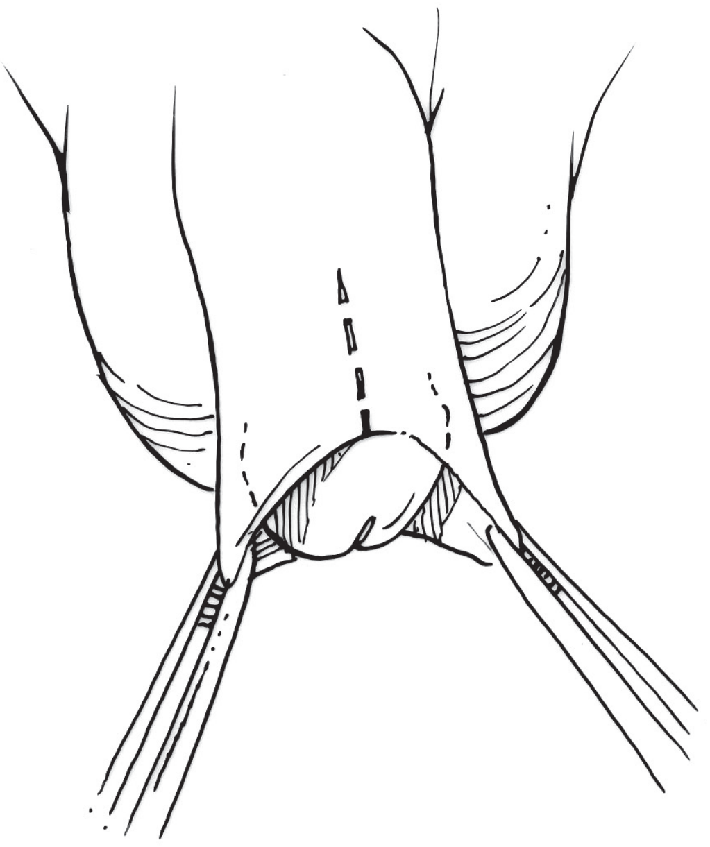
(Based on Hinman's Atlas of Urologic Surgery)
Step 1 - Preparation: Obtain informed consent, induce anesthesia, carefully dilate the preputial opening with a hemostat, and take down any adhesions between the inner foreskin and glans.
Step 2 - Mark the incision: Mark a vertical incision line at the 12 o'clock position extending approximately halfway down to the coronal sulcus (see Fig. 120.9 below).
Dorsal slit incision marking - vertical line on dorsal prepuce at 12 o'clock position
Fig. 120.9 - Hinman's Atlas: Marking of the vertical incision for the dorsal slit (dashed line shows the planned incision line at the dorsal midline of the phimotic prepuce)
Step 3 - Crush-clamp: Pick up the prepuce and apply a straight hemostat along the marked line. Leave the clamp in place for 10 seconds to crush the tissue (reduces bleeding).
Step 4 - Incision: Using sharp scissors or fine needle-point electrocautery, carefully cut along the crushed line. Guard and protect the underlying glans from inadvertent injury at all times.
Step 5 - Hemostasis and closure: Check hemostasis, then close the edges with 5-0 or 6-0 fine absorbable sutures in an interrupted fashion along the cut edges.
Step 6 - Aftercare: Apply bacitracin ointment to suture lines three to four times daily until sutures dissolve. Gently move the foreskin two to three times per day to prevent adhesion formation.

For Paraphimosis (Emergency Technique)
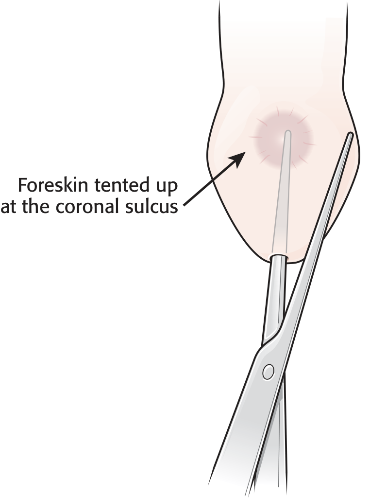
(Based on Hinman's Atlas + Roberts & Hedges)
Step 1 - Orientation: With the patient supine, clean and drape the penis. Use the meatus, scrotal raphe, and umbilicus as midline reference points (paraphimosis edema can distort landmarks).
Step 2 - Adhesion release: After anesthesia, carefully advance both jaws of a straight hemostat proximally between the inner foreskin layer and the glans penis to the level of the coronal sulcus, gently separating any preputial adhesions. Visualize or palpate the meatus at all times.
Step 3 - Single-jaw crush: Open the hemostat, reinsert only one jaw into the developed plane and advance again to the coronal sulcus. Close the hemostat, effectively crushing the interposed foreskin. Leave the hemostat in place for 3-5 minutes.
Step 4 - Incision: Remove the hemostat. Using straight scissors, cut longitudinally along the crushed tissue - this releases the constricting paraphimotic ring. The glans should now be easily reducible.
Step 5 - Managing the cut edges: The incised skin often separates into two layers - the outer foreskin and the inner foreskin. Absorbable chromic or Vicryl sutures (4-0 to 5-0 in children; 3-0 to 4-0 in adults) - running hemostatic sutures placed from the proximal apex of the slit distally, reapproximating the two leaves of foreskin. Standard antibiotic ointment is applied.
Step 6 - The "beagle ear" deformity: After the dorsal slit, the retracted foreskin edges fold laterally and ventrally, creating the characteristic "beagle ear" appearance. The patient and family should be counseled about this cosmetic result. A formal complete circumcision can be performed later once inflammation resolves, if desired.
Dorsal slit incision with foreskin retracted - showing the procedure steps with labeled anatomy including inner and outer foreskin layers and coronal sulcus

Dorsal Slit as Part of Standard Circumcision (Bailey & Love Technique)

(Bailey and Love's Short Practice of Surgery, 28th Ed, p. 1569)
In the standard surgical circumcision technique, the dorsal slit is performed as an integral preliminary step:
  1. The prepuce is held in artery forceps and put on gentle stretch
  2. A circumferential skin incision is made at the level of the corona using a knife
  3. The prepuce is then slit dorsally in the midline to within 1 cm of the corona - converting the foreskin into two flaps
  4. The undersurface of each flap is separated from the glans
  5. The inner layer of each flap is then marked and excised with a second circumferential incision, leaving ~0.5 cm of inner preputial skin
  6. Vessels are secured with bipolar diathermy or absorbable sutures; cut edges are approximated with interrupted sutures, ensuring the frenular vessels are ligated

Alternative Foreskin-Preserving Procedures

When the patient wishes to preserve the foreskin but relieve phimosis, alternatives include:

Triradiate Incisions

  • Multiple slits at 12, 4, and 8 o'clock positions around the foreskin circumference
  • Each cut increases the overall circumference of the preputial opening

Y-V Preputioplasty

  1. Retract the foreskin to identify the constricting band
  2. Make a full-thickness dorsal cut along the midline spanning the phimotic tight area
  3. Extend the cut laterally 1-2 cm to form a Y shape
  4. Advance the resulting triangular flap distally and close with fine absorbable sutures
  • Net result: converts a Y-shaped incision into a V-shaped closure, widening the preputial ring without visible circumcision appearance

Postoperative Considerations

  • Expect swelling and edematous edges for 2-4 weeks
  • The "beagle ear" cosmetic deformity from the laterally folded foreskin is expected and should be counseled upfront
  • In teenagers and adults, consider prescribing amyl nitrate or equivalent to suppress erections during recovery
  • After a dorsal slit for paraphimosis: "Postprocedural conscientious foreskin reduction to its anatomically correct position is encouraged to prevent further paraphimosis"
  • Formal circumcision may be performed after inflammation resolves if the patient desires

Complications

ComplicationNotes
BleedingManaged with hemostatic sutures; frenular vessels are highest risk
InfectionAntibiotic ointment prophylaxis
Beagle ear deformityExpected cosmetic result from lateral folding of foreskin
Penile edemaCan be significant; shown to be more pronounced with dorsal slit vs. clamp devices
Adhesion formationPrevented by regular gentle foreskin mobilization postoperatively
Incomplete releaseMay need extension of incision; rarely, formal circumcision
Glans injuryRisk during hemostat insertion if meatus not carefully guarded
The clinical photo below shows the difference between a device-based circumcision (Alisklamp, mild localized edema) and the dorsal slit technique (circumferential penile edema with venous congestion):
Comparison of postoperative edema - Alisklamp technique (mild) vs dorsal slit technique (severe circumferential edema)

Summary

The dorsal slit is a rapid, straightforward, foreskin-preserving procedure that can be performed under local anesthesia in emergency or office settings. Its main strength is accessibility - it requires no special devices and reliably resolves the emergency of an obstructed meatus or irreducible paraphimosis. Its cosmetic limitation (the beagle-ear deformity) makes it less desirable for elective, non-emergency phimosis where preputioplasty or formal circumcision is preferred.
Sources: Hinman's Atlas of Urologic Surgery (p. 878); Roberts and Hedges' Clinical Procedures in Emergency Medicine; Bailey and Love's Short Practice of Surgery, 28th Ed. (p. 1569); Pfenninger and Fowler's Procedures for Primary Care.
This is a shared conversation. Sign in to Orris to start your own chat.