
Dic Disseminated intravascular congestion
"disseminated intravascular coagulation"[MeSH Terms] AND diagnosis
disseminated intravascular coagulation pathophysiology diagram microthrombi
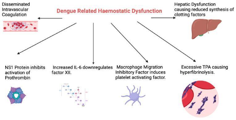
This medical pathophysiology diagram outlines the multifaceted mechanisms of 'Dengue Related Haemostatic Dysfunction'. The central heading branches into six key pathogenic pathways. On the left, a blood vessel illustration demonstrates Disseminated Intravascular Coagulation (DIC) with fibrin-platelet aggregates and trapped red blood cells. On the right, an anatomical liver illustration highlights hepatic dysfunction leading to reduced synthesis of clotting factors. The lower section details molecular and cellular interference: NS1 protein inhibiting prothrombin activation (shown as a hexameric molecular structure); increased IL-6 downregulating factor XII (illustrated via a signaling pathway map); Macrophage Migration Inhibitory Factor inducing platelet-activating factor (represented by an activated immune cell); and excessive Tissue Plasminogen Activator (TPA) causing hyperfibrinolysis, depicted as a microscopic view of fibrin degradation and clot dissolution. This infographic serves as an educational summary of how dengue virus proteins and host inflammatory responses collectively cause coagulopathy and thrombocytopenia.
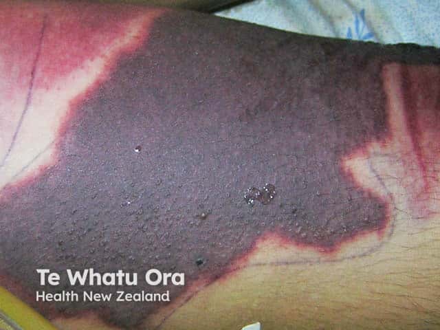
This is a clinical photograph illustrating extensive cutaneous involvement associated with disseminated intravascular coagulation (DIC). Imaging modality: Digital in vivo skin photography. Anatomical location: distal leg region with a large, nonblanching violaceous to dark purple patch showing irregular, geographic margins and a central necrotic area bordered by a pinkish-red rim. The lesion displays nonpalpable purpura with surrounding erythema and edema. Visual cues include mottled purple discoloration, ecchymosis-like zones, and tissue necrosis consistent with microvascular thrombosis and hemorrhage. Pathophysiology of DIC involves systemic activation of coagulation pathways with consumption coagulopathy and formation of dermal microthrombi leading to ischemic necrosis. This image provides a clinical correlate for severe coagulopathy in the setting of sepsis or hemorrhagic shock and aids differential diagnosis against purpura fulminans, meningococcemia, cutaneous vasculitis, necrotizing cellulitis, calciphylaxis, and stasis ulcers. Clinical significance includes urgent coagulation testing (PT, aPTT, platelet count, fibrinogen, D-dimer), blood cultures, sepsis evaluation, and immediate management: treating the underlying cause, hemodynamic support, and appropriate blood product transfusion with careful consideration of anticoagulation per protocol. Educational use: dermatology, hematology, emergency medicine, and pathology training in recognizing DIC cutaneous manifestations. Recognition of these lesions accelerates triage, sepsis workup, and multidisciplinary care.
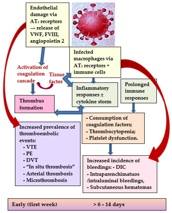
A pathophysiology diagram illustrating the dual pathways leading to thromboembolic and bleeding complications in COVID-19. The schematic begins with a SARS-CoV-2 virion triggering endothelial damage and macrophage infection via angiotensin-II (AT2) receptors. In the 'Early (first week)' phase, endothelial damage leads to the release of von Willebrand factor (VWF), factor VIII (FVIII), and angiopoietin-2, which activate the coagulation cascade and tissue factor, resulting in thrombus formation. This pathway culminates in an increased prevalence of thromboembolic events, including venous thromboembolism (VTE), pulmonary embolism (PE), deep vein thrombosis (DVT), in situ thrombosis, arterial thrombosis, and microthrombosis. Simultaneously, infected macrophages and immune cells trigger an inflammatory response (cytokine storm). In the later phase (>6-14 days), prolonged immune and inflammatory responses lead to the consumption of coagulation factors, thrombocytopenia, and platelet dysfunction. This secondary pathway results in an increased incidence of bleedings, such as disseminated intravascular coagulopathy (DIC), intraparenchymatous/intraluminal bleedings, and subcutaneous hematomas.
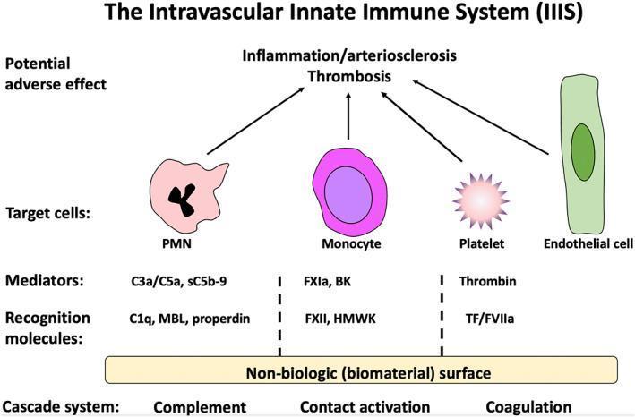
This medical pathophysiology diagram illustrates the Intravascular Innate Immune System (IIIS) response to biomaterial surfaces. The diagram is organized into four horizontal layers: Cascade Systems, Recognition Molecules, Mediators, and Target Cells. At the base, three cascade systems—Complement, Contact Activation, and Coagulation—are initiated upon contact with a 'Non-biologic (biomaterial) surface.' Specific recognition molecules are identified for each: C1q, MBL, and properdin for complement; FXII and HMWK for contact activation; and TF/FVIIa for coagulation. These activate soluble mediators, including anaphylatoxins (C3a/C5a, sC5b-9), FXIa, bradykinin (BK), and thrombin. These mediators target and activate blood and vascular cells: Polymorphonuclear leukocytes (PMN) with multi-lobed nuclei, Monocytes with kidney-shaped nuclei, star-shaped activated Platelets, and elongated Endothelial cells. Arrows indicate the final pathway where cellular and humoral activation converge to cause 'Potential adverse effects,' specifically Inflammation, Arteriosclerosis, and Thrombosis. This visual summarizes the mechanisms of bioincompatibility in extracorporeal therapies like hemodialysis.
Disseminated Intravascular Coagulation (DIC)
Terminology note: The correct term is "Coagulation," not "Congestion." DIC = Disseminated Intravascular Coagulation.
Definition
- Henry's Clinical Diagnosis and Management by Laboratory Methods, p. 954
Pathophysiology
- Trigger - An underlying disease activates the coagulation cascade (via tissue factor or endothelial damage), generating massive thrombin.
- Consumption - Platelets and coagulation factors are consumed, especially fibrinogen, factors V, VIII, and XIII.
- Fibrin deposition - Fibrin is deposited in small vessels of multiple organs, causing microthrombi and end-organ ischemia.
- Secondary fibrinolysis - The fibrinolytic system (via plasmin) lyses the fibrin, releasing fibrin degradation products (FDPs) including D-dimer.
- FDP interference - FDPs impair platelet function and inhibit fibrin polymerization, worsening bleeding.
- Inhibitor depletion - Antithrombin III, protein C, and tissue factor pathway inhibitor (TFPI) are depleted, removing the normal brakes on coagulation.
- Rosen's Emergency Medicine, p. 2477
Causes / Precipitants
| Category | Examples |
|---|---|
| Sepsis | Gram-negative most common; also gram-positive |
| Obstetric | Abruptio placentae, amniotic fluid embolism, HELLP syndrome, retained dead fetus |
| Malignancy | Especially AML (M3 - promyelocytic leukemia), solid tumors |
| Massive tissue injury | Trauma, burns, surgery |
| Vascular | Aortic aneurysm, giant hemangioma (Kasabach-Merritt) |
| Immunologic | Antiphospholipid antibody syndrome, SLE, transfusion reactions |
| Toxins | Envenomation (snake/arthropod) |
| Drugs | Long-term G-CSF therapy |
- Andrews' Diseases of the Skin; Henry's Clinical Diagnosis
Clinical Features
Hemorrhagic manifestations (dominant in acute DIC):
- Widespread petechiae, ecchymoses
- Bleeding from venipuncture/wound/surgical sites
- Mucosal bleeding (GI, GU)
- Hemorrhagic bullae
Thrombotic manifestations:
- Small-vessel obstruction → end-organ ischemia (kidney, brain, lung)
- Acral ischemia, digital gangrene
- Purpura fulminans - symmetric peripheral gangrene (severe)
Skin (present in up to 2/3 of patients):
Laboratory Diagnosis
ISTH Overt DIC Scoring System (score requires an underlying predisposing condition first):
| Test | Finding | Score |
|---|---|---|
| Platelet count | >100 k/μL | 0 |
| <100 k/μL | 1 | |
| <50 k/μL | 2 | |
| Elevated D-dimer | <0.4 μg/mL | 0 |
| 0.4-4.0 μg/mL | 2 | |
| >4.0 μg/mL | 3 | |
| PT prolongation | <3 sec prolonged | 0 |
| 3-6 sec | 1 | |
| >6 sec | 2 | |
| Fibrinogen | >100 mg/dL | 0 |
| <100 mg/dL | 1 |
Full Lab Profile:
| Test | Finding | Why |
|---|---|---|
| Peripheral smear | Thrombocytopenia + schistocytes/helmet cells | RBC fragmentation on fibrin strands (MAHA) |
| Platelet count | Low (<100,000/mm³) | Consumed in clotting |
| PT | Prolonged | Factors II, V consumed |
| PTT | Prolonged | Factors II, V, VIII consumed |
| Thrombin time | Prolonged | Factor II consumed, low fibrinogen |
| Fibrinogen | Low (but may be normal as acute-phase reactant) | Consumed |
| D-dimer / FDPs | Elevated | Secondary fibrinolysis |
| Creatinine / UA | May be abnormal | Renal microvascular fibrin deposition |
- Rosen's Emergency Medicine, Table 111.8, p. 2477
Differential Diagnosis
- Severe liver disease - PT/fibrinogen abnormal but factor VIII normal (not consumed), + jaundice/splenomegaly
- Primary fibrinolysis (rare) - affects fibrinogen/fibrin but preserves platelets, factor V, and factor VIII in low-normal range
- TTP/HUS - thrombocytopenia + MAHA but PT/PTT usually normal, no fibrinogen consumption
Management
Step 1: Treat the underlying cause (ALWAYS first priority)
Step 2: Specific management based on predominant picture
- Platelets - transfuse if <50,000/mm³ and actively bleeding
- Fresh Frozen Plasma (FFP) - correct coagulation factor deficiencies
- Cryoprecipitate - if fibrinogen <100-150 mg/dL (rich in fibrinogen and factor VIII)
- Monitor: slowing of bleeding, rise in platelet/fibrinogen, fall in FDPs
- Heparin (UFH or LMWH) - considered when fibrin deposition/microvascular thrombosis predominates (acral ischemia, purpura fulminans, venous/arterial thromboembolism)
- Heparin is NOT indicated in meningococcemia, abruptio placentae, severe liver disease, or trauma
- LMWH thromboprophylaxis until active bleeding or platelets <30 × 10⁹/L
-
Antithrombin III concentrate
-
Recombinant human soluble thrombomodulin (rTM) - growing evidence, especially in sepsis-DIC
-
Protein C concentrate (benefit in severe sepsis-DIC)
-
Tranexamic acid - considered in hyperfibrinolytic DIC
-
Recombinant factor VIIa, PCC - no proven improved outcome
-
Rosen's Emergency Medicine, p. 2477-2478; Andrews' Diseases of the Skin, p. 955
Acute vs. Chronic DIC
| Feature | Acute DIC | Chronic DIC |
|---|---|---|
| Onset | Explosive | Indolent |
| Predominant picture | Bleeding | Thrombosis |
| Example causes | Sepsis, abruption, trauma | Cancer, retained dead fetus, aortic aneurysm |
| Lab findings | Markedly abnormal | Mildly abnormal or compensated |
Recent Evidence (2025)
Memory Aid
DIC = "Death Is Coming" (mnemonic used in emergency medicine to signal severity)
- Sepsis
- Trauma
- Obstetric complications
- Pancreatitis / Placental abruption
- Malignancy
- Necrosis (massive tissue injury)
- Transfusion reactions