
Neuromuscular junction and myasthenia gravis
I'll start by reading the medical library skill, then search for content on neuromuscular junction and myasthenia gravis in parallel.
Reading File
Now let me search the library and PubMed in parallel:
Finding Sources
Finding Sources
Searching PubMed
"myasthenia gravis"[MeSH Terms] AND treatment
Excellent. Now let me read the most relevant blocks in parallel - Ganong's physiology for NMJ, and Bradley/Daroff's neurology for MG (the most comprehensive neurology textbook):
Reading File
Reading File
Reading File
Good content. Now let me read more of Bradley/Daroff's detailed MG section and the pharmacology section for treatment, plus fetch key images:
Reading File
Loading Image
Loading Image
Let me read the diagnosis and treatment sections of the Bradley/Daroff MG chapter and also get MG antibody content:
Reading File
Finding Sources
Excellent - I now have very rich content. Let me read more of the Bradley/Daroff treatment section:
Reading File
Loading Image
I have excellent content. Let me read the treatment and diagnostic testing sections:
Reading File
I now have comprehensive information. Let me compile the full answer:
Neuromuscular Junction & Myasthenia Gravis
PART 1 - THE NEUROMUSCULAR JUNCTION
Structure
The neuromuscular junction (NMJ) is the specialized synapse where a motor nerve terminates on a skeletal muscle fiber. As the motor axon approaches its target, it loses its myelin sheath and branches into multiple terminal boutons. Each terminal bouton:
- Contains thousands of synaptic vesicles loaded with acetylcholine (ACh)
- Sits within junctional folds (depressions in the motor endplate)
- Is separated from the postsynaptic muscle membrane by the synaptic cleft
The postsynaptic muscle membrane (sarcolemma) is heavily folded, and nicotinic ACh receptors (N_M) are concentrated at the tops of these junctional folds. Acetylcholinesterase (AChE) is anchored at the base of the folds and in the cleft.
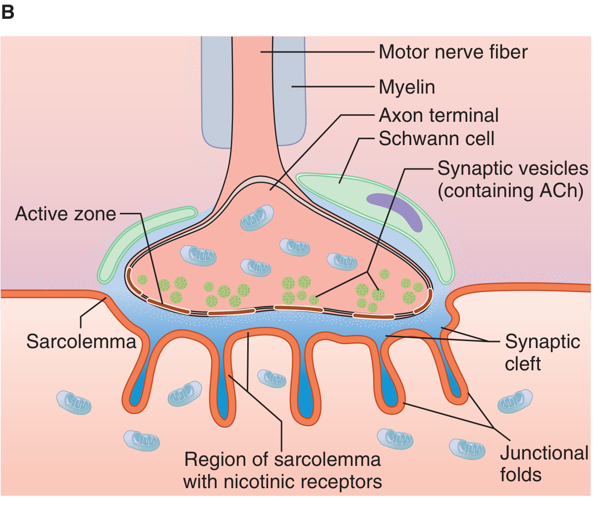
NMJ structure: motor nerve fiber, axon terminal, synaptic vesicles (ACh), synaptic cleft, junctional folds, and nicotinic receptors on the sarcolemma. (Ganong's Review of Medical Physiology, 26e)
Transmission Events (Step by Step)
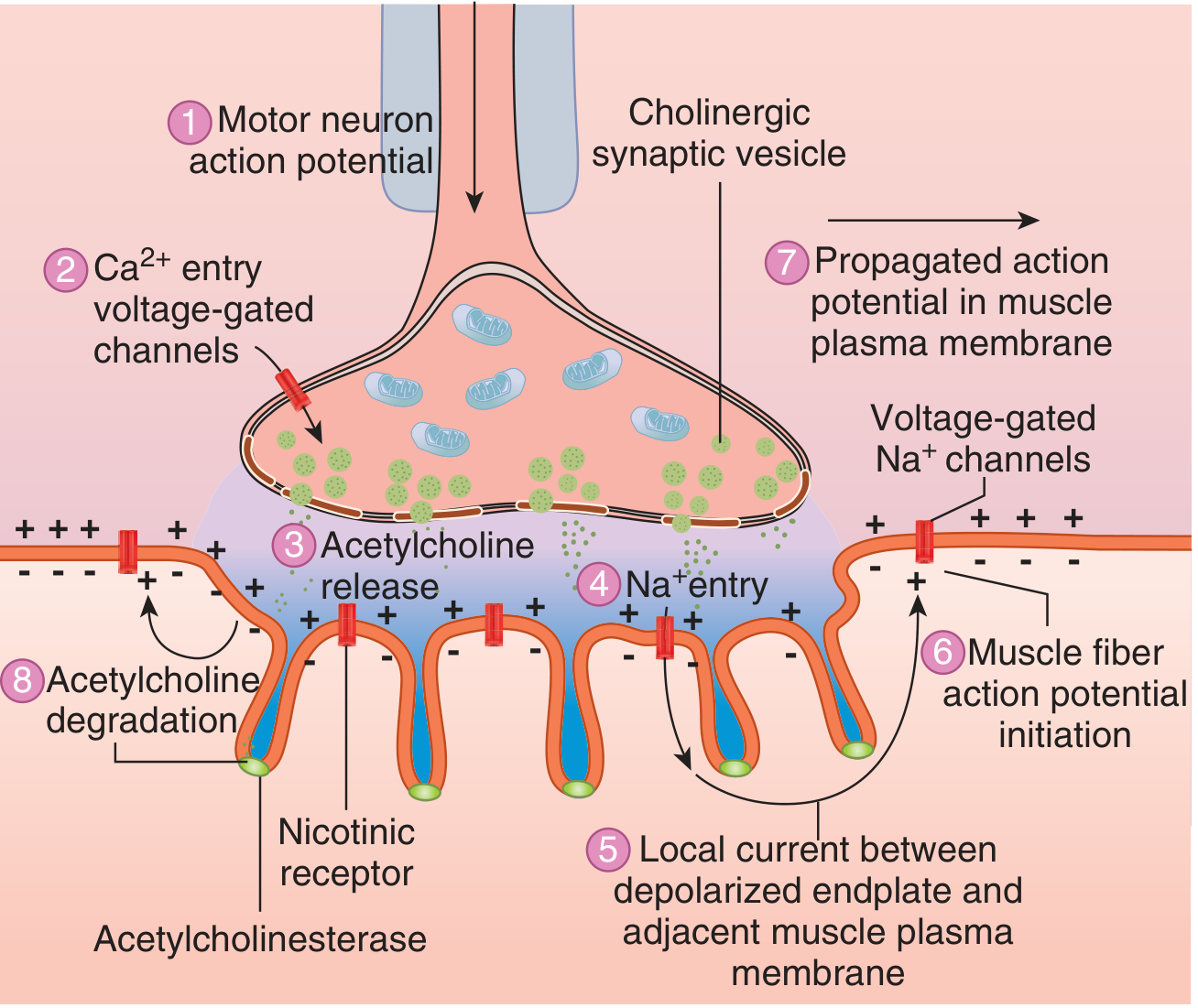
8 sequential events at the NMJ leading to muscle action potential. (Ganong's Review of Medical Physiology, 26e)
| Step | Event |
|---|---|
| 1 | Motor neuron action potential arrives at axon terminal |
| 2 | Voltage-gated Ca²+ channels open; Ca²+ floods into the terminal |
| 3 | Ca²+ triggers exocytosis of ACh-containing synaptic vesicles |
| 4 | ACh diffuses across the synaptic cleft and binds to postsynaptic nicotinic (N_M) receptors |
| 5 | Receptor binding increases Na⁺ and K⁺ conductance; net Na⁺ influx produces the endplate potential (EPP) |
| 6 | The current sink created by the EPP depolarizes adjacent muscle membrane to its firing threshold |
| 7 | Voltage-gated Na⁺ channels trigger a propagated action potential along the sarcolemma |
| 8 | ACh in the cleft is rapidly hydrolyzed by acetylcholinesterase (into choline + acetate); choline is recycled back into the nerve terminal |
- Ganong's Review of Medical Physiology, 26e
- Goodman & Gilman's Pharmacological Basis of Therapeutics
The Nicotinic AChR (N_M)
The muscle-type nicotinic receptor is a pentameric ligand-gated ion channel composed of α₁, α₁, β, δ, and ε (adult) or γ (fetal) subunits. Two ACh molecules must bind (one to each α subunit) to open the channel. This allows Na⁺ influx and K⁺ efflux, generating the EPP. The main immunogenic region (MIR) on the α subunit is the primary target of autoantibodies in MG.
PART 2 - MYASTHENIA GRAVIS
Definition
Acquired myasthenia gravis (MG) is the most common primary disorder of neuromuscular transmission. Autoantibodies - most commonly against the AChR - disrupt normal NMT, resulting in fatigable muscle weakness that worsens with activity and improves with rest. - Bradley and Daroff's Neurology in Clinical Practice
Epidemiology
- Prevalence ~20/100,000 (~60,000 US patients)
- Bimodal age distribution:
- Peak 1: Young women (< 40 years) - associated with thymic hyperplasia
- Peak 2: Older men (> 50 years) - may have thymoma
- Women are affected ~3x more than men before age 40; incidence is roughly equal after age 50
- Overall prevalence is rising (better ascertainment, improved survival, aging population)
- Bradley and Daroff's Neurology in Clinical Practice | Medical Physiology (Boron & Boulpaep)
Pathophysiology
Anti-AChR antibodies (present in ~80-85% of patients) act by three mechanisms:
- Complement-mediated destruction of junctional folds - loss of postsynaptic architecture
- Accelerated internalization and degradation of AChR
- Blocking of ACh-AChR binding (a smaller subset)
The net result is a reduced surface density of functional AChR, smaller endplate potentials, and failure to reach threshold at a significant fraction of NMJs during sustained activity - producing the characteristic fatigable weakness.
The amplitude of miniature endplate potentials (MEPPs) is reduced in MG, though quantal frequency is normal - pointing clearly to a postsynaptic defect. - Medical Physiology (Boron & Boulpaep)
The thymus plays a central role: thymic cells express AChR subunits (especially the α subunit), and aberrant thymic T cell activation initiates or perpetuates the autoimmune response.
Clinical Subtypes and Antibodies
| MG Subtype | Age at Onset | Thymic Histology | Autoantibodies | Notes |
|---|---|---|---|---|
| Ocular | Adult (US/EU); childhood (Asia) | Unknown | AChR (50%) | Weakness restricted to ocular muscles |
| Early Onset | < 50 years | Hyperplasia | AChR | Female > Male (1:3) |
| Late Onset | > 50 years | Normal | AChR, Titin | Male > Female |
| Thymoma | > 40 years | Neoplasia | AChR, Titin, Ryanodine | Anti-titin = severe disease |
| MuSK | < 40 years | Normal | MuSK | Oropharyngeal/facial/respiratory predominance; ChEI may worsen |
| LRP4 | 30-50 years | Unknown | LRP4 | Marked female predominance; IgG4 antibodies |
| Seronegative | Variable | Hyperplasia in some | Low-affinity AChR Abs, Agrin, Cortactin in some | Managed similarly to AChR-MG |
Bradley and Daroff's Neurology in Clinical Practice, Table 108.1
Clinical Presentation
Cardinal feature: Fatigable muscle weakness - worst at end of day or after exertion; improved by rest.
Initial symptoms:
- ~2/3: Ptosis or diplopia (ocular involvement) - the most common presentation
- ~1/6: Dysphagia, dysarthria, or jaw weakness
- ~10%: Limb weakness
- ~10-15%: Remain purely ocular (higher rate in Asian populations/children, up to 58%)
Key ocular signs:
- Ptosis (often asymmetric, shifts from eye to eye - virtually pathognomonic of MG)
- Ophthalmoplegia without pupillary involvement
- "Lid twitch" (Cogan's sign): after downgaze, brief lid overshoot on upgaze
- No pupillary abnormality (distinguishes from CN III palsy)
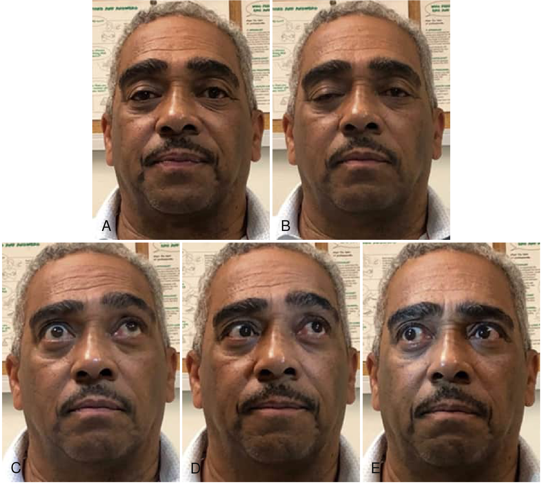
Ocular motility abnormalities in MG: ptosis, ophthalmoplegia, and asymmetric eye movements in various gaze directions. (Bradley and Daroff's Neurology in Clinical Practice)
Other affected muscle groups:
- Bulbar: jaw, facial, palatal, pharyngeal (nasal voice, regurgitation)
- Neck flexors (weaker than extensors)
- Proximal limbs: deltoids, hip flexors
- Respiratory muscles (myasthenic crisis)
Aggravating factors: Systemic illness, emotional stress, heat, thyroid disease, pregnancy, menstrual cycle, certain drugs (aminoglycosides, fluoroquinolones, beta-blockers, Mg²+, curare-type agents)
Diagnosis
1. Antibody testing:
- Anti-AChR binding antibodies: Positive in ~80-85% of generalized MG; ~50% of ocular MG. Virtually confirms the diagnosis when positive (false positives are rare but occur in lupus, autoimmune liver disease, ALS, thymoma without MG).
- Anti-MuSK antibodies: Present in ~50% of AChR-seronegative generalized MG
- Anti-LRP4 antibodies: A subset of double-seronegative patients
- Anti-titin/ryanodine antibodies: Predict thymoma; associated with severe disease
- AChR modulating antibodies: Detect ~10% of AChR binding Ab-negative patients
2. Edrophonium (Tensilon) test: IV edrophonium (short-acting AChEI) produces transient improvement in weakness (especially ptosis). Largely replaced by antibody testing due to false positives/negatives and cardiac risks.
3. Electrodiagnostic testing:
- Repetitive nerve stimulation (RNS): Characteristic decremental response (>10% decrement at 2-3 Hz)
- Single-fiber EMG (SFEMG): Most sensitive test - detects increased "jitter" (variability in NMJ transmission); abnormal in >95% of generalized MG
4. Chest CT/MRI: Mandatory in all MG patients to look for thymoma (found in ~10-15% of MG patients)
Treatment
Treatment is organized into four categories:
A. Symptomatic - Cholinesterase Inhibitors (ChEIs)
- Pyridostigmine (Mestinon): First-line symptomatic therapy; inhibits AChE, prolongs ACh action in the cleft. Dose: 30-60 mg every 4-6 hours. Overdose causes cholinergic crisis (muscarinic: excess secretions, bradycardia, diarrhea; nicotinic: muscle fasciculations, weakness).
- Caution in MuSK-MG: ChEIs may worsen symptoms, with profuse fasciculations.
B. Rapid, Short-Term Immunotherapy (Rescue)
- Plasmapheresis (PLEX): Removes circulating antibodies; rapid onset (days), lasts weeks. Used in myasthenic crisis or pre-operatively.
- IV immunoglobulin (IVIg): Comparable efficacy to PLEX; exact mechanism unclear (Fc receptor blockade, complement inhibition, anti-idiotype antibodies).
C. Long-Term Immunosuppression
- Corticosteroids (Prednisone): Most widely used. Initial transient worsening is common before improvement. Typically started low and escalated.
- Azathioprine: Steroid-sparing agent; onset 6-12 months. Monitor CBC and LFTs.
- Mycophenolate mofetil: Alternative steroid-sparing agent.
- Cyclosporine: Used in refractory cases.
- Rituximab (RTX): Particularly effective in MuSK-MG; recent Cochrane review (2025, PMID: 40607605) supports its use.
D. Novel Targeted Therapies
- FcRn inhibitors (efgartigimod, rozanolixizumab, nipocalimab): Reduce IgG (and hence autoantibody) levels by blocking neonatal Fc receptor-mediated IgG recycling. 2025 meta-analysis (PMID: 40288289) confirms efficacy and safety.
- Complement inhibitors (eculizumab, ravulizumab): Block the complement cascade that destroys junctional folds in AChR-ab MG.
E. Thymectomy
- Indicated for all thymoma patients (removes source of antigenic stimulation and/or neoplasm).
- Benefits patients with AChR-positive generalized MG, especially those with thymic hyperplasia (early-onset).
- Thymectomy leads to clinical improvement in ~75% of patients with thymoma. The MGTX randomized trial confirmed benefit in non-thymomatous AChR-positive generalized MG.
- Role in MuSK-MG is not yet established.
Myasthenic Crisis vs. Cholinergic Crisis
| Feature | Myasthenic Crisis | Cholinergic Crisis |
|---|---|---|
| Cause | Inadequate treatment / trigger | Excess ChEI |
| Muscle weakness | Yes | Yes |
| Pupils | Normal/dilated | Miotic |
| Secretions | Normal | Increased (SLUDGE) |
| Heart rate | Normal/fast | Bradycardia |
| Response to edrophonium | Improves weakness | Worsens weakness |
| Treatment | Intubation + PLEX/IVIg | Stop ChEI; atropine |
Differential Diagnosis
- Lambert-Eaton Myasthenic Syndrome (LEMS): Presynaptic disorder (anti-VGCC antibodies); proximal limb weakness, depressed reflexes that augment with exercise; often paraneoplastic (small cell lung cancer)
- Ocular MG vs. internuclear ophthalmoplegia (MS) - no fatigability in INO
- Botulism: Presynaptic; pupil involvement, descending paralysis
- Congenital myasthenic syndromes: No autoantibodies; present at birth or early childhood
- Miller Fisher syndrome (GBS variant): Ophthalmoplegia + ataxia + areflexia; anti-GQ1b antibodies
Key Points Summary
- The NMJ converts an electrical nerve signal into muscle contraction via Ca²+-triggered ACh release → EPP → muscle action potential
- MG is an autoimmune disease targeting postsynaptic AChR (80-85%), MuSK, or LRP4
- Cardinal feature: fatigable, fluctuating weakness - worse with activity, better with rest
- Hallmark diagnostic features: positive AChR-ab, decremental RNS response, increased SFEMG jitter
- First-line symptomatic treatment: pyridostigmine
- Long-term: corticosteroids ± steroid-sparing immunosuppressants
- Always image the chest for thymoma
- New biologic agents (FcRn inhibitors, complement inhibitors, rituximab) are transforming management of refractory MG
Sources: Ganong's Review of Medical Physiology, 26e | Bradley and Daroff's Neurology in Clinical Practice | Medical Physiology (Boron & Boulpaep) | Goodman & Gilman's Pharmacological Basis of Therapeutics | Recent PubMed: FcRn inhibitor meta-analysis PMID 40288289, Rituximab in MG Cochrane review PMID 40607605
This is a shared conversation. Sign in to Orris to start your own chat.